


Rachael Ott
@rachaelott.bsky.social
Neurobiologist @UofSC 🔬 Super-resolution #microscopy, adipocytes, cytoskeleton, and #neurodegeneration 🦠
Pinned
Rachael Ott
@rachaelott.bsky.social
· Jan 17

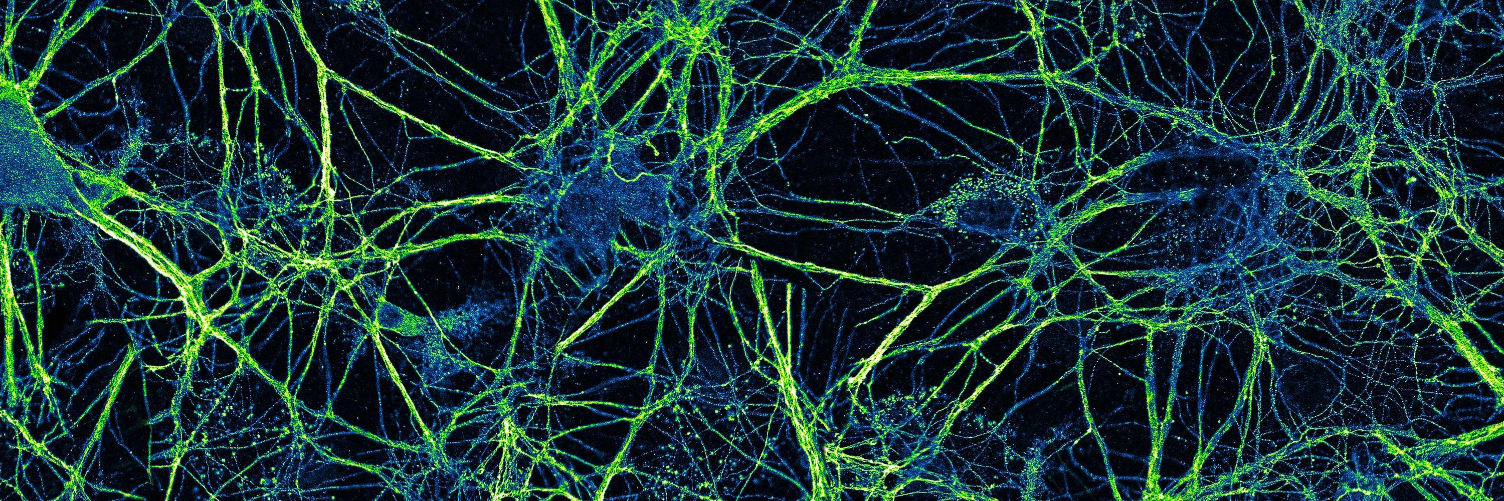
For #FluorescenceFriday, tau (blue) and long loops of neurofilament proteins are shown in a primary hippocampal neuron 🔬 #Neuroscience #Microscopy
Reposted by Rachael Ott
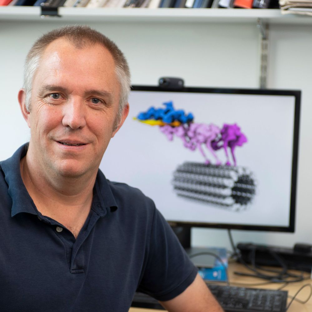
Latest work from the lab by Aude Nommick et al., in which we propose a "size-scaling" model for microtubule force exertion that regulates centrosome centration vs decentration during embryo development!
@ijmonod.bsky.social
www.biorxiv.org/content/10.6...
@ijmonod.bsky.social
www.biorxiv.org/content/10.6...
January 26, 2026 at 3:45 PM
Latest work from the lab by Aude Nommick et al., in which we propose a "size-scaling" model for microtubule force exertion that regulates centrosome centration vs decentration during embryo development!
@ijmonod.bsky.social
www.biorxiv.org/content/10.6...
@ijmonod.bsky.social
www.biorxiv.org/content/10.6...
Reposted by Rachael Ott
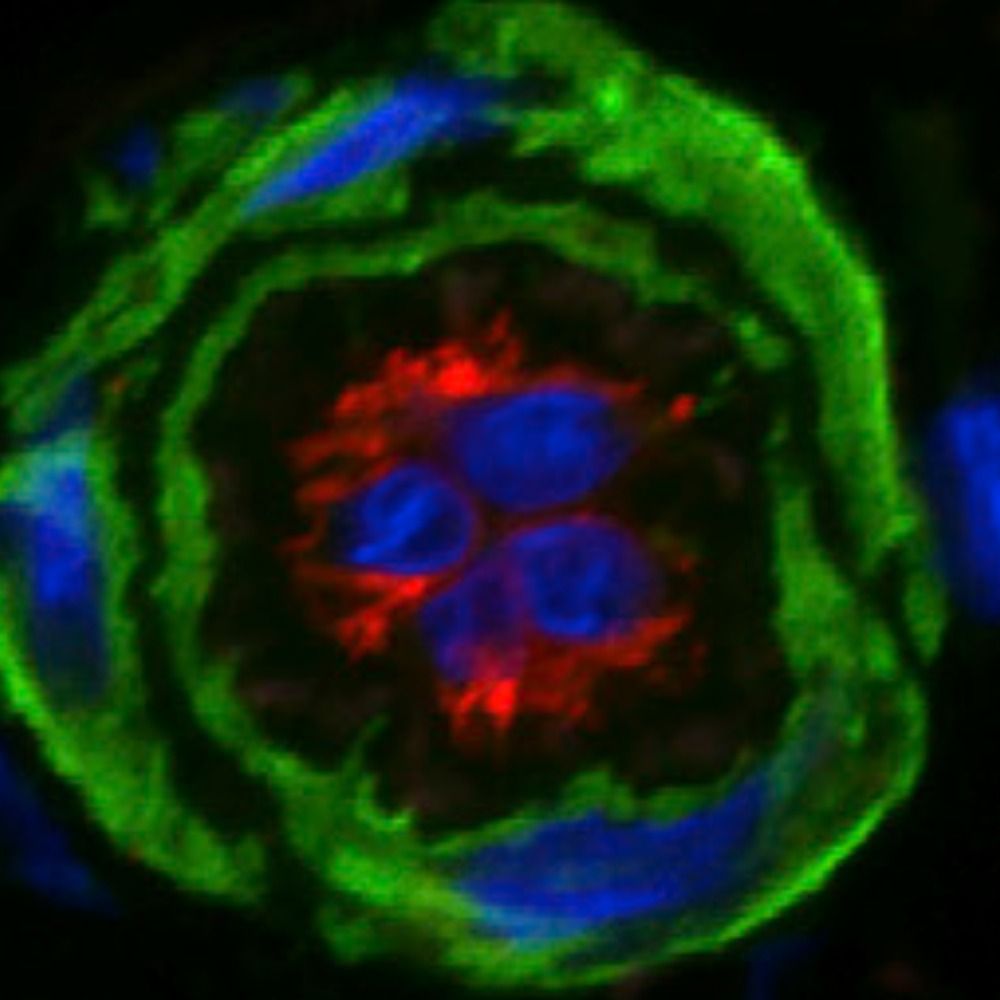
So many tuft cells (yellow/orange) for #TuftTuesday. Immune cells are in green.

January 20, 2026 at 10:03 PM
So many tuft cells (yellow/orange) for #TuftTuesday. Immune cells are in green.
Reposted by Rachael Ott
Beautiful images from @christlet.bsky.social labelling tubulin in neurons #journalclub
www.biorxiv.org/content/10.6...
www.biorxiv.org/content/10.6...

January 19, 2026 at 9:29 AM
Beautiful images from @christlet.bsky.social labelling tubulin in neurons #journalclub
www.biorxiv.org/content/10.6...
www.biorxiv.org/content/10.6...
Reposted by Rachael Ott

A protein called Uninflatable acts as a switch between glial cells growing and glial cells wrapping themselves around axons.
🔗 buff.ly/WRdybO3
🔗 buff.ly/WRdybO3

January 17, 2026 at 11:01 AM
A protein called Uninflatable acts as a switch between glial cells growing and glial cells wrapping themselves around axons.
🔗 buff.ly/WRdybO3
🔗 buff.ly/WRdybO3
Reposted by Rachael Ott
Excited to share Alison's @alisonkickuth.bsky.social paper from the lab out in @nature.com this week! We uncovered how a mechanical ratchet mechanism drives cytokinesis in early #zebrafish embryos. Read more in this thread 🧵 and at www.nature.com/articles/s41... 🤩
@poldresden.bsky.social @mpi-cbg.de
@poldresden.bsky.social @mpi-cbg.de
January 7, 2026 at 4:02 PM
Excited to share Alison's @alisonkickuth.bsky.social paper from the lab out in @nature.com this week! We uncovered how a mechanical ratchet mechanism drives cytokinesis in early #zebrafish embryos. Read more in this thread 🧵 and at www.nature.com/articles/s41... 🤩
@poldresden.bsky.social @mpi-cbg.de
@poldresden.bsky.social @mpi-cbg.de
Reposted by Rachael Ott
Diffusion first, Binding second
Organ-scale uniform immunostaining in 1 DAY🤠, via temporally controlling antibody reaction strength
CuRVE + eFLASH
🐭👤Brain
62 Ab tested
👉deoxycholic acid + pH
👉compatible with transgenic fluorescent proteins
#NatBiotech 2025
www.nature.com/articles/s41...
Organ-scale uniform immunostaining in 1 DAY🤠, via temporally controlling antibody reaction strength
CuRVE + eFLASH
🐭👤Brain
62 Ab tested
👉deoxycholic acid + pH
👉compatible with transgenic fluorescent proteins
#NatBiotech 2025
www.nature.com/articles/s41...

January 3, 2026 at 1:16 PM
Diffusion first, Binding second
Organ-scale uniform immunostaining in 1 DAY🤠, via temporally controlling antibody reaction strength
CuRVE + eFLASH
🐭👤Brain
62 Ab tested
👉deoxycholic acid + pH
👉compatible with transgenic fluorescent proteins
#NatBiotech 2025
www.nature.com/articles/s41...
Organ-scale uniform immunostaining in 1 DAY🤠, via temporally controlling antibody reaction strength
CuRVE + eFLASH
🐭👤Brain
62 Ab tested
👉deoxycholic acid + pH
👉compatible with transgenic fluorescent proteins
#NatBiotech 2025
www.nature.com/articles/s41...
Reposted by Rachael Ott
New paper out!
We show how mechanosensitive adherens junction proteins link actomyosin contractility to actin assembly using in vitro reconstitution.
Huge congrats to Aurélie Favarin, Rayan Said, & all authors!
In Science Advances: www.science.org/doi/10.1126/...
#actin #myosin #mechanobiology
We show how mechanosensitive adherens junction proteins link actomyosin contractility to actin assembly using in vitro reconstitution.
Huge congrats to Aurélie Favarin, Rayan Said, & all authors!
In Science Advances: www.science.org/doi/10.1126/...
#actin #myosin #mechanobiology

January 2, 2026 at 4:00 PM
New paper out!
We show how mechanosensitive adherens junction proteins link actomyosin contractility to actin assembly using in vitro reconstitution.
Huge congrats to Aurélie Favarin, Rayan Said, & all authors!
In Science Advances: www.science.org/doi/10.1126/...
#actin #myosin #mechanobiology
We show how mechanosensitive adherens junction proteins link actomyosin contractility to actin assembly using in vitro reconstitution.
Huge congrats to Aurélie Favarin, Rayan Said, & all authors!
In Science Advances: www.science.org/doi/10.1126/...
#actin #myosin #mechanobiology
Reposted by Rachael Ott
Hoping everyone's new year fluorescence is clear and bright. Happy #FluorescenceFriday.

January 3, 2026 at 4:52 AM
Hoping everyone's new year fluorescence is clear and bright. Happy #FluorescenceFriday.
Reposted by Rachael Ott
New Perspective from myself, Sarah Heilbronner and @myoo.bsky.social . “Rethinking the centrality of brain areas in understanding functional organization” in Nature Neuroscience. 🧵
rdcu.be/eVZ1A
rdcu.be/eVZ1A

Rethinking the centrality of brain areas in understanding functional organization
Nature Neuroscience - Parcellation of the cortex into functionally modular brain areas is foundational to neuroscience. Here, Hayden, Heilbronner and Yoo question the central status of brain areas...
rdcu.be
December 23, 2025 at 1:02 PM
New Perspective from myself, Sarah Heilbronner and @myoo.bsky.social . “Rethinking the centrality of brain areas in understanding functional organization” in Nature Neuroscience. 🧵
rdcu.be/eVZ1A
rdcu.be/eVZ1A
Reposted by Rachael Ott
I don't always get pretty, isolated, AND transfected primary neurons in culture, but when I do I take advantage.... Rat hippocampal neuron overexpressing ThymosinB4-mScarlet and imaged for 16hr on a @zeiss-microscopy.bsky.social LSM880 with Airyscan. #FluorescenceFriday #Microscopy
December 19, 2025 at 2:01 PM
I don't always get pretty, isolated, AND transfected primary neurons in culture, but when I do I take advantage.... Rat hippocampal neuron overexpressing ThymosinB4-mScarlet and imaged for 16hr on a @zeiss-microscopy.bsky.social LSM880 with Airyscan. #FluorescenceFriday #Microscopy
Reposted by Rachael Ott
Felt a little festive at the microscope this morning for #FluorescenceFriday 🎄
Here’s the nervous system of a juvenile sea star ⭐️
Green = acetylated tubulin, red = nuclei
Happy holidays!
Here’s the nervous system of a juvenile sea star ⭐️
Green = acetylated tubulin, red = nuclei
Happy holidays!

December 19, 2025 at 5:32 PM
Felt a little festive at the microscope this morning for #FluorescenceFriday 🎄
Here’s the nervous system of a juvenile sea star ⭐️
Green = acetylated tubulin, red = nuclei
Happy holidays!
Here’s the nervous system of a juvenile sea star ⭐️
Green = acetylated tubulin, red = nuclei
Happy holidays!
Reposted by Rachael Ott
✨ Blinking #nanobodies that work for single-molecule localization 🔬
Our new preprint shows that the self-blinking dye JF635b restores robust, buffer-free blinking in #nanobodies, enabling reliable #dSTORM, #MINFLUX, and more, without chemical-switching buffers. Opening new possibilities for #ExM!
Our new preprint shows that the self-blinking dye JF635b restores robust, buffer-free blinking in #nanobodies, enabling reliable #dSTORM, #MINFLUX, and more, without chemical-switching buffers. Opening new possibilities for #ExM!
December 22, 2025 at 10:45 AM
✨ Blinking #nanobodies that work for single-molecule localization 🔬
Our new preprint shows that the self-blinking dye JF635b restores robust, buffer-free blinking in #nanobodies, enabling reliable #dSTORM, #MINFLUX, and more, without chemical-switching buffers. Opening new possibilities for #ExM!
Our new preprint shows that the self-blinking dye JF635b restores robust, buffer-free blinking in #nanobodies, enabling reliable #dSTORM, #MINFLUX, and more, without chemical-switching buffers. Opening new possibilities for #ExM!
Reposted by Rachael Ott
Network of somatosensory axons and immune cells in the adult zebrafish skin. Credit to @errricpeterman.bsky.social. #ZebrafishZunday 🧪

December 7, 2025 at 11:49 AM
Network of somatosensory axons and immune cells in the adult zebrafish skin. Credit to @errricpeterman.bsky.social. #ZebrafishZunday 🧪
Reposted by Rachael Ott

I am presenting at the Science and Art Minisymposium at #CellBio2025 today! The line up is great this year. I am hands down the least qualified person presenting. It is a weird feeling but I am excited! Here is my title slide.
Room 122 4pm
Room 122 4pm

December 7, 2025 at 4:49 PM
I am presenting at the Science and Art Minisymposium at #CellBio2025 today! The line up is great this year. I am hands down the least qualified person presenting. It is a weird feeling but I am excited! Here is my title slide.
Room 122 4pm
Room 122 4pm
Reposted by Rachael Ott
since you made it all the way to then end - we've already made transgenic lines with the mSG from Ando et al - it's amazing! here's a movie from our Cell Observatory project at the Advanced BioImaging Center 7/7
December 5, 2025 at 9:20 PM
since you made it all the way to then end - we've already made transgenic lines with the mSG from Ando et al - it's amazing! here's a movie from our Cell Observatory project at the Advanced BioImaging Center 7/7
Reposted by Rachael Ott
Our Science paper is out!
Huge congratulations to @huabin-zhou.bsky.social, Mike Rosen, and the brilliant @janhuemar.bsky.social @juliamaristany.bsky.social and @kieran-russell.bsky.social from our group
News: bit.ly/4avnkAr and bit.ly/3XBGVHS
Great perspective by @vram142.bsky.social +K Zhang
Huge congratulations to @huabin-zhou.bsky.social, Mike Rosen, and the brilliant @janhuemar.bsky.social @juliamaristany.bsky.social and @kieran-russell.bsky.social from our group
News: bit.ly/4avnkAr and bit.ly/3XBGVHS
Great perspective by @vram142.bsky.social +K Zhang
December 5, 2025 at 9:47 AM
Our Science paper is out!
Huge congratulations to @huabin-zhou.bsky.social, Mike Rosen, and the brilliant @janhuemar.bsky.social @juliamaristany.bsky.social and @kieran-russell.bsky.social from our group
News: bit.ly/4avnkAr and bit.ly/3XBGVHS
Great perspective by @vram142.bsky.social +K Zhang
Huge congratulations to @huabin-zhou.bsky.social, Mike Rosen, and the brilliant @janhuemar.bsky.social @juliamaristany.bsky.social and @kieran-russell.bsky.social from our group
News: bit.ly/4avnkAr and bit.ly/3XBGVHS
Great perspective by @vram142.bsky.social +K Zhang
Reposted by Rachael Ott

For #FluorescenceFriday - RNAscope marking subfields of the hippocampus in a section of postmortem human 🧠 tissue. Image credit to the Imaging Development team @lieberinstitute.bsky.social 🔬🧪👩🔬

December 5, 2025 at 2:01 PM
For #FluorescenceFriday - RNAscope marking subfields of the hippocampus in a section of postmortem human 🧠 tissue. Image credit to the Imaging Development team @lieberinstitute.bsky.social 🔬🧪👩🔬
Reposted by Rachael Ott
This distal gill tip is giving full branchial art vibes for #FluorescenceFriday 🦓🐟
— who knew oxygen exchange could be this pretty.
🔴 kdrl:mCherry highlights the vasculature
⚪ fgf10b:nEOS marks the pillar cells
#Zebrafish #DevBio #SciArt
— who knew oxygen exchange could be this pretty.
🔴 kdrl:mCherry highlights the vasculature
⚪ fgf10b:nEOS marks the pillar cells
#Zebrafish #DevBio #SciArt

December 5, 2025 at 1:39 PM
This distal gill tip is giving full branchial art vibes for #FluorescenceFriday 🦓🐟
— who knew oxygen exchange could be this pretty.
🔴 kdrl:mCherry highlights the vasculature
⚪ fgf10b:nEOS marks the pillar cells
#Zebrafish #DevBio #SciArt
— who knew oxygen exchange could be this pretty.
🔴 kdrl:mCherry highlights the vasculature
⚪ fgf10b:nEOS marks the pillar cells
#Zebrafish #DevBio #SciArt
Reposted by Rachael Ott

It's #FluorescenceFriday, and this is a reminder that I'm coordinating a collection on the Brain-Immune-Vascular interface in Dementia for @natureportfolio.nature.com, and we have several APC (Article Processing Charge) waivers available! Check out more here:
www.nature.com/collections/...
www.nature.com/collections/...

December 5, 2025 at 5:11 PM
It's #FluorescenceFriday, and this is a reminder that I'm coordinating a collection on the Brain-Immune-Vascular interface in Dementia for @natureportfolio.nature.com, and we have several APC (Article Processing Charge) waivers available! Check out more here:
www.nature.com/collections/...
www.nature.com/collections/...
Reposted by Rachael Ott
New preprint! Do you like ocean waves? We found similar waves on bacterial colonies! We found that this collective behavior, known as rippling, is nothing but surface waves on an active nematic. @princeton.edu @mpipks.bsky.social @ub.edu @icreacommunity.bsky.social
www.biorxiv.org/content/10.1...
www.biorxiv.org/content/10.1...
December 2, 2025 at 8:02 PM
New preprint! Do you like ocean waves? We found similar waves on bacterial colonies! We found that this collective behavior, known as rippling, is nothing but surface waves on an active nematic. @princeton.edu @mpipks.bsky.social @ub.edu @icreacommunity.bsky.social
www.biorxiv.org/content/10.1...
www.biorxiv.org/content/10.1...
Reposted by Rachael Ott
Exciting new #Zebrafish research from the #WeinsteinLab, led by Jong Park!
“Specialized gas-exchange endothelium of the zebrafish gill” —
www.biorxiv.org/content/10.6...
Amazing to see red blood cells moving through the gills! Don’t forget to check out the supplemental movies ;-)
“Specialized gas-exchange endothelium of the zebrafish gill” —
www.biorxiv.org/content/10.6...
Amazing to see red blood cells moving through the gills! Don’t forget to check out the supplemental movies ;-)
December 2, 2025 at 10:48 PM
Exciting new #Zebrafish research from the #WeinsteinLab, led by Jong Park!
“Specialized gas-exchange endothelium of the zebrafish gill” —
www.biorxiv.org/content/10.6...
Amazing to see red blood cells moving through the gills! Don’t forget to check out the supplemental movies ;-)
“Specialized gas-exchange endothelium of the zebrafish gill” —
www.biorxiv.org/content/10.6...
Amazing to see red blood cells moving through the gills! Don’t forget to check out the supplemental movies ;-)
Reposted by Rachael Ott

New must-read review article for all lovers of #centrioles and #centrosomes 😍👇

November 28, 2025 at 3:01 PM
New must-read review article for all lovers of #centrioles and #centrosomes 😍👇
Reposted by Rachael Ott
One-day-old transgenic zebrafish embryo. Credit to Dr. Kazuhide Shaun Okuda. #ZebrafishZunday 🧪

November 30, 2025 at 7:04 PM
One-day-old transgenic zebrafish embryo. Credit to Dr. Kazuhide Shaun Okuda. #ZebrafishZunday 🧪
Reposted by Rachael Ott

Cells can form patterns within themselves just like embryos do. How? Connie Yan's new preprint shows how the anterior-posterior cytoskeleton pattern in Stentor is dictated by regionalized scaffolding proteins
www.biorxiv.org/content/10.1...
www.biorxiv.org/content/10.1...

August 19, 2025 at 8:42 AM
Cells can form patterns within themselves just like embryos do. How? Connie Yan's new preprint shows how the anterior-posterior cytoskeleton pattern in Stentor is dictated by regionalized scaffolding proteins
www.biorxiv.org/content/10.1...
www.biorxiv.org/content/10.1...