


Kevin Terretaz
@kwolbachia.bsky.social
Cells, microscopy, colorful LUTs and ImageJ
https://github.com/kwolbachia
For work I study Wolbachia symbiosis in Montpellier
https://github.com/kwolbachia
For work I study Wolbachia symbiosis in Montpellier
Pinned

My new ImageJ / Fiji toolkit is out 🔥! The goal is to make image handling & visualization easy, with an intuitive interface! Install it on Fiji with the "Image Viewer" update site
#microscopy #ImageJ #FluorescenceFriday #microscopyMonday
imagej.net/plugins/imag...
#microscopy #ImageJ #FluorescenceFriday #microscopyMonday
imagej.net/plugins/imag...
I updated the Image Viewer and the wiki page with many small bug fixes and features! If you haven't tried it yet, now it's even better ;)
#microscopy #ImageJ #FluorescenceFriday #microscopyMonday
#microscopy #ImageJ #FluorescenceFriday #microscopyMonday
My new ImageJ / Fiji toolkit is out 🔥! The goal is to make image handling & visualization easy, with an intuitive interface! Install it on Fiji with the "Image Viewer" update site
#microscopy #ImageJ #FluorescenceFriday #microscopyMonday
imagej.net/plugins/imag...
#microscopy #ImageJ #FluorescenceFriday #microscopyMonday
imagej.net/plugins/imag...
November 16, 2025 at 10:03 PM
I updated the Image Viewer and the wiki page with many small bug fixes and features! If you haven't tried it yet, now it's even better ;)
#microscopy #ImageJ #FluorescenceFriday #microscopyMonday
#microscopy #ImageJ #FluorescenceFriday #microscopyMonday
Reposted by Kevin Terretaz

This week's #FluorescenceFriday, we'll be treated with one of the classical models of #devbio, the neural crest cells. Here is a beautiful video of neural crest cells with GFP-tagged focal adhesion kinase (🔵) and LifeAct-RFP (🟣) migrating on a Fibronectin matrix.
📹: Adam Shellard
📹: Adam Shellard
November 14, 2025 at 5:45 PM
This week's #FluorescenceFriday, we'll be treated with one of the classical models of #devbio, the neural crest cells. Here is a beautiful video of neural crest cells with GFP-tagged focal adhesion kinase (🔵) and LifeAct-RFP (🟣) migrating on a Fibronectin matrix.
📹: Adam Shellard
📹: Adam Shellard
Reposted by Kevin Terretaz

I guess it never gets old...bc we love #CellMigration
ATP has many roles: energy currency, hydrotrope, and *danger signal*
Immune cells are faster after ATP exposure, F-actin (cyan) goes to the rear for nitro boost!
www.science.org/doi/full/10....
@focalplane.bsky.social #fluorescencefriday 🧪🔬
ATP has many roles: energy currency, hydrotrope, and *danger signal*
Immune cells are faster after ATP exposure, F-actin (cyan) goes to the rear for nitro boost!
www.science.org/doi/full/10....
@focalplane.bsky.social #fluorescencefriday 🧪🔬
November 14, 2025 at 8:38 PM
I guess it never gets old...bc we love #CellMigration
ATP has many roles: energy currency, hydrotrope, and *danger signal*
Immune cells are faster after ATP exposure, F-actin (cyan) goes to the rear for nitro boost!
www.science.org/doi/full/10....
@focalplane.bsky.social #fluorescencefriday 🧪🔬
ATP has many roles: energy currency, hydrotrope, and *danger signal*
Immune cells are faster after ATP exposure, F-actin (cyan) goes to the rear for nitro boost!
www.science.org/doi/full/10....
@focalplane.bsky.social #fluorescencefriday 🧪🔬
Reposted by Kevin Terretaz
🔬🍀
A timelapse movie (unpublished data) from the lab.
In the movie, the nucleus is moving towards the base of the polarized root hair cell.
A timelapse movie (unpublished data) from the lab.
In the movie, the nucleus is moving towards the base of the polarized root hair cell.
November 11, 2025 at 6:01 PM
🔬🍀
A timelapse movie (unpublished data) from the lab.
In the movie, the nucleus is moving towards the base of the polarized root hair cell.
A timelapse movie (unpublished data) from the lab.
In the movie, the nucleus is moving towards the base of the polarized root hair cell.
Reposted by Kevin Terretaz

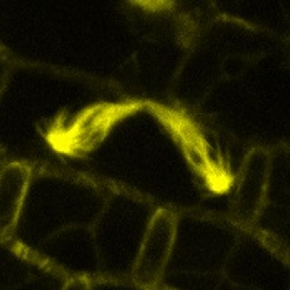
During my postdoc, I looked at hundreds of images like this 🤩
These are two apical caulonemal cells from moss Physcomitrium patens stained with MDY64 (shown in shades of orange). The natural autofluorescence of chlorophyll is in cyan.
#microscopymonday #moss #plantcells #plantmicroscopy
These are two apical caulonemal cells from moss Physcomitrium patens stained with MDY64 (shown in shades of orange). The natural autofluorescence of chlorophyll is in cyan.
#microscopymonday #moss #plantcells #plantmicroscopy

November 10, 2025 at 3:33 PM
During my postdoc, I looked at hundreds of images like this 🤩
These are two apical caulonemal cells from moss Physcomitrium patens stained with MDY64 (shown in shades of orange). The natural autofluorescence of chlorophyll is in cyan.
#microscopymonday #moss #plantcells #plantmicroscopy
These are two apical caulonemal cells from moss Physcomitrium patens stained with MDY64 (shown in shades of orange). The natural autofluorescence of chlorophyll is in cyan.
#microscopymonday #moss #plantcells #plantmicroscopy
Reposted by Kevin Terretaz

This image by MDI Bio Lab's Travis Carney is a #drosophila larval brain. Neural stem cells and neurons are marked, including axons that project into the brain. The flare in the center of each lobe is part of a learning and memory center in flies.
ZEISS Microscopy #microscopymonday 🧪 🤝
ZEISS Microscopy #microscopymonday 🧪 🤝

November 10, 2025 at 3:01 PM
This image by MDI Bio Lab's Travis Carney is a #drosophila larval brain. Neural stem cells and neurons are marked, including axons that project into the brain. The flare in the center of each lobe is part of a learning and memory center in flies.
ZEISS Microscopy #microscopymonday 🧪 🤝
ZEISS Microscopy #microscopymonday 🧪 🤝
Reposted by Kevin Terretaz

Happy #FluorescenceFriday from these primary astrocytes grown on glass slides and robotically stained on the Biocare Oncore Pro X! 🧠🔬🧪
Cyan = GFAP
Royal Purple = ALDH1L1
Salmon = Nuclear Counterstain
Cyan = GFAP
Royal Purple = ALDH1L1
Salmon = Nuclear Counterstain

November 7, 2025 at 7:26 PM
Happy #FluorescenceFriday from these primary astrocytes grown on glass slides and robotically stained on the Biocare Oncore Pro X! 🧠🔬🧪
Cyan = GFAP
Royal Purple = ALDH1L1
Salmon = Nuclear Counterstain
Cyan = GFAP
Royal Purple = ALDH1L1
Salmon = Nuclear Counterstain
Reposted by Kevin Terretaz
My new ImageJ / Fiji toolkit is out 🔥! The goal is to make image handling & visualization easy, with an intuitive interface! Install it on Fiji with the "Image Viewer" update site
#microscopy #ImageJ #FluorescenceFriday #microscopyMonday
imagej.net/plugins/imag...
#microscopy #ImageJ #FluorescenceFriday #microscopyMonday
imagej.net/plugins/imag...
November 4, 2025 at 10:40 PM
My new ImageJ / Fiji toolkit is out 🔥! The goal is to make image handling & visualization easy, with an intuitive interface! Install it on Fiji with the "Image Viewer" update site
#microscopy #ImageJ #FluorescenceFriday #microscopyMonday
imagej.net/plugins/imag...
#microscopy #ImageJ #FluorescenceFriday #microscopyMonday
imagej.net/plugins/imag...
Reposted by Kevin Terretaz

Two neurons went into a bar...

November 6, 2025 at 11:52 AM
Two neurons went into a bar...
Reposted by Kevin Terretaz

HL60 cells take on so many fun shapes as they migrate! This #InsightFromImaging data features cells prepared by Leanna and imaged on the @aicjanelia.bsky.social LLSM by @cmhobson.bsky.social
November 4, 2025 at 10:36 PM
HL60 cells take on so many fun shapes as they migrate! This #InsightFromImaging data features cells prepared by Leanna and imaged on the @aicjanelia.bsky.social LLSM by @cmhobson.bsky.social
My new ImageJ / Fiji toolkit is out 🔥! The goal is to make image handling & visualization easy, with an intuitive interface! Install it on Fiji with the "Image Viewer" update site
#microscopy #ImageJ #FluorescenceFriday #microscopyMonday
imagej.net/plugins/imag...
#microscopy #ImageJ #FluorescenceFriday #microscopyMonday
imagej.net/plugins/imag...
November 4, 2025 at 10:40 PM
My new ImageJ / Fiji toolkit is out 🔥! The goal is to make image handling & visualization easy, with an intuitive interface! Install it on Fiji with the "Image Viewer" update site
#microscopy #ImageJ #FluorescenceFriday #microscopyMonday
imagej.net/plugins/imag...
#microscopy #ImageJ #FluorescenceFriday #microscopyMonday
imagej.net/plugins/imag...
Reposted by Kevin Terretaz

Congratulations @centriolelab.bsky.social @dudinlab.bsky.social and @gautamdey.bsky.social for your publication in @cp-cell.bsky.social: Charting the landscape of #cytoskeletal diversity in #microbial eukaryotes #Expansion #Microscopy @sciencesunige.bsky.social
www.unige.ch/medias/en/20...
Congratulations @centriolelab.bsky.social @dudinlab.bsky.social and @gautamdey.bsky.social for your publication in @cp-cell.bsky.social: Charting the landscape of #cytoskeletal diversity in #microbial eukaryotes #Expansion #Microscopy @sciencesunige.bsky.social
www.unige.ch/medias/en/20...

November 3, 2025 at 1:34 PM
Congratulations @centriolelab.bsky.social @dudinlab.bsky.social and @gautamdey.bsky.social for your publication in @cp-cell.bsky.social: Charting the landscape of #cytoskeletal diversity in #microbial eukaryotes #Expansion #Microscopy @sciencesunige.bsky.social
www.unige.ch/medias/en/20...
Congratulations @centriolelab.bsky.social @dudinlab.bsky.social and @gautamdey.bsky.social for your publication in @cp-cell.bsky.social: Charting the landscape of #cytoskeletal diversity in #microbial eukaryotes #Expansion #Microscopy @sciencesunige.bsky.social
www.unige.ch/medias/en/20...
Reposted by Kevin Terretaz
In the spirit 👻 of Freaky Friday 🎃, here is a beautiful yet eery video of zebrafish retina development showing horizontal cells (🔵) finding their way out of the crowded amacrine (🟠) layer to settle beneath the photoreceptors. Happy #Halloween!
🎥: PhD student Rae Wong from the @nordenlab.bsky.social
🎥: PhD student Rae Wong from the @nordenlab.bsky.social
October 31, 2025 at 2:29 PM
In the spirit 👻 of Freaky Friday 🎃, here is a beautiful yet eery video of zebrafish retina development showing horizontal cells (🔵) finding their way out of the crowded amacrine (🟠) layer to settle beneath the photoreceptors. Happy #Halloween!
🎥: PhD student Rae Wong from the @nordenlab.bsky.social
🎥: PhD student Rae Wong from the @nordenlab.bsky.social
Reposted by Kevin Terretaz

An iPSC heart muscle cell forming sarcomeres videoed through a microscope by Emma Koory for 20 h before it dies most dramatically. This was our first try on a new microscope, and Emma now has the imaging parameters dialed in so her cells do not go POP! Alpha-actinin-2 is shown. #CellBiology
October 31, 2025 at 2:46 PM
An iPSC heart muscle cell forming sarcomeres videoed through a microscope by Emma Koory for 20 h before it dies most dramatically. This was our first try on a new microscope, and Emma now has the imaging parameters dialed in so her cells do not go POP! Alpha-actinin-2 is shown. #CellBiology
Reposted by Kevin Terretaz
I don’t always manage to get the cochlea out… But I love it when I do! #FluorescenceFriday


October 31, 2025 at 12:55 PM
I don’t always manage to get the cochlea out… But I love it when I do! #FluorescenceFriday
Reposted by Kevin Terretaz

Happy (and spooky) #FluorescenceFriday
Switching from frogs (which are underrated btw) to zebrafish has really made me appreciate transparent tissues! 🐟
28 hpf, ⚪ nuclei, 🔴 F-actin
Switching from frogs (which are underrated btw) to zebrafish has really made me appreciate transparent tissues! 🐟
28 hpf, ⚪ nuclei, 🔴 F-actin

October 31, 2025 at 3:20 PM
Happy (and spooky) #FluorescenceFriday
Switching from frogs (which are underrated btw) to zebrafish has really made me appreciate transparent tissues! 🐟
28 hpf, ⚪ nuclei, 🔴 F-actin
Switching from frogs (which are underrated btw) to zebrafish has really made me appreciate transparent tissues! 🐟
28 hpf, ⚪ nuclei, 🔴 F-actin
Reposted by Kevin Terretaz
For this #FluorescenceFriday I wanted to share this spooky eye from a squid embryo! 👁️🦑

October 31, 2025 at 4:18 PM
For this #FluorescenceFriday I wanted to share this spooky eye from a squid embryo! 👁️🦑
Reposted by Kevin Terretaz
When #halloween meets #FluorescenceFriday 🎃

October 31, 2025 at 4:21 PM
When #halloween meets #FluorescenceFriday 🎃
Reposted by Kevin Terretaz
Can't believe it — my first‑author paper is out and my image graces the cover of @dev-journal.bsky.social 🎉
Here, we reveal how early developmental programs shape and maintain #zebrafish gill architecture throughout life
🔗 journals.biologists.com/dev/issue/15...
#FluorescenceFriday #LifelongDevSI
Here, we reveal how early developmental programs shape and maintain #zebrafish gill architecture throughout life
🔗 journals.biologists.com/dev/issue/15...
#FluorescenceFriday #LifelongDevSI

October 31, 2025 at 10:55 AM
Can't believe it — my first‑author paper is out and my image graces the cover of @dev-journal.bsky.social 🎉
Here, we reveal how early developmental programs shape and maintain #zebrafish gill architecture throughout life
🔗 journals.biologists.com/dev/issue/15...
#FluorescenceFriday #LifelongDevSI
Here, we reveal how early developmental programs shape and maintain #zebrafish gill architecture throughout life
🔗 journals.biologists.com/dev/issue/15...
#FluorescenceFriday #LifelongDevSI
Reposted by Kevin Terretaz

Brain microscopy with annotations for better understanding ☺️! What if we take a deep dive into the cerebral sulcus? The grey matter has the neurons' cell bodies🔵, and while microglia🟣are more evenly distributed, astrocytes🟡 shape changes in the white matter. #Neuroscience #Microscopy 🧠🔬

October 30, 2025 at 4:59 AM
Brain microscopy with annotations for better understanding ☺️! What if we take a deep dive into the cerebral sulcus? The grey matter has the neurons' cell bodies🔵, and while microglia🟣are more evenly distributed, astrocytes🟡 shape changes in the white matter. #Neuroscience #Microscopy 🧠🔬
Reposted by Kevin Terretaz

Ascidians (Ciona intestinalis)🪸Our distant chordate cousins! 🌊 Simple sea squirts that reveal how vertebrate body plans evolved 🧬 A key model for notochord formation, neural induction, and cell lineage mapping 📸 Video by MBL Embryology 2019 #ModelMonday #DevBio #EvoDevo
October 28, 2025 at 12:46 AM
Ascidians (Ciona intestinalis)🪸Our distant chordate cousins! 🌊 Simple sea squirts that reveal how vertebrate body plans evolved 🧬 A key model for notochord formation, neural induction, and cell lineage mapping 📸 Video by MBL Embryology 2019 #ModelMonday #DevBio #EvoDevo
Reposted by Kevin Terretaz


Reposted by Kevin Terretaz

What looks like art is actually biology — a rat liver cell seen under the microscope.
Those glowing strands are actin and microtubules, the scaffolding that lets the liver filter blood, detoxify chemicals, and even regrow itself.
Credit to Dr. Francisco Lázaro-Diéguez!
#Science #Microscopy
Those glowing strands are actin and microtubules, the scaffolding that lets the liver filter blood, detoxify chemicals, and even regrow itself.
Credit to Dr. Francisco Lázaro-Diéguez!
#Science #Microscopy

October 17, 2025 at 4:08 PM
What looks like art is actually biology — a rat liver cell seen under the microscope.
Those glowing strands are actin and microtubules, the scaffolding that lets the liver filter blood, detoxify chemicals, and even regrow itself.
Credit to Dr. Francisco Lázaro-Diéguez!
#Science #Microscopy
Those glowing strands are actin and microtubules, the scaffolding that lets the liver filter blood, detoxify chemicals, and even regrow itself.
Credit to Dr. Francisco Lázaro-Diéguez!
#Science #Microscopy
Have a nice #FluorescenceFriday!
This is pollen from pine tree, imaged from auto-fluorescence by confocal microscopy
the LUT is "KTZ bw DarkGold" 🔥
This is pollen from pine tree, imaged from auto-fluorescence by confocal microscopy
the LUT is "KTZ bw DarkGold" 🔥

October 17, 2025 at 4:29 PM
Have a nice #FluorescenceFriday!
This is pollen from pine tree, imaged from auto-fluorescence by confocal microscopy
the LUT is "KTZ bw DarkGold" 🔥
This is pollen from pine tree, imaged from auto-fluorescence by confocal microscopy
the LUT is "KTZ bw DarkGold" 🔥
Reposted by Kevin Terretaz
For this #FluorescenceFriday 🔬
a cryosection of a dissected zebrafish gill, stained with phalloidin (orange) and DAPI (blue-ish).
#Zebrafish #DevBio #Microscopy
a cryosection of a dissected zebrafish gill, stained with phalloidin (orange) and DAPI (blue-ish).
#Zebrafish #DevBio #Microscopy

October 17, 2025 at 3:15 PM
For this #FluorescenceFriday 🔬
a cryosection of a dissected zebrafish gill, stained with phalloidin (orange) and DAPI (blue-ish).
#Zebrafish #DevBio #Microscopy
a cryosection of a dissected zebrafish gill, stained with phalloidin (orange) and DAPI (blue-ish).
#Zebrafish #DevBio #Microscopy