


Ivan Radin
@radinbio.bsky.social
🌱🪴🌿🔬🏳️🌈
Lover of plants and microscopy.
Assistant Professor, Department of Plant and Microbial Biology, Univerisity of Minnesota.
Lab: radinlab.org/
Instagram: radinbio
Lover of plants and microscopy.
Assistant Professor, Department of Plant and Microbial Biology, Univerisity of Minnesota.
Lab: radinlab.org/
Instagram: radinbio
Pinned
Ivan Radin
@radinbio.bsky.social
· Oct 1

Imaging Workshop Webinar Series: FIJI Basics for Visualizing and Quantifying Plant Images
YouTube video by Plant Cell Atlas
www.youtube.com
In case you missed it, our workshop on Fiji Basics for Visualization and Quantification of Plant Images is now available on YouTube:
www.youtube.com/watch?v=4TMJ...
We give a lot of practical tips on how to get started with Fiji.
Keep an eye out for announcements on follow-up workshops.
www.youtube.com/watch?v=4TMJ...
We give a lot of practical tips on how to get started with Fiji.
Keep an eye out for announcements on follow-up workshops.
These Brewer's yeast cells were in a hurry!
This was captured with a point-scanning confocal microscope, which scans one point (pixel) at a time, moving from left to right and top to bottom. As a consequence, cells moving faster than the scan speed appear as streaks or exhibit distortions.
This was captured with a point-scanning confocal microscope, which scans one point (pixel) at a time, moving from left to right and top to bottom. As a consequence, cells moving faster than the scan speed appear as streaks or exhibit distortions.

January 26, 2026 at 4:44 PM
These Brewer's yeast cells were in a hurry!
This was captured with a point-scanning confocal microscope, which scans one point (pixel) at a time, moving from left to right and top to bottom. As a consequence, cells moving faster than the scan speed appear as streaks or exhibit distortions.
This was captured with a point-scanning confocal microscope, which scans one point (pixel) at a time, moving from left to right and top to bottom. As a consequence, cells moving faster than the scan speed appear as streaks or exhibit distortions.
Reposted by Ivan Radin

Our featured image shows epidermal cells of a tobacco leaf expressing cytosolic GFP. The image was acquired by Ivan Radin @radinbio.bsky.social.
You can read more about the image and Ivan’s research in our post ⤵️
focalplane.biologists.com/2026/01/16/f...
#FluorescenceFriday
You can read more about the image and Ivan’s research in our post ⤵️
focalplane.biologists.com/2026/01/16/f...
#FluorescenceFriday

Featured image with Ivan Radin - FocalPlane
Featured image with Ivan Radin - News
focalplane.biologists.com
January 16, 2026 at 11:13 AM
Our featured image shows epidermal cells of a tobacco leaf expressing cytosolic GFP. The image was acquired by Ivan Radin @radinbio.bsky.social.
You can read more about the image and Ivan’s research in our post ⤵️
focalplane.biologists.com/2026/01/16/f...
#FluorescenceFriday
You can read more about the image and Ivan’s research in our post ⤵️
focalplane.biologists.com/2026/01/16/f...
#FluorescenceFriday
I absolutely love this paper from @stuartmcdaniel.bsky.social
It is a part of a must-read list for all new members of my lab.
Bryophytes are not early diverging land plants - McDaniel - 2021 - New Phytologist - Wiley Online Library nph.onlinelibrary.wiley.com/doi/10.1111/...
It is a part of a must-read list for all new members of my lab.
Bryophytes are not early diverging land plants - McDaniel - 2021 - New Phytologist - Wiley Online Library nph.onlinelibrary.wiley.com/doi/10.1111/...

Bryophytes are not early diverging land plants
Phylogenetic trees have permeated biology. However, an understanding of how to interpret phylogenies has lagged behind, notably in publications outside of evolutionary biology. Here I argue that some...
nph.onlinelibrary.wiley.com
January 6, 2026 at 4:49 PM
I absolutely love this paper from @stuartmcdaniel.bsky.social
It is a part of a must-read list for all new members of my lab.
Bryophytes are not early diverging land plants - McDaniel - 2021 - New Phytologist - Wiley Online Library nph.onlinelibrary.wiley.com/doi/10.1111/...
It is a part of a must-read list for all new members of my lab.
Bryophytes are not early diverging land plants - McDaniel - 2021 - New Phytologist - Wiley Online Library nph.onlinelibrary.wiley.com/doi/10.1111/...
Happy New Year, everyone. For this #microscopymonday, here is a moss (Physcomitrium patens) protoplast (wall-less cell) transformed with a nuclear marker (in magenta). The much brighter area is the nucleolus. The chlorophyll autofluorescence is in green.
#moss #plantcells
#moss #plantcells

January 5, 2026 at 4:19 PM
Happy New Year, everyone. For this #microscopymonday, here is a moss (Physcomitrium patens) protoplast (wall-less cell) transformed with a nuclear marker (in magenta). The much brighter area is the nucleolus. The chlorophyll autofluorescence is in green.
#moss #plantcells
#moss #plantcells
Reposted by Ivan Radin

Today I made a small but momentous start to work in 2026 by changing a single number.
I renamed the file “Papers to write and submit in 2025” to “Papers to write and submit in 2026”.
Stay tuned for more file updates on 1st January 2027.
I renamed the file “Papers to write and submit in 2025” to “Papers to write and submit in 2026”.
Stay tuned for more file updates on 1st January 2027.
January 1, 2026 at 4:26 PM
Today I made a small but momentous start to work in 2026 by changing a single number.
I renamed the file “Papers to write and submit in 2025” to “Papers to write and submit in 2026”.
Stay tuned for more file updates on 1st January 2027.
I renamed the file “Papers to write and submit in 2025” to “Papers to write and submit in 2026”.
Stay tuned for more file updates on 1st January 2027.
Reposted by Ivan Radin

December 28, 2025 at 4:37 AM
Reposted by Ivan Radin

🔬 EXPERT VIEW 🔬
In this review, Cox & Czymmek cover the recent developments of expansion microscopy techniques in plant systems and provides examples of their applications in plant biology research.
🔗 doi.org/10.1093/jxb/...
#PlantScience 🧪 @kcox-bioguy.bsky.social
In this review, Cox & Czymmek cover the recent developments of expansion microscopy techniques in plant systems and provides examples of their applications in plant biology research.
🔗 doi.org/10.1093/jxb/...
#PlantScience 🧪 @kcox-bioguy.bsky.social

December 27, 2025 at 4:45 PM
🔬 EXPERT VIEW 🔬
In this review, Cox & Czymmek cover the recent developments of expansion microscopy techniques in plant systems and provides examples of their applications in plant biology research.
🔗 doi.org/10.1093/jxb/...
#PlantScience 🧪 @kcox-bioguy.bsky.social
In this review, Cox & Czymmek cover the recent developments of expansion microscopy techniques in plant systems and provides examples of their applications in plant biology research.
🔗 doi.org/10.1093/jxb/...
#PlantScience 🧪 @kcox-bioguy.bsky.social
Reposted by Ivan Radin
Our #VirtualIssue on Plant terrestrialization focuses on piecing together #streptophyte trait #evolution and curating research that has contributed to advances in the evolutionary inference of (early) land plant form and function 👇
📚 nph.onlinelibrary.wiley.com/doi/toc/10.1...
#PlantScience
🧵1/2
📚 nph.onlinelibrary.wiley.com/doi/toc/10.1...
#PlantScience
🧵1/2

December 28, 2025 at 12:01 PM
Our #VirtualIssue on Plant terrestrialization focuses on piecing together #streptophyte trait #evolution and curating research that has contributed to advances in the evolutionary inference of (early) land plant form and function 👇
📚 nph.onlinelibrary.wiley.com/doi/toc/10.1...
#PlantScience
🧵1/2
📚 nph.onlinelibrary.wiley.com/doi/toc/10.1...
#PlantScience
🧵1/2
Reposted by Ivan Radin

A crucial step towards understanding tip growth in plants. Ivan Radin @radinbio.bsky.social (University of Minnesota) highlights work from Ryken et al. of the Bezanilla lab (rupress.org/jcb/article/...) in Spotlight: rupress.org/jcb/article/...
#CellSignaling #PlantBiology #PlantCellBiology
#CellSignaling #PlantBiology #PlantCellBiology

December 24, 2025 at 4:15 PM
A crucial step towards understanding tip growth in plants. Ivan Radin @radinbio.bsky.social (University of Minnesota) highlights work from Ryken et al. of the Bezanilla lab (rupress.org/jcb/article/...) in Spotlight: rupress.org/jcb/article/...
#CellSignaling #PlantBiology #PlantCellBiology
#CellSignaling #PlantBiology #PlantCellBiology
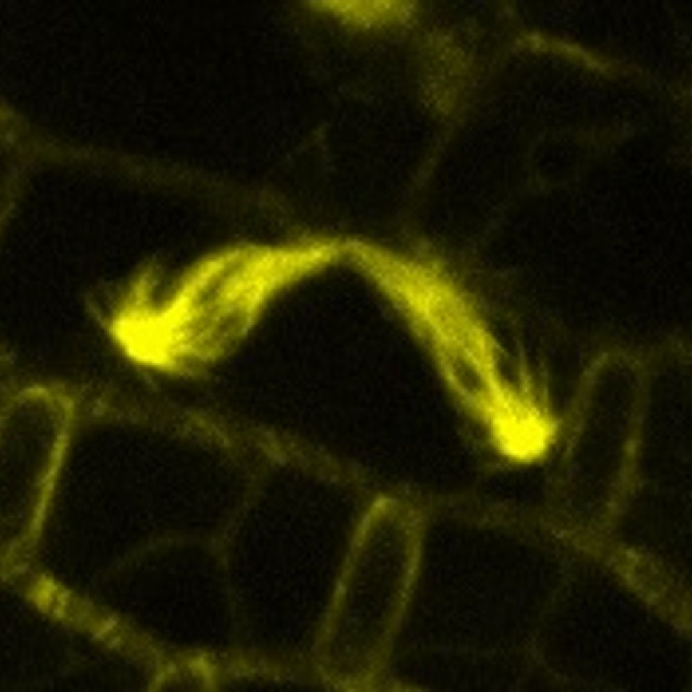
These close-ups of mucilage-covered tentacle tips from the leaf of a carnivorous sundew plant (Drosera spatulata) definitely have a strong holiday vibe, so I am posting them just in time for the holidays. Happy Holidays.
#microscopymonday #carnivorousplants
#microscopymonday #carnivorousplants


December 22, 2025 at 4:57 PM
These close-ups of mucilage-covered tentacle tips from the leaf of a carnivorous sundew plant (Drosera spatulata) definitely have a strong holiday vibe, so I am posting them just in time for the holidays. Happy Holidays.
#microscopymonday #carnivorousplants
#microscopymonday #carnivorousplants
Reposted by Ivan Radin
Spotlight: Ivan Radin discusses new work from Ryken and colleagues (rupress.org/jcb/article/...) which shows how localization and function of autoinhibitory calcium ATPases maintains the strength of the tip-focus Ca2²⁺ gradient during polarized plant growth. rupress.org/jcb/article/...
#PlantBiology
#PlantBiology

December 17, 2025 at 5:15 PM
Spotlight: Ivan Radin discusses new work from Ryken and colleagues (rupress.org/jcb/article/...) which shows how localization and function of autoinhibitory calcium ATPases maintains the strength of the tip-focus Ca2²⁺ gradient during polarized plant growth. rupress.org/jcb/article/...
#PlantBiology
#PlantBiology
This is what happens to the moss (Physcomitrium patens) chloroplasts when we grow cells on β-lactam antibiotics. β-lactams inhibit the synthesis of the peptidoglycan in the chloroplast envelope, leading to their dramatic expansion.
#microscopymonday #moss #plantcells #plantmicroscopy
1/3
#microscopymonday #moss #plantcells #plantmicroscopy
1/3

December 15, 2025 at 4:32 PM
This is what happens to the moss (Physcomitrium patens) chloroplasts when we grow cells on β-lactam antibiotics. β-lactams inhibit the synthesis of the peptidoglycan in the chloroplast envelope, leading to their dramatic expansion.
#microscopymonday #moss #plantcells #plantmicroscopy
1/3
#microscopymonday #moss #plantcells #plantmicroscopy
1/3
Reposted by Ivan Radin

Please RT‼️ #TenureTrack position @zmbh.uni-heidelberg.de one of the best #proteostasis research centres in the world. www.nature.com/naturecareer...

Tenure Track Professorship (W1 with Tenure Track to W3) in “Molecular Biology" (f/m/d) - Heidelberg job with Universität Heidelberg | 12850668
The Center for Molecular Biology of Heidelberg University (ZMBH) and the Faculty of Biosciences invite applications for a Tenure Track Professors...
www.nature.com
December 10, 2025 at 2:14 PM
Please RT‼️ #TenureTrack position @zmbh.uni-heidelberg.de one of the best #proteostasis research centres in the world. www.nature.com/naturecareer...
The Arabidopsis thaliana cells can have different types of plastids. In this image, plastids are labeled with the stroma-targeted fluorescence marker RecARed. RecARed fluorescence is false-colored yellow/orange, while the chlorophyll autofluorescence is in grey.
#microscopymonday #plantcells
#microscopymonday #plantcells

December 1, 2025 at 4:47 PM
The Arabidopsis thaliana cells can have different types of plastids. In this image, plastids are labeled with the stroma-targeted fluorescence marker RecARed. RecARed fluorescence is false-colored yellow/orange, while the chlorophyll autofluorescence is in grey.
#microscopymonday #plantcells
#microscopymonday #plantcells
During my postdoc, I looked at hundreds of images like this 🤩
These are two apical caulonemal cells from moss Physcomitrium patens stained with MDY64 (shown in shades of orange). The natural autofluorescence of chlorophyll is in cyan.
#microscopymonday #moss #plantcells #plantmicroscopy
These are two apical caulonemal cells from moss Physcomitrium patens stained with MDY64 (shown in shades of orange). The natural autofluorescence of chlorophyll is in cyan.
#microscopymonday #moss #plantcells #plantmicroscopy

November 10, 2025 at 3:33 PM
During my postdoc, I looked at hundreds of images like this 🤩
These are two apical caulonemal cells from moss Physcomitrium patens stained with MDY64 (shown in shades of orange). The natural autofluorescence of chlorophyll is in cyan.
#microscopymonday #moss #plantcells #plantmicroscopy
These are two apical caulonemal cells from moss Physcomitrium patens stained with MDY64 (shown in shades of orange). The natural autofluorescence of chlorophyll is in cyan.
#microscopymonday #moss #plantcells #plantmicroscopy
Check out this new paper on the Arabidopsis MLS8 channel and its importance for oscillatory growth
and cell wall dynamics in pollen tubes!
link.springer.com/article/10.1...
and cell wall dynamics in pollen tubes!
link.springer.com/article/10.1...

Mechanosensitive ion channel MSL8 is required for oscillatory growth and cell wall dynamics in Arabidopsis pollen tubes - Plant Reproduction
The male gametophyte in flowering plants, pollen, both performs the critical role of fertilization and represents a unique and accessible system for interrogating plant cell mechanics. A key component...
link.springer.com
November 7, 2025 at 4:20 PM
Check out this new paper on the Arabidopsis MLS8 channel and its importance for oscillatory growth
and cell wall dynamics in pollen tubes!
link.springer.com/article/10.1...
and cell wall dynamics in pollen tubes!
link.springer.com/article/10.1...
In preparation for Halloween, these chloroplasts had a seance! 👻🎃
These five Arabidopsis chloroplasts were extending their stromules (protrusions of the chloroplast envelope and stroma) towards each other.
#microscopymonday #Arabidopsis #plantcells #plantmicroscopy
These five Arabidopsis chloroplasts were extending their stromules (protrusions of the chloroplast envelope and stroma) towards each other.
#microscopymonday #Arabidopsis #plantcells #plantmicroscopy

October 27, 2025 at 3:44 PM
In preparation for Halloween, these chloroplasts had a seance! 👻🎃
These five Arabidopsis chloroplasts were extending their stromules (protrusions of the chloroplast envelope and stroma) towards each other.
#microscopymonday #Arabidopsis #plantcells #plantmicroscopy
These five Arabidopsis chloroplasts were extending their stromules (protrusions of the chloroplast envelope and stroma) towards each other.
#microscopymonday #Arabidopsis #plantcells #plantmicroscopy
Reposted by Ivan Radin
Genetically engineered color-changing Arabidopsis 🧬📷- attempt #3
I think I finally nailed it with this one.
I think I finally nailed it with this one.
October 27, 2025 at 11:53 AM
Genetically engineered color-changing Arabidopsis 🧬📷- attempt #3
I think I finally nailed it with this one.
I think I finally nailed it with this one.
This image of moss Physcomitrium patens beautifully demonstrates what we mean when we say that this cell type is characterized by oblique cell walls.
You can see that the cell walls between cells are at an angle and not perpendicular to the cell axis.
#microscopymonday #moss #plantcells
You can see that the cell walls between cells are at an angle and not perpendicular to the cell axis.
#microscopymonday #moss #plantcells

October 20, 2025 at 1:51 PM
This image of moss Physcomitrium patens beautifully demonstrates what we mean when we say that this cell type is characterized by oblique cell walls.
You can see that the cell walls between cells are at an angle and not perpendicular to the cell axis.
#microscopymonday #moss #plantcells
You can see that the cell walls between cells are at an angle and not perpendicular to the cell axis.
#microscopymonday #moss #plantcells
Here are some “leaf” or phyllid cells from moss Physcomitrium patens gametophore. Cells are expressing a green fluorescence marker that labels the vacuolar membrane. Chloroplasts' autofluorescence is in magenta.
Can you spot dividing chloroplasts?
#microscopymonday #moss #plantcells
Can you spot dividing chloroplasts?
#microscopymonday #moss #plantcells

October 13, 2025 at 2:40 PM
Here are some “leaf” or phyllid cells from moss Physcomitrium patens gametophore. Cells are expressing a green fluorescence marker that labels the vacuolar membrane. Chloroplasts' autofluorescence is in magenta.
Can you spot dividing chloroplasts?
#microscopymonday #moss #plantcells
Can you spot dividing chloroplasts?
#microscopymonday #moss #plantcells
Reposted by Ivan Radin

I started to work on MscS-Like proteins 20 years ago, dreaming that they were involved in plant mechanotransduction. And it is becoming more & more clear that they are!
#plantscience 🧪
"DmMSL10 is crucial for mechanosensing, facilitating AP firing by generating a receptor potential (RP) amplitude."
#plantscience 🧪
"DmMSL10 is crucial for mechanosensing, facilitating AP firing by generating a receptor potential (RP) amplitude."
🍀🔬
MSL10 is a high-sensitivity mechanosensor in the tactile sense of the Venus flytrap @natcomms.nature.com from Toyota lab.
www.nature.com/articles/s41...
MSL10 is a high-sensitivity mechanosensor in the tactile sense of the Venus flytrap @natcomms.nature.com from Toyota lab.
www.nature.com/articles/s41...
October 7, 2025 at 4:48 PM
I started to work on MscS-Like proteins 20 years ago, dreaming that they were involved in plant mechanotransduction. And it is becoming more & more clear that they are!
#plantscience 🧪
"DmMSL10 is crucial for mechanosensing, facilitating AP firing by generating a receptor potential (RP) amplitude."
#plantscience 🧪
"DmMSL10 is crucial for mechanosensing, facilitating AP firing by generating a receptor potential (RP) amplitude."
Reposted by Ivan Radin

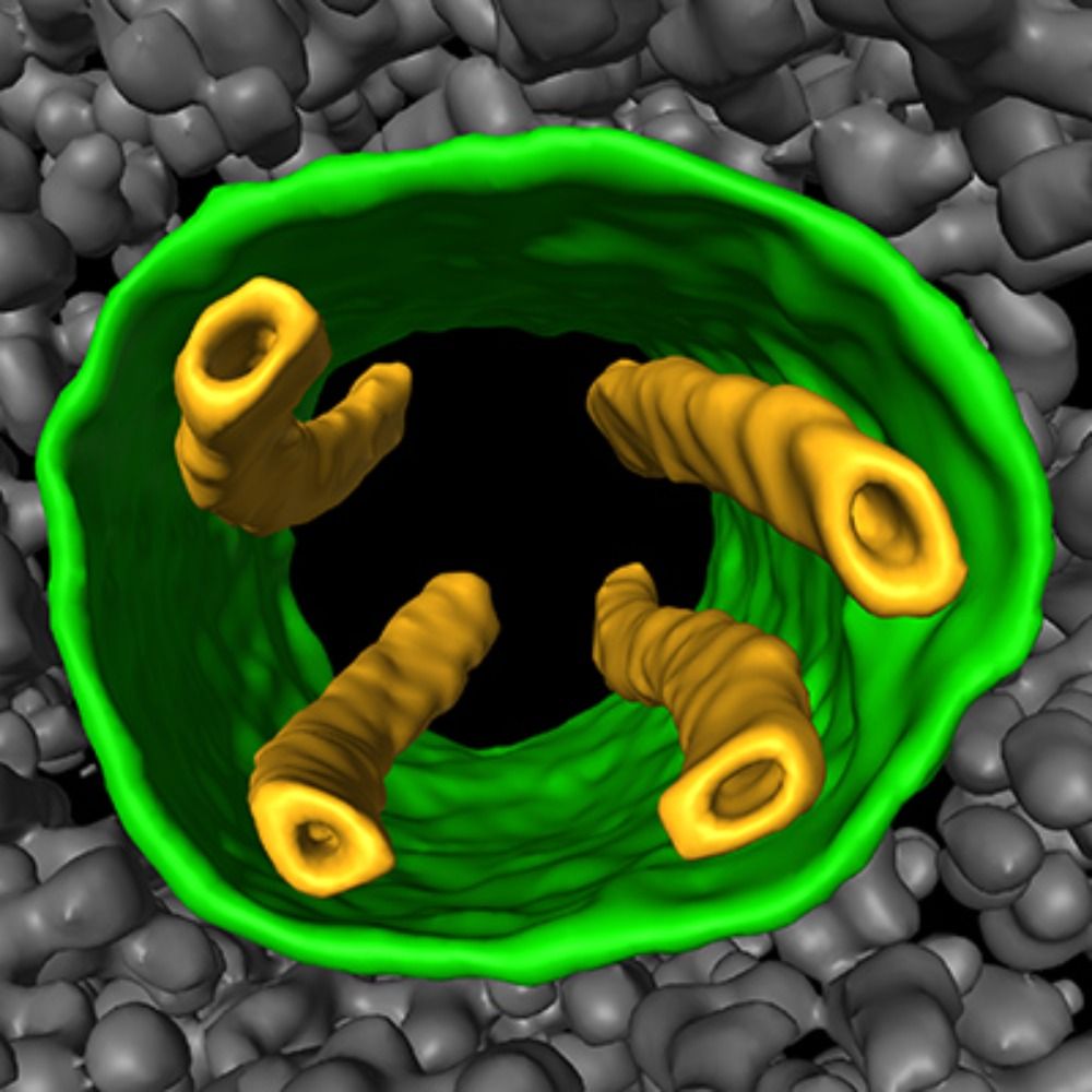
Inside a thylakoid membrane
The molecular architecture of the thylakoid membrane in a vascular plant has been determined with single-molecule precision.
buff.ly/U9TzrOh
The molecular architecture of the thylakoid membrane in a vascular plant has been determined with single-molecule precision.
buff.ly/U9TzrOh

October 8, 2025 at 10:02 AM
Inside a thylakoid membrane
The molecular architecture of the thylakoid membrane in a vascular plant has been determined with single-molecule precision.
buff.ly/U9TzrOh
The molecular architecture of the thylakoid membrane in a vascular plant has been determined with single-molecule precision.
buff.ly/U9TzrOh
This is a section of the cotyledon (first leaf) surface from the flowering plant Arabidopsis thaliana, which is expressing the green fluorescent protein vacuolar membrane marker (vac-GB). Chloroplasts are in magenta, visible due to the autofluorescence of the chlorophyll.
#microscopymonday
#microscopymonday

October 6, 2025 at 3:55 PM
This is a section of the cotyledon (first leaf) surface from the flowering plant Arabidopsis thaliana, which is expressing the green fluorescent protein vacuolar membrane marker (vac-GB). Chloroplasts are in magenta, visible due to the autofluorescence of the chlorophyll.
#microscopymonday
#microscopymonday
In case you missed it, our workshop on Fiji Basics for Visualization and Quantification of Plant Images is now available on YouTube:
www.youtube.com/watch?v=4TMJ...
We give a lot of practical tips on how to get started with Fiji.
Keep an eye out for announcements on follow-up workshops.
www.youtube.com/watch?v=4TMJ...
We give a lot of practical tips on how to get started with Fiji.
Keep an eye out for announcements on follow-up workshops.
Imaging Workshop Webinar Series: FIJI Basics for Visualizing and Quantifying Plant Images
YouTube video by Plant Cell Atlas
www.youtube.com
October 1, 2025 at 3:17 PM
In case you missed it, our workshop on Fiji Basics for Visualization and Quantification of Plant Images is now available on YouTube:
www.youtube.com/watch?v=4TMJ...
We give a lot of practical tips on how to get started with Fiji.
Keep an eye out for announcements on follow-up workshops.
www.youtube.com/watch?v=4TMJ...
We give a lot of practical tips on how to get started with Fiji.
Keep an eye out for announcements on follow-up workshops.
Reposted by Ivan Radin

In Two Days!
Attendees: we encourage you to download and install FIJI in advance of the workshop if you wish to follow along on your own computer. Keep an eye on your email & visit:
hpc.nih.gov/apps/Fiji and fiji.sc
Register Now! bit.ly/45zJE7V
Attendees: we encourage you to download and install FIJI in advance of the workshop if you wish to follow along on your own computer. Keep an eye on your email & visit:
hpc.nih.gov/apps/Fiji and fiji.sc
Register Now! bit.ly/45zJE7V

September 22, 2025 at 3:28 PM
In Two Days!
Attendees: we encourage you to download and install FIJI in advance of the workshop if you wish to follow along on your own computer. Keep an eye on your email & visit:
hpc.nih.gov/apps/Fiji and fiji.sc
Register Now! bit.ly/45zJE7V
Attendees: we encourage you to download and install FIJI in advance of the workshop if you wish to follow along on your own computer. Keep an eye on your email & visit:
hpc.nih.gov/apps/Fiji and fiji.sc
Register Now! bit.ly/45zJE7V