


Ivan Radin
@radinbio.bsky.social
🌱🪴🌿🔬🏳️🌈
Lover of plants and microscopy.
Assistant Professor, Department of Plant and Microbial Biology, Univerisity of Minnesota.
Lab: radinlab.org/
Instagram: radinbio
Lover of plants and microscopy.
Assistant Professor, Department of Plant and Microbial Biology, Univerisity of Minnesota.
Lab: radinlab.org/
Instagram: radinbio
During my postdoc, I looked at hundreds of images like this 🤩
These are two apical caulonemal cells from moss Physcomitrium patens stained with MDY64 (shown in shades of orange). The natural autofluorescence of chlorophyll is in cyan.
#microscopymonday #moss #plantcells #plantmicroscopy
These are two apical caulonemal cells from moss Physcomitrium patens stained with MDY64 (shown in shades of orange). The natural autofluorescence of chlorophyll is in cyan.
#microscopymonday #moss #plantcells #plantmicroscopy

November 10, 2025 at 3:33 PM
During my postdoc, I looked at hundreds of images like this 🤩
These are two apical caulonemal cells from moss Physcomitrium patens stained with MDY64 (shown in shades of orange). The natural autofluorescence of chlorophyll is in cyan.
#microscopymonday #moss #plantcells #plantmicroscopy
These are two apical caulonemal cells from moss Physcomitrium patens stained with MDY64 (shown in shades of orange). The natural autofluorescence of chlorophyll is in cyan.
#microscopymonday #moss #plantcells #plantmicroscopy
In preparation for Halloween, these chloroplasts had a seance! 👻🎃
These five Arabidopsis chloroplasts were extending their stromules (protrusions of the chloroplast envelope and stroma) towards each other.
#microscopymonday #Arabidopsis #plantcells #plantmicroscopy
These five Arabidopsis chloroplasts were extending their stromules (protrusions of the chloroplast envelope and stroma) towards each other.
#microscopymonday #Arabidopsis #plantcells #plantmicroscopy

October 27, 2025 at 3:44 PM
In preparation for Halloween, these chloroplasts had a seance! 👻🎃
These five Arabidopsis chloroplasts were extending their stromules (protrusions of the chloroplast envelope and stroma) towards each other.
#microscopymonday #Arabidopsis #plantcells #plantmicroscopy
These five Arabidopsis chloroplasts were extending their stromules (protrusions of the chloroplast envelope and stroma) towards each other.
#microscopymonday #Arabidopsis #plantcells #plantmicroscopy
This image of moss Physcomitrium patens beautifully demonstrates what we mean when we say that this cell type is characterized by oblique cell walls.
You can see that the cell walls between cells are at an angle and not perpendicular to the cell axis.
#microscopymonday #moss #plantcells
You can see that the cell walls between cells are at an angle and not perpendicular to the cell axis.
#microscopymonday #moss #plantcells

October 20, 2025 at 1:51 PM
This image of moss Physcomitrium patens beautifully demonstrates what we mean when we say that this cell type is characterized by oblique cell walls.
You can see that the cell walls between cells are at an angle and not perpendicular to the cell axis.
#microscopymonday #moss #plantcells
You can see that the cell walls between cells are at an angle and not perpendicular to the cell axis.
#microscopymonday #moss #plantcells
Here are some “leaf” or phyllid cells from moss Physcomitrium patens gametophore. Cells are expressing a green fluorescence marker that labels the vacuolar membrane. Chloroplasts' autofluorescence is in magenta.
Can you spot dividing chloroplasts?
#microscopymonday #moss #plantcells
Can you spot dividing chloroplasts?
#microscopymonday #moss #plantcells

October 13, 2025 at 2:40 PM
Here are some “leaf” or phyllid cells from moss Physcomitrium patens gametophore. Cells are expressing a green fluorescence marker that labels the vacuolar membrane. Chloroplasts' autofluorescence is in magenta.
Can you spot dividing chloroplasts?
#microscopymonday #moss #plantcells
Can you spot dividing chloroplasts?
#microscopymonday #moss #plantcells
This is a section of the cotyledon (first leaf) surface from the flowering plant Arabidopsis thaliana, which is expressing the green fluorescent protein vacuolar membrane marker (vac-GB). Chloroplasts are in magenta, visible due to the autofluorescence of the chlorophyll.
#microscopymonday
#microscopymonday

October 6, 2025 at 3:55 PM
This is a section of the cotyledon (first leaf) surface from the flowering plant Arabidopsis thaliana, which is expressing the green fluorescent protein vacuolar membrane marker (vac-GB). Chloroplasts are in magenta, visible due to the autofluorescence of the chlorophyll.
#microscopymonday
#microscopymonday
This is what happens when you remove the wall from plant cells. They become perfectly spherical and are called protoplasts.
These protoplasts were isolated from Arabidopsis and transformed with a vector to express cytosolic GFP (in orange). The chloroplast autofluorescence is in grey.
#microscopy
These protoplasts were isolated from Arabidopsis and transformed with a vector to express cytosolic GFP (in orange). The chloroplast autofluorescence is in grey.
#microscopy

September 22, 2025 at 7:36 PM
This is what happens when you remove the wall from plant cells. They become perfectly spherical and are called protoplasts.
These protoplasts were isolated from Arabidopsis and transformed with a vector to express cytosolic GFP (in orange). The chloroplast autofluorescence is in grey.
#microscopy
These protoplasts were isolated from Arabidopsis and transformed with a vector to express cytosolic GFP (in orange). The chloroplast autofluorescence is in grey.
#microscopy
Sundews need to be very sensitive to touch, to recognize when their prey has been captured.
This project was a great collaboration with Carl Procko from @salkinstitute.bsky.social. Videos were captured with the THUNDER Imager Model Organism dissecting scope from @leicamicrosystems.bsky.social
This project was a great collaboration with Carl Procko from @salkinstitute.bsky.social. Videos were captured with the THUNDER Imager Model Organism dissecting scope from @leicamicrosystems.bsky.social
September 15, 2025 at 2:26 PM
Sundews need to be very sensitive to touch, to recognize when their prey has been captured.
This project was a great collaboration with Carl Procko from @salkinstitute.bsky.social. Videos were captured with the THUNDER Imager Model Organism dissecting scope from @leicamicrosystems.bsky.social
This project was a great collaboration with Carl Procko from @salkinstitute.bsky.social. Videos were captured with the THUNDER Imager Model Organism dissecting scope from @leicamicrosystems.bsky.social
These sundews (Drosera spatulata) plants are expressing a cytosolic calcium biosensor. The video is false-colored, where the color corresponds to the calcium concentration in the cytosol of the cells. Blue = lowest, green = medium, and yellow = highest calcium levels
doi: 10.1073/pnas.2206433119
doi: 10.1073/pnas.2206433119
September 15, 2025 at 2:26 PM
These sundews (Drosera spatulata) plants are expressing a cytosolic calcium biosensor. The video is false-colored, where the color corresponds to the calcium concentration in the cytosol of the cells. Blue = lowest, green = medium, and yellow = highest calcium levels
doi: 10.1073/pnas.2206433119
doi: 10.1073/pnas.2206433119
Do you know what happens when you touch a carnivorous sundew plant?
If the touch is strong and large enough, a cytosolic calcium wave will spread from the site of touch throughout the whole plant, but if you only touch one tentacle (see post below), the calcium wave will be local and less intense.
If the touch is strong and large enough, a cytosolic calcium wave will spread from the site of touch throughout the whole plant, but if you only touch one tentacle (see post below), the calcium wave will be local and less intense.
September 15, 2025 at 2:26 PM
Do you know what happens when you touch a carnivorous sundew plant?
If the touch is strong and large enough, a cytosolic calcium wave will spread from the site of touch throughout the whole plant, but if you only touch one tentacle (see post below), the calcium wave will be local and less intense.
If the touch is strong and large enough, a cytosolic calcium wave will spread from the site of touch throughout the whole plant, but if you only touch one tentacle (see post below), the calcium wave will be local and less intense.
While the first image showed the outside edge of surface mature Arabidopsis root cell, this second image from the same experiment shows the border between two root cells. The large plastid (blue due to chlorophyll autofluorescence) is squashed between the two membranes.
#arabidopsis #cellbiology
#arabidopsis #cellbiology

September 8, 2025 at 4:22 PM
While the first image showed the outside edge of surface mature Arabidopsis root cell, this second image from the same experiment shows the border between two root cells. The large plastid (blue due to chlorophyll autofluorescence) is squashed between the two membranes.
#arabidopsis #cellbiology
#arabidopsis #cellbiology
This might be one of the simplest images I captured, but very fascinating nonetheless, as it shows how tight the cytoplasm in a plant cell is. The cytosol and all other organelles are squished between the plasma membrane (magenta) and the central vacuole (its membrane is green)
#MicroscopyMonday
#MicroscopyMonday

September 8, 2025 at 4:22 PM
This might be one of the simplest images I captured, but very fascinating nonetheless, as it shows how tight the cytoplasm in a plant cell is. The cytosol and all other organelles are squished between the plasma membrane (magenta) and the central vacuole (its membrane is green)
#MicroscopyMonday
#MicroscopyMonday
We love working with our moss, Physcomitrium (formerly Physcomitrella) patens.
In this dissecting microscope image, you can see the juvenile protonema filaments spread away from the main plant as they explore the new areas.
#MicroscopyMonday #Moss #Plants
In this dissecting microscope image, you can see the juvenile protonema filaments spread away from the main plant as they explore the new areas.
#MicroscopyMonday #Moss #Plants

September 1, 2025 at 5:16 PM
We love working with our moss, Physcomitrium (formerly Physcomitrella) patens.
In this dissecting microscope image, you can see the juvenile protonema filaments spread away from the main plant as they explore the new areas.
#MicroscopyMonday #Moss #Plants
In this dissecting microscope image, you can see the juvenile protonema filaments spread away from the main plant as they explore the new areas.
#MicroscopyMonday #Moss #Plants
The video shows the 3D reconstruction of the Z-stack (the image above is Z-maximum projection), and it's clear that the small green plastids are on the surface of the tissue, while the larger magenta ones are deeper in the tissue.
Here we imaged the bottom side of the Arabidopsis thaliana leaf.
Here we imaged the bottom side of the Arabidopsis thaliana leaf.
August 25, 2025 at 2:20 PM
The video shows the 3D reconstruction of the Z-stack (the image above is Z-maximum projection), and it's clear that the small green plastids are on the surface of the tissue, while the larger magenta ones are deeper in the tissue.
Here we imaged the bottom side of the Arabidopsis thaliana leaf.
Here we imaged the bottom side of the Arabidopsis thaliana leaf.
Did you know that plants can have different types of plastids/chloroplasts? The small green plastids are from the surface (epidermal) cells of the Arabidopsis leaf, while the much bigger magenta plastids are from the deeper spongy tissue. See video below.
#MicroscopyMonday #cellbiology #plastids
#MicroscopyMonday #cellbiology #plastids

August 25, 2025 at 2:20 PM
Did you know that plants can have different types of plastids/chloroplasts? The small green plastids are from the surface (epidermal) cells of the Arabidopsis leaf, while the much bigger magenta plastids are from the deeper spongy tissue. See video below.
#MicroscopyMonday #cellbiology #plastids
#MicroscopyMonday #cellbiology #plastids
A fun experiment! I injected the bacteria Agrobacterium tumefaciens into the tobacco leaf, Nicotiana benthamiana. The bacteria transferred some DNA into plant cells, forcing them to make GFP (green fluorescent protein). I used blue light to excite GFP.
Guess which leaf was infiltrated! #fluorescence
Guess which leaf was infiltrated! #fluorescence

August 18, 2025 at 1:50 PM
A fun experiment! I injected the bacteria Agrobacterium tumefaciens into the tobacco leaf, Nicotiana benthamiana. The bacteria transferred some DNA into plant cells, forcing them to make GFP (green fluorescent protein). I used blue light to excite GFP.
Guess which leaf was infiltrated! #fluorescence
Guess which leaf was infiltrated! #fluorescence
Not all microscopy images turn out great. Nevertheless, they can still be cool. This one definitely has an abstract art vibe. 🖼️
#MicroscopyMonday #Fluoresence #CellBiology
#MicroscopyMonday #Fluoresence #CellBiology

August 11, 2025 at 2:03 PM
Not all microscopy images turn out great. Nevertheless, they can still be cool. This one definitely has an abstract art vibe. 🖼️
#MicroscopyMonday #Fluoresence #CellBiology
#MicroscopyMonday #Fluoresence #CellBiology
This is what you see when you zoom in on fungus, growing uninvitedly on our "sterile" moss plates. What a beautiful hyphal network. #Fungi #Beatifulcontamination #Imaging #Microscopymonday
August 4, 2025 at 1:38 PM
This is what you see when you zoom in on fungus, growing uninvitedly on our "sterile" moss plates. What a beautiful hyphal network. #Fungi #Beatifulcontamination #Imaging #Microscopymonday
The image above is a Z-maximum projection, and here is a single slice from the same stack, where it's easy to see how closely vacuole and chloroplasts interact.
I used Olympus FV3000 confocal and 60X/1.2W objective from Evident to make this Z stack, which was also subjected to deconvolution.
I used Olympus FV3000 confocal and 60X/1.2W objective from Evident to make this Z stack, which was also subjected to deconvolution.


July 28, 2025 at 11:53 AM
The image above is a Z-maximum projection, and here is a single slice from the same stack, where it's easy to see how closely vacuole and chloroplasts interact.
I used Olympus FV3000 confocal and 60X/1.2W objective from Evident to make this Z stack, which was also subjected to deconvolution.
I used Olympus FV3000 confocal and 60X/1.2W objective from Evident to make this Z stack, which was also subjected to deconvolution.
I love this image of moss (Physcomitrium patens) chloronema cells, as you can see how chloroplasts (in magenta 🟣) indent the vacuole to make room for themselves. The vacuolar membrane is labeled with a GFP marker (green 🟢).
#microscopymonday #cellbiology #plantmicroscopy #plant #moss
#microscopymonday #cellbiology #plantmicroscopy #plant #moss

July 28, 2025 at 11:53 AM
I love this image of moss (Physcomitrium patens) chloronema cells, as you can see how chloroplasts (in magenta 🟣) indent the vacuole to make room for themselves. The vacuolar membrane is labeled with a GFP marker (green 🟢).
#microscopymonday #cellbiology #plantmicroscopy #plant #moss
#microscopymonday #cellbiology #plantmicroscopy #plant #moss
I think our Chlamy culture is haunted 😱👻😅
This is a Z-stack through a chloroplast from a single Chlamydomonas reinhardtii cell. Signal = chlorophyll autofluorescence. The image was captured on AXR confocal from @healthcare.nikon.com with 100x/1.4 oil objective
#cellbiology #algae #microscopymonday
This is a Z-stack through a chloroplast from a single Chlamydomonas reinhardtii cell. Signal = chlorophyll autofluorescence. The image was captured on AXR confocal from @healthcare.nikon.com with 100x/1.4 oil objective
#cellbiology #algae #microscopymonday
July 21, 2025 at 4:50 PM
I think our Chlamy culture is haunted 😱👻😅
This is a Z-stack through a chloroplast from a single Chlamydomonas reinhardtii cell. Signal = chlorophyll autofluorescence. The image was captured on AXR confocal from @healthcare.nikon.com with 100x/1.4 oil objective
#cellbiology #algae #microscopymonday
This is a Z-stack through a chloroplast from a single Chlamydomonas reinhardtii cell. Signal = chlorophyll autofluorescence. The image was captured on AXR confocal from @healthcare.nikon.com with 100x/1.4 oil objective
#cellbiology #algae #microscopymonday
Last week, I uploaded a picture of a moss gametophore taken with a bright-field microscope. For today’s #microscopymonday, we have an image of the same moss structure, this time captured with a confocal scope. We are only seeing autofluorescence of numerous chloroplasts filling the cells. #moss
July 14, 2025 at 11:24 AM
Last week, I uploaded a picture of a moss gametophore taken with a bright-field microscope. For today’s #microscopymonday, we have an image of the same moss structure, this time captured with a confocal scope. We are only seeing autofluorescence of numerous chloroplasts filling the cells. #moss
Last Monday, we saw baby moss plants, and this is how the adult stage looks like.
This is a single gametophore with the accompanying rhizoids from moss Physcomitrium patens. The image was made with the Olympus SZX7 dissecting scope with a DP71 camera from Evident/Olympus.
#microscopymonday #moss
This is a single gametophore with the accompanying rhizoids from moss Physcomitrium patens. The image was made with the Olympus SZX7 dissecting scope with a DP71 camera from Evident/Olympus.
#microscopymonday #moss

July 7, 2025 at 2:03 PM
Last Monday, we saw baby moss plants, and this is how the adult stage looks like.
This is a single gametophore with the accompanying rhizoids from moss Physcomitrium patens. The image was made with the Olympus SZX7 dissecting scope with a DP71 camera from Evident/Olympus.
#microscopymonday #moss
This is a single gametophore with the accompanying rhizoids from moss Physcomitrium patens. The image was made with the Olympus SZX7 dissecting scope with a DP71 camera from Evident/Olympus.
#microscopymonday #moss
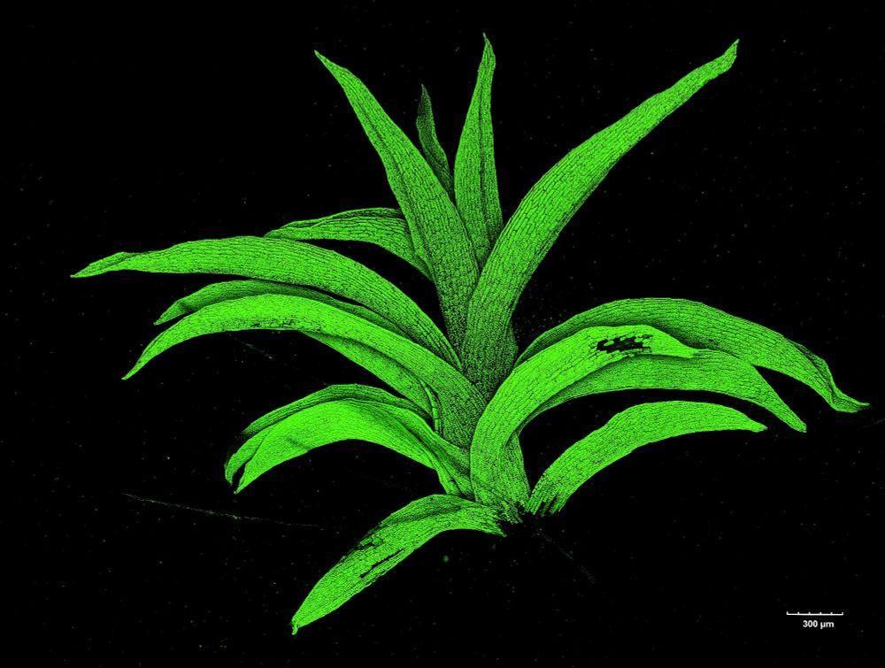
Are these little aliens 👽 or baby moss plants🪴? 😄
.
.
.
This collage is a great example of the power of plant regeneration. Each of these individual 7-day-old moss (Physcomitrium patens) plants was regenerated from a single protoplast (a plant cell with removed cell walls).
#microscopymonday #moss
.
.
.
This collage is a great example of the power of plant regeneration. Each of these individual 7-day-old moss (Physcomitrium patens) plants was regenerated from a single protoplast (a plant cell with removed cell walls).
#microscopymonday #moss

June 30, 2025 at 2:04 PM
Are these little aliens 👽 or baby moss plants🪴? 😄
.
.
.
This collage is a great example of the power of plant regeneration. Each of these individual 7-day-old moss (Physcomitrium patens) plants was regenerated from a single protoplast (a plant cell with removed cell walls).
#microscopymonday #moss
.
.
.
This collage is a great example of the power of plant regeneration. Each of these individual 7-day-old moss (Physcomitrium patens) plants was regenerated from a single protoplast (a plant cell with removed cell walls).
#microscopymonday #moss
This is a deconvolved maximum Z projection image made on an Evident/Olympus FV3000 confocal microscope using a 60x/1.2 water objective.
And as a bonus, here is a 3D rendering of that Z-stack #3Dvolume
And as a bonus, here is a 3D rendering of that Z-stack #3Dvolume
June 23, 2025 at 1:42 PM
This is a deconvolved maximum Z projection image made on an Evident/Olympus FV3000 confocal microscope using a 60x/1.2 water objective.
And as a bonus, here is a 3D rendering of that Z-stack #3Dvolume
And as a bonus, here is a 3D rendering of that Z-stack #3Dvolume
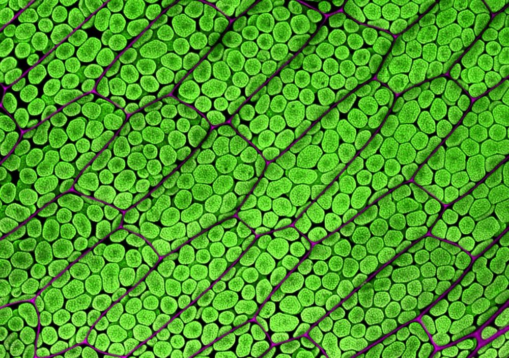
Plant cells can be filled with chloroplasts, as shown in this image of moss gametophore phyllid ("leaf") cells. These cells have not been stained with fluorescent dyes; we only observe the autofluorescence of chloroplasts (green 🟢) and of the cell walls (magenta 🟣)
#microscopymonday #plantcell #moss
#microscopymonday #plantcell #moss
June 23, 2025 at 1:42 PM
Plant cells can be filled with chloroplasts, as shown in this image of moss gametophore phyllid ("leaf") cells. These cells have not been stained with fluorescent dyes; we only observe the autofluorescence of chloroplasts (green 🟢) and of the cell walls (magenta 🟣)
#microscopymonday #plantcell #moss
#microscopymonday #plantcell #moss