


Dr. rer. nat. Ina Klockner🧬👩🔬🔬
@inaklockner.bsky.social
Scientist | former Postdoc at The University of North Carolina at Chapel Hill (USA) | now at UKM Münster (Germany) | Gene Therapy & Post-Transcriptional Gene Regulation & Molecular Genetics | she/her | 🇩🇪> 🇺🇲>🇩🇪
Pinned

Compact Calm1 promoter enables AAV mediated neuron-targeted expression in human iPSC-derived brain organoids - Scientific Reports
Scientific Reports - Compact Calm1 promoter enables AAV mediated neuron-targeted expression in human iPSC-derived brain organoids
www.nature.com
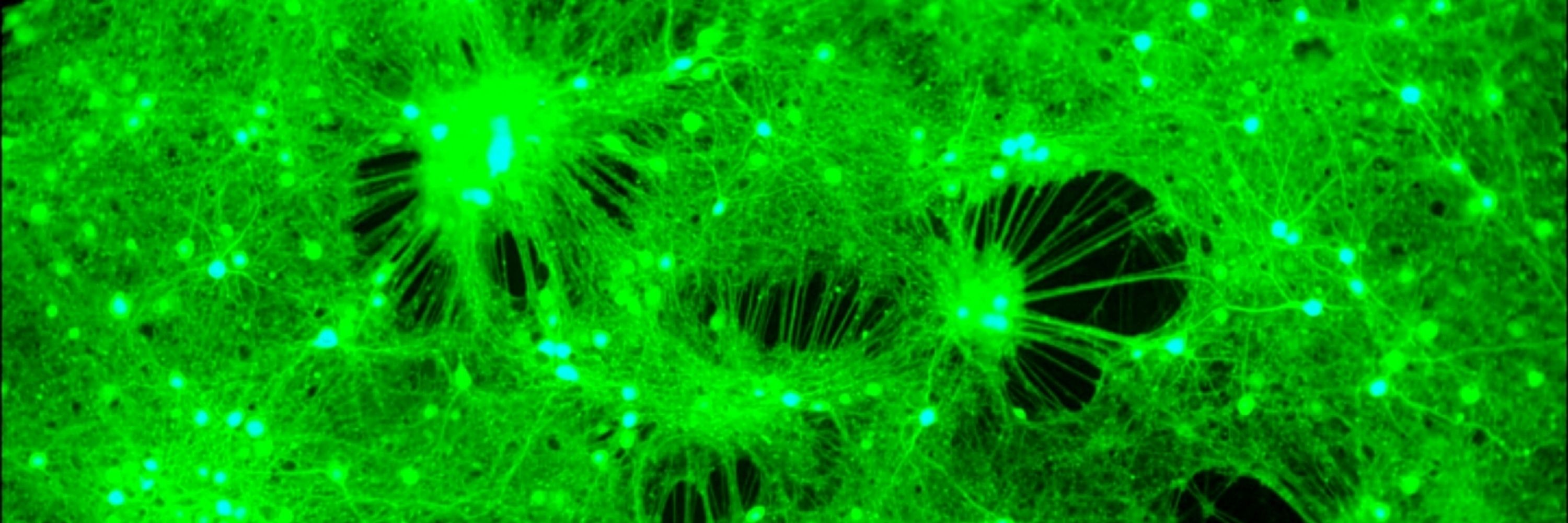
New paper out 🔥!
Small promoter, big potential🧬 We show that the ultra-compact Calm1 promoter efficiently targets human neurons in brain organoids and is a promising alternative to hSyn for AAV gene delivery.
Huge thanks to everyone who contributed to this work!
🧠 📄: www.nature.com/articles/s41...
Small promoter, big potential🧬 We show that the ultra-compact Calm1 promoter efficiently targets human neurons in brain organoids and is a promising alternative to hSyn for AAV gene delivery.
Huge thanks to everyone who contributed to this work!
🧠 📄: www.nature.com/articles/s41...
New biosensor💥
🎯precisely measures endogenous UBE3A activity with super high sensitivity
📌providing a powerful tool for studying Angelman syndrome & advancing therapeutic development for UBE3A-related disorders
link to study:
www.sciencedirect.com/science/arti...
🎯precisely measures endogenous UBE3A activity with super high sensitivity
📌providing a powerful tool for studying Angelman syndrome & advancing therapeutic development for UBE3A-related disorders
link to study:
www.sciencedirect.com/science/arti...

A luminescence-based biosensor to measure endogenous UBE3A activity
Loss- and gain-of-function (LOF and GOF) mutations in the E3 ubiquitin ligase UBE3A cause distinct neurodevelopmental disorders. The AZUL domain of UB…
www.sciencedirect.com
January 12, 2026 at 5:03 PM
New biosensor💥
🎯precisely measures endogenous UBE3A activity with super high sensitivity
📌providing a powerful tool for studying Angelman syndrome & advancing therapeutic development for UBE3A-related disorders
link to study:
www.sciencedirect.com/science/arti...
🎯precisely measures endogenous UBE3A activity with super high sensitivity
📌providing a powerful tool for studying Angelman syndrome & advancing therapeutic development for UBE3A-related disorders
link to study:
www.sciencedirect.com/science/arti...
Reposted by Dr. rer. nat. Ina Klockner🧬👩🔬🔬

Excited to share @rbrianroome.bsky.social ‘s beautiful paper on development of the dorsal horn of the mouse spinal cord @science.org
This is how the anatomical organization and cell types that process pain, touch, body position and more are laid down.
www.science.org/doi/10.1126/...
This is how the anatomical organization and cell types that process pain, touch, body position and more are laid down.
www.science.org/doi/10.1126/...

Ontogeny of the spinal cord dorsal horn
The dorsal horn of the mammalian spinal cord is organized into laminae where each layer is populated by different neuron types, has distinctive circuit connections, and plays specialized roles in beha...
www.science.org
January 8, 2026 at 7:59 PM
Excited to share @rbrianroome.bsky.social ‘s beautiful paper on development of the dorsal horn of the mouse spinal cord @science.org
This is how the anatomical organization and cell types that process pain, touch, body position and more are laid down.
www.science.org/doi/10.1126/...
This is how the anatomical organization and cell types that process pain, touch, body position and more are laid down.
www.science.org/doi/10.1126/...
Reposted by Dr. rer. nat. Ina Klockner🧬👩🔬🔬
I am happy our recent study on the human sperm head-tail junction is now online. We find that SUN5 form an hexagonal lattice at the nuclear membrane, rationalising how mutations in SUN5 lead to infertility. Great work of @cryonas.bsky.social !
www.biorxiv.org/content/10.1...
www.biorxiv.org/content/10.1...

SUN5 forms a regular protein lattice reinforcing the sperm head-tail junction
Linker of nucleo- and cytoskeleton (LINC) complexes reside in the nuclear envelope, the double-membrane surrounding the nucleus, where they establish a physical bridge between nucleus and cytoplasm. L...
www.biorxiv.org
July 31, 2025 at 3:07 PM
I am happy our recent study on the human sperm head-tail junction is now online. We find that SUN5 form an hexagonal lattice at the nuclear membrane, rationalising how mutations in SUN5 lead to infertility. Great work of @cryonas.bsky.social !
www.biorxiv.org/content/10.1...
www.biorxiv.org/content/10.1...
Reposted by Dr. rer. nat. Ina Klockner🧬👩🔬🔬

There is still over a week left to apply.
Please send your documents to maik.bischoff@uni-muenster.de
Please send your documents to maik.bischoff@uni-muenster.de
Join us to study developmental cell biology with a focus on cell migration and morphogenesis.
More info about our research:
www.bischofflab.com
Job posting and application details:
focalplane.biologists.com/cell-biology...
#Science #CellBio #DevBio #AcademicJobs #PhDJobs #PhDPosition #Drosophila
More info about our research:
www.bischofflab.com
Job posting and application details:
focalplane.biologists.com/cell-biology...
#Science #CellBio #DevBio #AcademicJobs #PhDJobs #PhDPosition #Drosophila
January 6, 2026 at 12:43 PM
There is still over a week left to apply.
Please send your documents to maik.bischoff@uni-muenster.de
Please send your documents to maik.bischoff@uni-muenster.de
Reposted by Dr. rer. nat. Ina Klockner🧬👩🔬🔬

#eNeuro: Ringelberg et al. identify a key role for excitatory neuron loss of UBE3A in motor, innate, and sleep behavioral phenotypes of Angelman syndrome model mice.
https://doi.org/10.1523/ENEURO.0453-24.2025
https://doi.org/10.1523/ENEURO.0453-24.2025

November 11, 2025 at 11:32 AM
#eNeuro: Ringelberg et al. identify a key role for excitatory neuron loss of UBE3A in motor, innate, and sleep behavioral phenotypes of Angelman syndrome model mice.
https://doi.org/10.1523/ENEURO.0453-24.2025
https://doi.org/10.1523/ENEURO.0453-24.2025
Reposted by Dr. rer. nat. Ina Klockner🧬👩🔬🔬

Hong Cheng and collaborators show how alternative polyadenylation can facilitate muscle differentiation and regeneration: 3’UTR shortening of differentiation-gene mRNAs alleviates their repression by myomiR muscle-specific miRNAs
link.springer.com/article/10.1...
link.springer.com/article/10.1...

3′UTR shortening alleviates miRNA repression of mRNAs critical for muscle stem cell differentiation - The EMBO Journal
Alternative polyadenylation (APA) modulates gene expression by altering 3′ untranslated region (3′UTR) length. Although 3′UTR lengthening typically accompanies cell differentiation, we unexpectedly ob...
link.springer.com
December 22, 2025 at 12:13 PM
Hong Cheng and collaborators show how alternative polyadenylation can facilitate muscle differentiation and regeneration: 3’UTR shortening of differentiation-gene mRNAs alleviates their repression by myomiR muscle-specific miRNAs
link.springer.com/article/10.1...
link.springer.com/article/10.1...
New paper out 🔥!
Small promoter, big potential🧬 We show that the ultra-compact Calm1 promoter efficiently targets human neurons in brain organoids and is a promising alternative to hSyn for AAV gene delivery.
Huge thanks to everyone who contributed to this work!
🧠 📄: www.nature.com/articles/s41...
Small promoter, big potential🧬 We show that the ultra-compact Calm1 promoter efficiently targets human neurons in brain organoids and is a promising alternative to hSyn for AAV gene delivery.
Huge thanks to everyone who contributed to this work!
🧠 📄: www.nature.com/articles/s41...
Compact Calm1 promoter enables AAV mediated neuron-targeted expression in human iPSC-derived brain organoids - Scientific Reports
Scientific Reports - Compact Calm1 promoter enables AAV mediated neuron-targeted expression in human iPSC-derived brain organoids
www.nature.com
January 5, 2026 at 12:37 PM
New paper out 🔥!
Small promoter, big potential🧬 We show that the ultra-compact Calm1 promoter efficiently targets human neurons in brain organoids and is a promising alternative to hSyn for AAV gene delivery.
Huge thanks to everyone who contributed to this work!
🧠 📄: www.nature.com/articles/s41...
Small promoter, big potential🧬 We show that the ultra-compact Calm1 promoter efficiently targets human neurons in brain organoids and is a promising alternative to hSyn for AAV gene delivery.
Huge thanks to everyone who contributed to this work!
🧠 📄: www.nature.com/articles/s41...
Reposted by Dr. rer. nat. Ina Klockner🧬👩🔬🔬
1/2 A special #FluorescenceFriday for me! 🌈
Live muscle precursors on a pupal fly testis (Lifeact-eGFP, 🔥-depth-coded).
It's special because this is on the poster for the @sfb1348.bsky.social Meeting in May — my first time being featured as a speaker on a poster! 😀
Feeling very grateful & excited!
Live muscle precursors on a pupal fly testis (Lifeact-eGFP, 🔥-depth-coded).
It's special because this is on the poster for the @sfb1348.bsky.social Meeting in May — my first time being featured as a speaker on a poster! 😀
Feeling very grateful & excited!


November 28, 2025 at 8:34 AM
1/2 A special #FluorescenceFriday for me! 🌈
Live muscle precursors on a pupal fly testis (Lifeact-eGFP, 🔥-depth-coded).
It's special because this is on the poster for the @sfb1348.bsky.social Meeting in May — my first time being featured as a speaker on a poster! 😀
Feeling very grateful & excited!
Live muscle precursors on a pupal fly testis (Lifeact-eGFP, 🔥-depth-coded).
It's special because this is on the poster for the @sfb1348.bsky.social Meeting in May — my first time being featured as a speaker on a poster! 😀
Feeling very grateful & excited!
Reposted by Dr. rer. nat. Ina Klockner🧬👩🔬🔬
So excited to share this as a new junior PI:
My brand-new lab website! 🎉🪰🌀
www.bischofflab.com
Please pass it on to young, motivated researchers looking for PhD positions 😊
And for the #FluorescenceFriday community: don’t miss the SciArt Gallery!
#CellBio #DevBio #PhDjob #PhDposition #Science
My brand-new lab website! 🎉🪰🌀
www.bischofflab.com
Please pass it on to young, motivated researchers looking for PhD positions 😊
And for the #FluorescenceFriday community: don’t miss the SciArt Gallery!
#CellBio #DevBio #PhDjob #PhDposition #Science


November 27, 2025 at 8:26 AM
So excited to share this as a new junior PI:
My brand-new lab website! 🎉🪰🌀
www.bischofflab.com
Please pass it on to young, motivated researchers looking for PhD positions 😊
And for the #FluorescenceFriday community: don’t miss the SciArt Gallery!
#CellBio #DevBio #PhDjob #PhDposition #Science
My brand-new lab website! 🎉🪰🌀
www.bischofflab.com
Please pass it on to young, motivated researchers looking for PhD positions 😊
And for the #FluorescenceFriday community: don’t miss the SciArt Gallery!
#CellBio #DevBio #PhDjob #PhDposition #Science
Amazing paper with gorgeous imaging 🔥
from @mathpreu.bsky.social
journals.biologists.com/dev/article/...
from @mathpreu.bsky.social
journals.biologists.com/dev/article/...

A developmental atlas of zebrafish gills links early vascular patterning to adult architecture
Summary: An atlas of zebrafish gill vasculature reveals developmental asymmetries and shows how early vascular patterning shapes filaments and lamellae and is reflected in adult gill architecture.
journals.biologists.com
October 31, 2025 at 9:19 PM
Amazing paper with gorgeous imaging 🔥
from @mathpreu.bsky.social
journals.biologists.com/dev/article/...
from @mathpreu.bsky.social
journals.biologists.com/dev/article/...
Reposted by Dr. rer. nat. Ina Klockner🧬👩🔬🔬
Can't believe it — my first‑author paper is out and my image graces the cover of @dev-journal.bsky.social 🎉
Here, we reveal how early developmental programs shape and maintain #zebrafish gill architecture throughout life
🔗 journals.biologists.com/dev/issue/15...
#FluorescenceFriday #LifelongDevSI
Here, we reveal how early developmental programs shape and maintain #zebrafish gill architecture throughout life
🔗 journals.biologists.com/dev/issue/15...
#FluorescenceFriday #LifelongDevSI

October 31, 2025 at 10:55 AM
Can't believe it — my first‑author paper is out and my image graces the cover of @dev-journal.bsky.social 🎉
Here, we reveal how early developmental programs shape and maintain #zebrafish gill architecture throughout life
🔗 journals.biologists.com/dev/issue/15...
#FluorescenceFriday #LifelongDevSI
Here, we reveal how early developmental programs shape and maintain #zebrafish gill architecture throughout life
🔗 journals.biologists.com/dev/issue/15...
#FluorescenceFriday #LifelongDevSI
Reposted by Dr. rer. nat. Ina Klockner🧬👩🔬🔬
A developmental atlas of zebrafish gills links early vascular patterning to adult architecture
Read this #LifelongDevSI #OA Research Article by Mathieu Preußner @mathpreu.bsky.social, Virginie Lecaudey @vlecaudey.bsky.social and colleagues:
journals.biologists.com/dev/article/...
Read this #LifelongDevSI #OA Research Article by Mathieu Preußner @mathpreu.bsky.social, Virginie Lecaudey @vlecaudey.bsky.social and colleagues:
journals.biologists.com/dev/article/...
October 21, 2025 at 12:36 PM
A developmental atlas of zebrafish gills links early vascular patterning to adult architecture
Read this #LifelongDevSI #OA Research Article by Mathieu Preußner @mathpreu.bsky.social, Virginie Lecaudey @vlecaudey.bsky.social and colleagues:
journals.biologists.com/dev/article/...
Read this #LifelongDevSI #OA Research Article by Mathieu Preußner @mathpreu.bsky.social, Virginie Lecaudey @vlecaudey.bsky.social and colleagues:
journals.biologists.com/dev/article/...
Reposted by Dr. rer. nat. Ina Klockner🧬👩🔬🔬
My entry for today’s #FluorescenceFriday: a pupal #Drosophila testis with muscles expressing
🔵 lifeact &
🔴 RFP-nls
Honored & grateful to receive an honorable mention at @healthcare.nikon.com Nikon Small World 🌍🔬✨
🔗 www.nikonsmallworld.com/galleries/20...
#NikonSmallWorld #Microscopy #ScienceArt
🔵 lifeact &
🔴 RFP-nls
Honored & grateful to receive an honorable mention at @healthcare.nikon.com Nikon Small World 🌍🔬✨
🔗 www.nikonsmallworld.com/galleries/20...
#NikonSmallWorld #Microscopy #ScienceArt
October 3, 2025 at 9:06 AM
My entry for today’s #FluorescenceFriday: a pupal #Drosophila testis with muscles expressing
🔵 lifeact &
🔴 RFP-nls
Honored & grateful to receive an honorable mention at @healthcare.nikon.com Nikon Small World 🌍🔬✨
🔗 www.nikonsmallworld.com/galleries/20...
#NikonSmallWorld #Microscopy #ScienceArt
🔵 lifeact &
🔴 RFP-nls
Honored & grateful to receive an honorable mention at @healthcare.nikon.com Nikon Small World 🌍🔬✨
🔗 www.nikonsmallworld.com/galleries/20...
#NikonSmallWorld #Microscopy #ScienceArt
Reposted by Dr. rer. nat. Ina Klockner🧬👩🔬🔬
1/12 A very special moment for me! 🎉
My first paper as corresponding author—a @jcb.org Perspective on how contact-based decision-making in collective cell migration can itself encode blueprints for complex patterns and shapes.
rupress.org/jcb/article/...
#cellbio #devbio #science
🧵for details
My first paper as corresponding author—a @jcb.org Perspective on how contact-based decision-making in collective cell migration can itself encode blueprints for complex patterns and shapes.
rupress.org/jcb/article/...
#cellbio #devbio #science
🧵for details

September 24, 2025 at 1:06 PM
1/12 A very special moment for me! 🎉
My first paper as corresponding author—a @jcb.org Perspective on how contact-based decision-making in collective cell migration can itself encode blueprints for complex patterns and shapes.
rupress.org/jcb/article/...
#cellbio #devbio #science
🧵for details
My first paper as corresponding author—a @jcb.org Perspective on how contact-based decision-making in collective cell migration can itself encode blueprints for complex patterns and shapes.
rupress.org/jcb/article/...
#cellbio #devbio #science
🧵for details
Reposted by Dr. rer. nat. Ina Klockner🧬👩🔬🔬

New perspective by @maikbischoff.bsky.social & @mayorlab.bsky.social about how mesenchymal cells use contact-dependent rules to generate swarm-like behaviors, patterns & organ forms. rupress.org/jcb/article/...
#CellBio #CellMigration #Morphogenesis #Development #Science
#CellBio #CellMigration #Morphogenesis #Development #Science

September 23, 2025 at 6:45 PM
New perspective by @maikbischoff.bsky.social & @mayorlab.bsky.social about how mesenchymal cells use contact-dependent rules to generate swarm-like behaviors, patterns & organ forms. rupress.org/jcb/article/...
#CellBio #CellMigration #Morphogenesis #Development #Science
#CellBio #CellMigration #Morphogenesis #Development #Science
Reposted by Dr. rer. nat. Ina Klockner🧬👩🔬🔬

We’re excited to announce the launch of our new website at angelman.org! 🎉 Our updated site features easy navigation and filters that help you find the resources, clinics, research, and support you need on any device. Check it out!➡️ angelman.org

June 26, 2025 at 9:40 PM
We’re excited to announce the launch of our new website at angelman.org! 🎉 Our updated site features easy navigation and filters that help you find the resources, clinics, research, and support you need on any device. Check it out!➡️ angelman.org
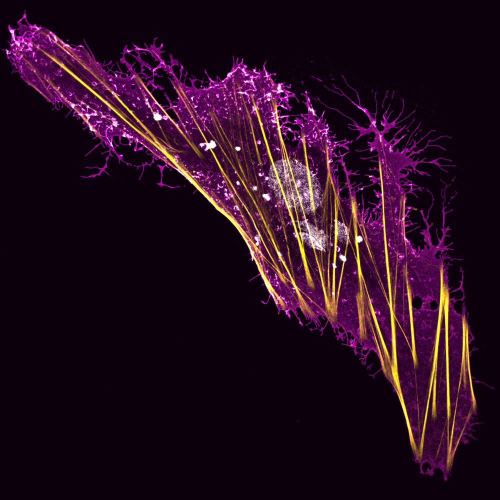
Reposted by Dr. rer. nat. Ina Klockner🧬👩🔬🔬

A Boa Constrictor brain-derived cell expressing a viral glycoprotein (magenta) and stained for actin (yellow) and DNA (white).
#FluorescenceFriday
#FluorescenceFriday
June 27, 2025 at 11:20 AM
A Boa Constrictor brain-derived cell expressing a viral glycoprotein (magenta) and stained for actin (yellow) and DNA (white).
#FluorescenceFriday
#FluorescenceFriday
Reposted by Dr. rer. nat. Ina Klockner🧬👩🔬🔬
FINALLY out in #Science Advances! 🎉
3 years of hard work, a fantastic team — and I’m proud to be first and co-corresponding author!
It's about Plexin signaling driving collective cell migration & organ sculpting. Check out the videos 😀
www.science.org/doi/10.1126/...
#DevBio #CellBio #Microscopy
3 years of hard work, a fantastic team — and I’m proud to be first and co-corresponding author!
It's about Plexin signaling driving collective cell migration & organ sculpting. Check out the videos 😀
www.science.org/doi/10.1126/...
#DevBio #CellBio #Microscopy
June 18, 2025 at 7:06 PM
FINALLY out in #Science Advances! 🎉
3 years of hard work, a fantastic team — and I’m proud to be first and co-corresponding author!
It's about Plexin signaling driving collective cell migration & organ sculpting. Check out the videos 😀
www.science.org/doi/10.1126/...
#DevBio #CellBio #Microscopy
3 years of hard work, a fantastic team — and I’m proud to be first and co-corresponding author!
It's about Plexin signaling driving collective cell migration & organ sculpting. Check out the videos 😀
www.science.org/doi/10.1126/...
#DevBio #CellBio #Microscopy
Reposted by Dr. rer. nat. Ina Klockner🧬👩🔬🔬

For #fluorescencefriday the Mesoscopic Integrated NeuroImaging Data (MIND) platform @westernu.bsky.social presents abeta plaques and blood vessels in the 🧠 of our ApoE4 mouse model of AD. 🧪 #clearing #lightsheet 🔬 Courtesy of Arash Salahinejad, Luke Smolders and @shahrzadbahrampour.bsky.social
June 13, 2025 at 5:35 PM
For #fluorescencefriday the Mesoscopic Integrated NeuroImaging Data (MIND) platform @westernu.bsky.social presents abeta plaques and blood vessels in the 🧠 of our ApoE4 mouse model of AD. 🧪 #clearing #lightsheet 🔬 Courtesy of Arash Salahinejad, Luke Smolders and @shahrzadbahrampour.bsky.social
Stunning images!
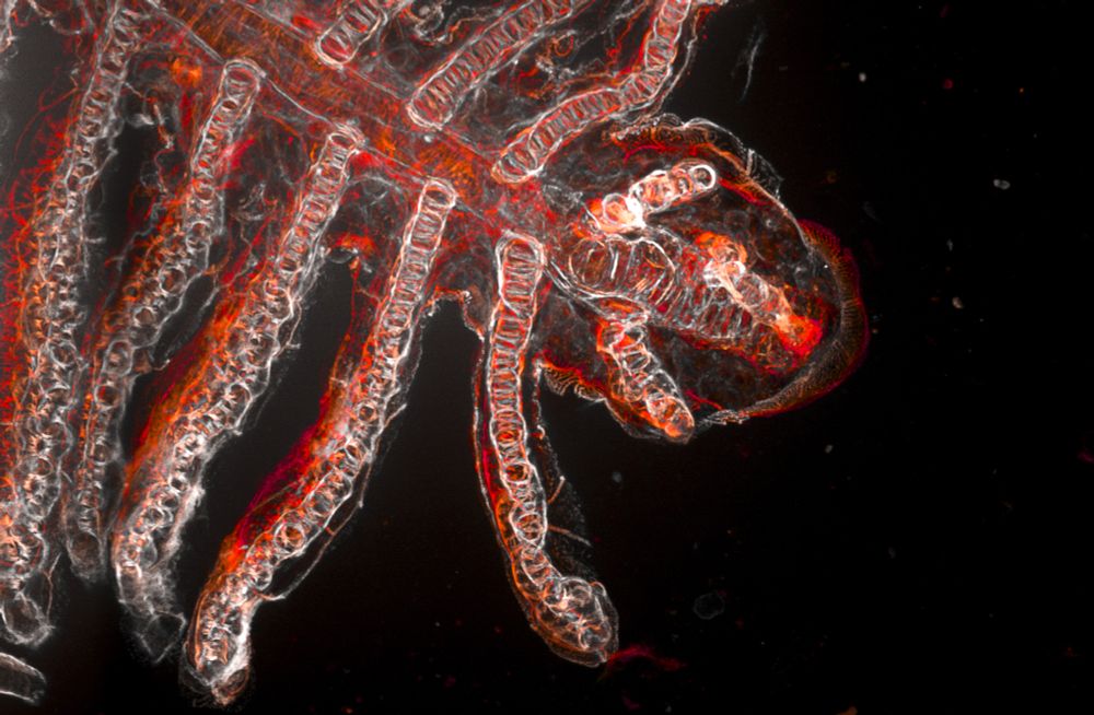
1)
Branching morphogenesis is fundamental to animal development, shaping complex organs like lungs, kidneys, and vascular networks. Gills are another highly branched organ. Yet, the developmental processes behind gill formation in zebrafish remain poorly understood.
#zebrafish #gills #microscopy
Branching morphogenesis is fundamental to animal development, shaping complex organs like lungs, kidneys, and vascular networks. Gills are another highly branched organ. Yet, the developmental processes behind gill formation in zebrafish remain poorly understood.
#zebrafish #gills #microscopy
June 6, 2025 at 5:09 PM
Stunning images!
Reposted by Dr. rer. nat. Ina Klockner🧬👩🔬🔬
1)
Branching morphogenesis is fundamental to animal development, shaping complex organs like lungs, kidneys, and vascular networks. Gills are another highly branched organ. Yet, the developmental processes behind gill formation in zebrafish remain poorly understood.
#zebrafish #gills #microscopy
Branching morphogenesis is fundamental to animal development, shaping complex organs like lungs, kidneys, and vascular networks. Gills are another highly branched organ. Yet, the developmental processes behind gill formation in zebrafish remain poorly understood.
#zebrafish #gills #microscopy
June 6, 2025 at 4:12 PM
1)
Branching morphogenesis is fundamental to animal development, shaping complex organs like lungs, kidneys, and vascular networks. Gills are another highly branched organ. Yet, the developmental processes behind gill formation in zebrafish remain poorly understood.
#zebrafish #gills #microscopy
Branching morphogenesis is fundamental to animal development, shaping complex organs like lungs, kidneys, and vascular networks. Gills are another highly branched organ. Yet, the developmental processes behind gill formation in zebrafish remain poorly understood.
#zebrafish #gills #microscopy
Reposted by Dr. rer. nat. Ina Klockner🧬👩🔬🔬
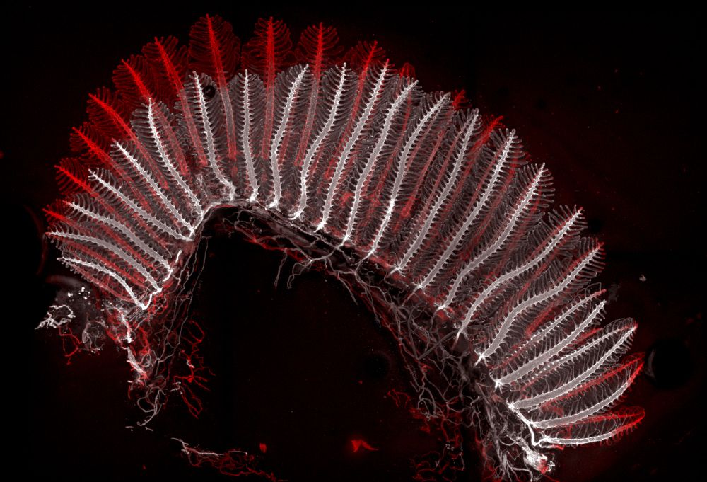
Gills just wanna have #FluorescenceFriday 🎵
Phalloidin reveals the beauty of the filament's distal tip in cleared gills of adult 🦓🐟
#microscopy #devbio #sciart
Phalloidin reveals the beauty of the filament's distal tip in cleared gills of adult 🦓🐟
#microscopy #devbio #sciart
April 11, 2025 at 4:20 PM
Gills just wanna have #FluorescenceFriday 🎵
Phalloidin reveals the beauty of the filament's distal tip in cleared gills of adult 🦓🐟
#microscopy #devbio #sciart
Phalloidin reveals the beauty of the filament's distal tip in cleared gills of adult 🦓🐟
#microscopy #devbio #sciart
Reposted by Dr. rer. nat. Ina Klockner🧬👩🔬🔬
Reposted by Dr. rer. nat. Ina Klockner🧬👩🔬🔬
I know, it's a bit late for #FluorescenceFriday but I want to share my new banner :D
#cellmigration #cellbio #devbio #sciart #microcosm #science
#cellmigration #cellbio #devbio #sciart #microcosm #science
March 28, 2025 at 9:39 PM
I know, it's a bit late for #FluorescenceFriday but I want to share my new banner :D
#cellmigration #cellbio #devbio #sciart #microcosm #science
#cellmigration #cellbio #devbio #sciart #microcosm #science
Reposted by Dr. rer. nat. Ina Klockner🧬👩🔬🔬

I begrudgingly admit that actin is pretty cool 🤩
🎥 Primary mouse myotubes labeled with Lifeact-mScarlet-I3 (cyan) and H2B-mStayGreen (red). #FluorescenceFriday
🎥 Primary mouse myotubes labeled with Lifeact-mScarlet-I3 (cyan) and H2B-mStayGreen (red). #FluorescenceFriday
March 22, 2025 at 5:15 AM
I begrudgingly admit that actin is pretty cool 🤩
🎥 Primary mouse myotubes labeled with Lifeact-mScarlet-I3 (cyan) and H2B-mStayGreen (red). #FluorescenceFriday
🎥 Primary mouse myotubes labeled with Lifeact-mScarlet-I3 (cyan) and H2B-mStayGreen (red). #FluorescenceFriday