

George Campbell
@geobellward.bsky.social
Light microscopy facility staff with interest in learning and sharing information about sample preparation, image acquisition, and image display best practices. Keen interest in Expansion Microscopy.
Reposted by George Campbell
Ever wondered if analyzing your images by using intensity Z-projections impacts your data?
Happy to announce my guest article in Microscopy & Analysis on that topic. It's a very short read but worth for everyone dealing with image analysis!
buff.ly/KJ147tI
#Microscopy #ZProjection #ImageAnalysis
Happy to announce my guest article in Microscopy & Analysis on that topic. It's a very short read but worth for everyone dealing with image analysis!
buff.ly/KJ147tI
#Microscopy #ZProjection #ImageAnalysis


November 10, 2025 at 4:05 PM
Ever wondered if analyzing your images by using intensity Z-projections impacts your data?
Happy to announce my guest article in Microscopy & Analysis on that topic. It's a very short read but worth for everyone dealing with image analysis!
buff.ly/KJ147tI
#Microscopy #ZProjection #ImageAnalysis
Happy to announce my guest article in Microscopy & Analysis on that topic. It's a very short read but worth for everyone dealing with image analysis!
buff.ly/KJ147tI
#Microscopy #ZProjection #ImageAnalysis
Reposted by George Campbell

My new ImageJ / Fiji toolkit is out 🔥! The goal is to make image handling & visualization easy, with an intuitive interface! Install it on Fiji with the "Image Viewer" update site
#microscopy #ImageJ #FluorescenceFriday #microscopyMonday
imagej.net/plugins/imag...
#microscopy #ImageJ #FluorescenceFriday #microscopyMonday
imagej.net/plugins/imag...

November 4, 2025 at 10:40 PM
My new ImageJ / Fiji toolkit is out 🔥! The goal is to make image handling & visualization easy, with an intuitive interface! Install it on Fiji with the "Image Viewer" update site
#microscopy #ImageJ #FluorescenceFriday #microscopyMonday
imagej.net/plugins/imag...
#microscopy #ImageJ #FluorescenceFriday #microscopyMonday
imagej.net/plugins/imag...
Reposted by George Campbell
One of the most useful resources for anyone who uses fluorescence methods.

The FPbase spectra viewer just had it's biggest software refactor in 6 years. It *should* be largely invisible to the end user (hopefully just faster and less buggy). Please let me know if you find any issues as you use it!
www.fpbase.org/spectra/
www.fpbase.org/spectra/

FPbase Fluorescence Spectra Viewer
An interactive fluorescence spectra viewer to evaluate the spectral properties of fluorescent proteins, organic dyes, filters, and detectors.
www.fpbase.org
November 4, 2025 at 5:01 PM
One of the most useful resources for anyone who uses fluorescence methods.
Reposted by George Campbell
Do you like building novel optical microscopes to answer complex biological questions? How about doing that as part of one of the world’s most unique and vibrant research and education communities, which is incidentally located in a beautiful seaside New England village? 🐙🌊🔬

We are hiring an Imaging Scientis. Apply here: go.mbl.edu/AS1887

October 29, 2025 at 1:19 PM
Do you like building novel optical microscopes to answer complex biological questions? How about doing that as part of one of the world’s most unique and vibrant research and education communities, which is incidentally located in a beautiful seaside New England village? 🐙🌊🔬
Reposted by George Campbell

Not all colormaps are created equal. Notice the nonlinear brightness scaling in two very popular ImageJ LUTs! (Glow & GFB)

October 20, 2025 at 3:59 PM
Not all colormaps are created equal. Notice the nonlinear brightness scaling in two very popular ImageJ LUTs! (Glow & GFB)
Reposted by George Campbell
Another aspect to consider is how much colors are saturated, depending on the LUT, the screen type and settings it can make a huge difference on data readability. Green Fire Blue is popular but really saturated

October 21, 2025 at 3:45 PM
Another aspect to consider is how much colors are saturated, depending on the LUT, the screen type and settings it can make a huge difference on data readability. Green Fire Blue is popular but really saturated
Reposted by George Campbell
Periodic reminder that the fluorescent compound in mammalian cell culture media, such as DMEM, is riboflavin (not phenol red)

October 14, 2025 at 7:51 PM
Periodic reminder that the fluorescent compound in mammalian cell culture media, such as DMEM, is riboflavin (not phenol red)
Reposted by George Campbell

Does anyone know a database where let say I have 2 proteins: Integrin and FAK, I can find out what motif/sequence of amino acids mediate their interaction? Thank you.
#questions #academia
#questions #academia
October 8, 2025 at 10:07 PM
Does anyone know a database where let say I have 2 proteins: Integrin and FAK, I can find out what motif/sequence of amino acids mediate their interaction? Thank you.
#questions #academia
#questions #academia
Reposted by George Campbell
I’m happy to share some plugins I’ve been developping this summer: "Channels and Contrast" and LUTs Manager!
I can’t find new bugs and ideas by now so I need your help to please test them in your machines and report bugs, feedbacks and ideas! forum.image.sc/t/looking-fo...
I can’t find new bugs and ideas by now so I need your help to please test them in your machines and report bugs, feedbacks and ideas! forum.image.sc/t/looking-fo...

October 4, 2025 at 11:50 AM
I’m happy to share some plugins I’ve been developping this summer: "Channels and Contrast" and LUTs Manager!
I can’t find new bugs and ideas by now so I need your help to please test them in your machines and report bugs, feedbacks and ideas! forum.image.sc/t/looking-fo...
I can’t find new bugs and ideas by now so I need your help to please test them in your machines and report bugs, feedbacks and ideas! forum.image.sc/t/looking-fo...
Reposted by George Campbell
Bio-Imaging with Shakespeare: pseudocoloring (LUTs) — reveal hidden imaging issues and highlight important data.
✔ Spot noise, shading & saturation
✔ Map data for spatial insight
✔ Make color accessible to all
✔ Why to use a calibration bar
Full guide, tools & tutorial inside 👇
buff.ly/8x7Shiq
✔ Spot noise, shading & saturation
✔ Map data for spatial insight
✔ Make color accessible to all
✔ Why to use a calibration bar
Full guide, tools & tutorial inside 👇
buff.ly/8x7Shiq

September 24, 2025 at 8:15 AM
Bio-Imaging with Shakespeare: pseudocoloring (LUTs) — reveal hidden imaging issues and highlight important data.
✔ Spot noise, shading & saturation
✔ Map data for spatial insight
✔ Make color accessible to all
✔ Why to use a calibration bar
Full guide, tools & tutorial inside 👇
buff.ly/8x7Shiq
✔ Spot noise, shading & saturation
✔ Map data for spatial insight
✔ Make color accessible to all
✔ Why to use a calibration bar
Full guide, tools & tutorial inside 👇
buff.ly/8x7Shiq
Reposted by George Campbell
#GEF25 the expansion microscopy community would benefit from high NA, long working distance water objectives. Who's working on these? What's out there?
September 28, 2025 at 2:48 PM
#GEF25 the expansion microscopy community would benefit from high NA, long working distance water objectives. Who's working on these? What's out there?
Reposted by George Campbell

Now out on bioRxiv. 🥳My research on #cytokinesis, averaging thousands of #ExM images🔬, creating a dynamic atlas of cytokinesis 🦠⏳. Here's an animated sneak peek of what we found. Better resolution on bioRxiv😄 #PSFoftheGIF
September 28, 2025 at 2:15 PM
Now out on bioRxiv. 🥳My research on #cytokinesis, averaging thousands of #ExM images🔬, creating a dynamic atlas of cytokinesis 🦠⏳. Here's an animated sneak peek of what we found. Better resolution on bioRxiv😄 #PSFoftheGIF
Reposted by George Campbell
#FluorescenceFriday: getting ready for #GEF25, playing with Ciarán's stunning ExM data
September 26, 2025 at 11:48 AM
#FluorescenceFriday: getting ready for #GEF25, playing with Ciarán's stunning ExM data
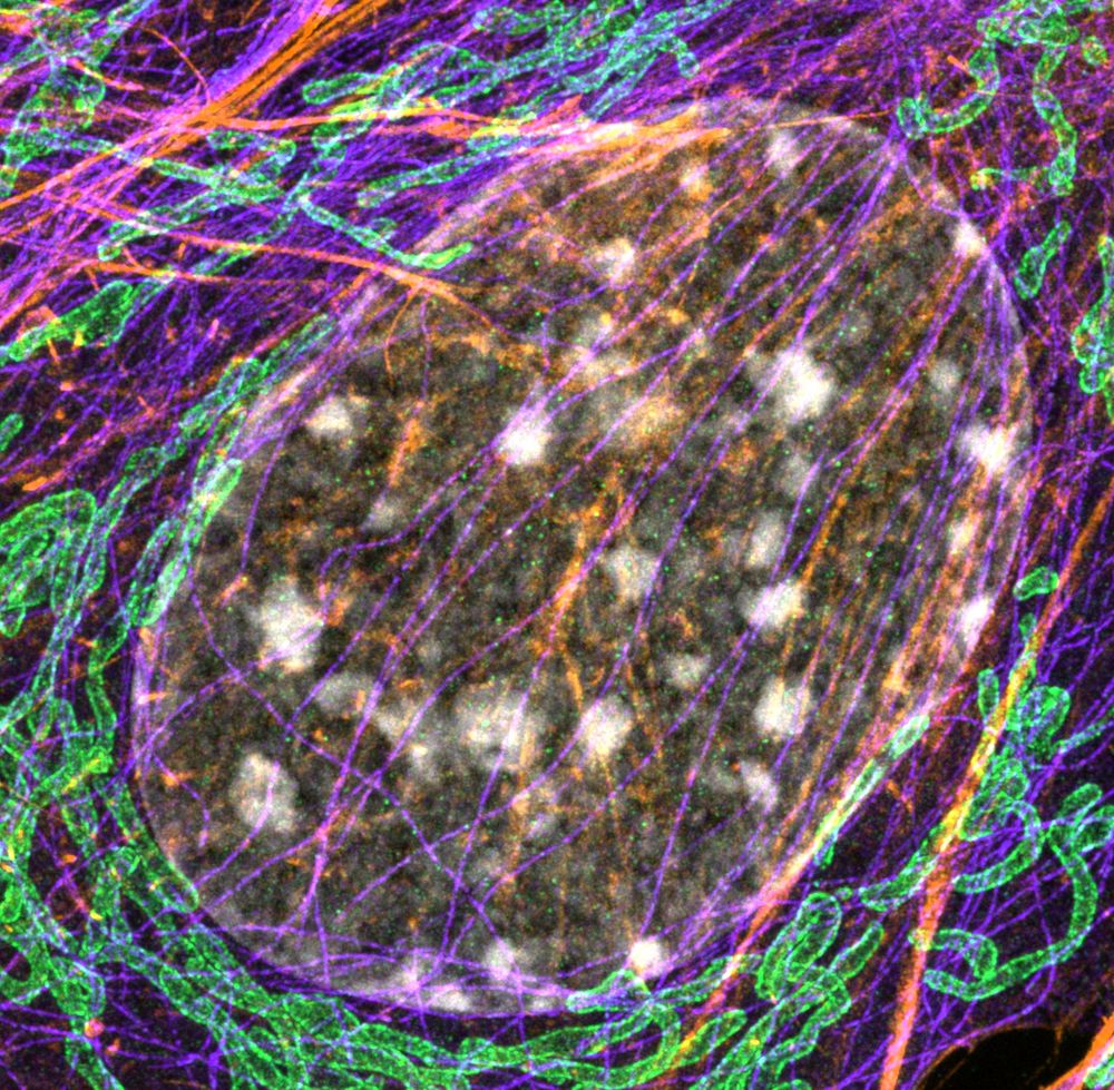
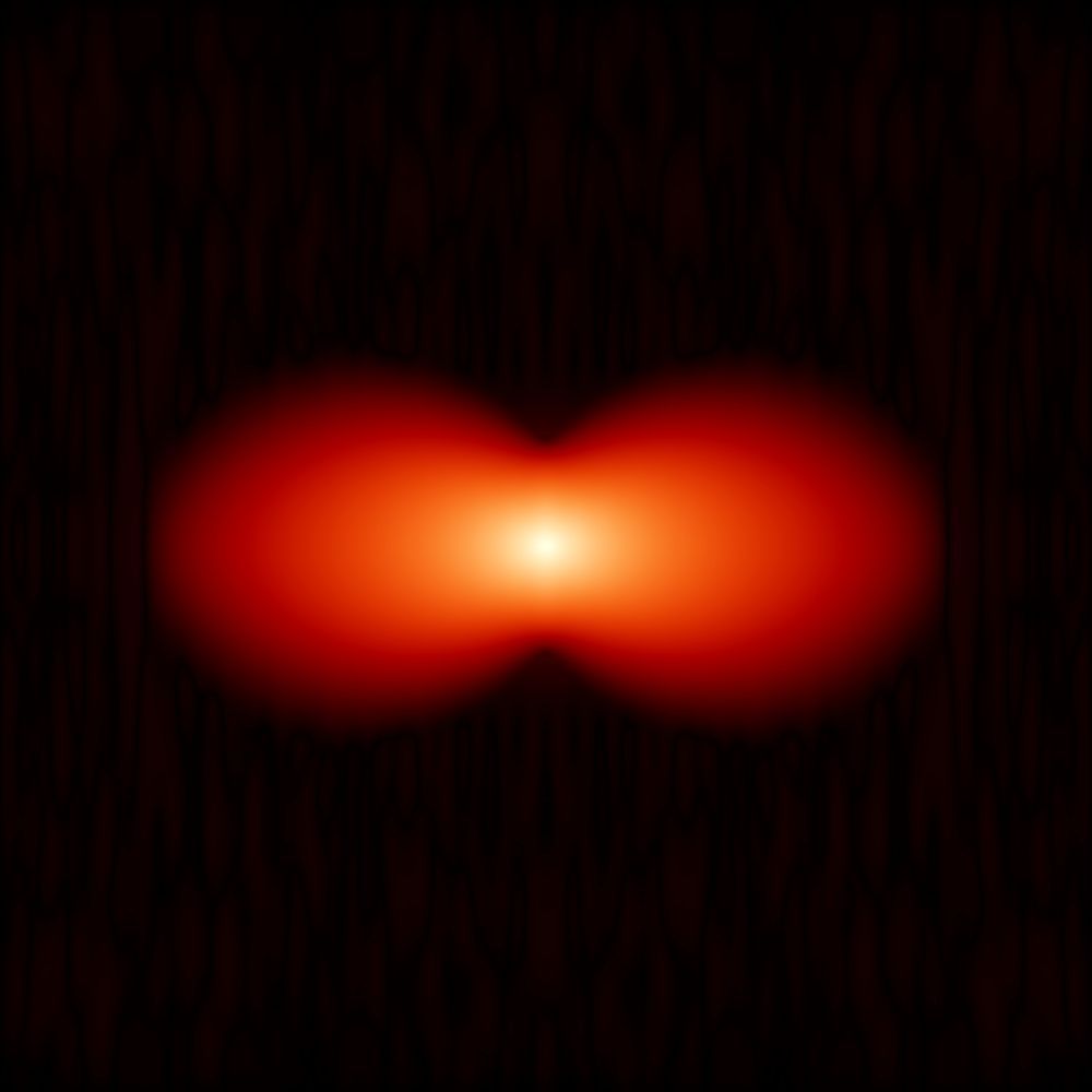
Bringing some more ExM to #FluorescenceFriday with what appears to be a frustrated cell division in fibroblasts.
More details in Alt-text.
More details in Alt-text.
September 26, 2025 at 2:08 PM
Bringing some more ExM to #FluorescenceFriday with what appears to be a frustrated cell division in fibroblasts.
More details in Alt-text.
More details in Alt-text.
Reposted by George Campbell
The WB-ExM protocol described here works with.... every sample we tested! Here a 3do quail embryo (white= pan-protein; red=MF20) www.biorxiv.org/content/10.1...

September 19, 2025 at 7:39 PM
The WB-ExM protocol described here works with.... every sample we tested! Here a 3do quail embryo (white= pan-protein; red=MF20) www.biorxiv.org/content/10.1...
Reposted by George Campbell
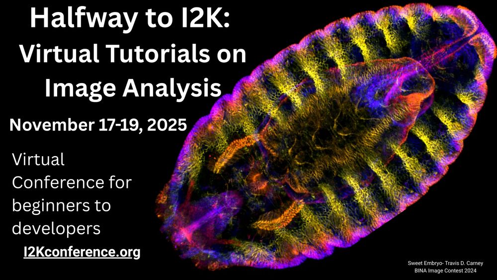
Halfway to I2K is BACK, friends of all kinds! Last year, 650 people attended 30+ TOTALLY FREE image analysis workshops of all kinds, across many timezones.
If you make image analysis software and want to teach it, workshop submissions are open now! We'd love to have your tool highlighted.
If you make image analysis software and want to teach it, workshop submissions are open now! We'd love to have your tool highlighted.

#HappyFluorescenceFriday!
#microscopycommunity- want to learn open source image analysis or share your knowledge to help others? We’ve got a FREE virtual workshop Nov 17-19! Now accepting workshop session applications!
Learn more & sign up: buff.ly/esGIotD
#microscopycommunity- want to learn open source image analysis or share your knowledge to help others? We’ve got a FREE virtual workshop Nov 17-19! Now accepting workshop session applications!
Learn more & sign up: buff.ly/esGIotD
September 23, 2025 at 3:48 PM
Halfway to I2K is BACK, friends of all kinds! Last year, 650 people attended 30+ TOTALLY FREE image analysis workshops of all kinds, across many timezones.
If you make image analysis software and want to teach it, workshop submissions are open now! We'd love to have your tool highlighted.
If you make image analysis software and want to teach it, workshop submissions are open now! We'd love to have your tool highlighted.
Reposted by George Campbell

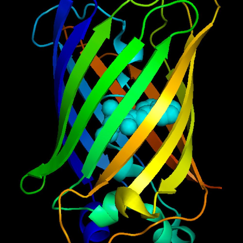
@joachimgoedhart.bsky.social and Flurescent protein colleagues--help! With advice from imaging freinds, we tagged a protein of interest with "tagRFP", which we now know from its protein sequence and digging is a derivative of
eqFP578 from Entacmaea quadricolor. We want an antibody to it but 1/n
eqFP578 from Entacmaea quadricolor. We want an antibody to it but 1/n
September 19, 2025 at 6:55 PM
@joachimgoedhart.bsky.social and Flurescent protein colleagues--help! With advice from imaging freinds, we tagged a protein of interest with "tagRFP", which we now know from its protein sequence and digging is a derivative of
eqFP578 from Entacmaea quadricolor. We want an antibody to it but 1/n
eqFP578 from Entacmaea quadricolor. We want an antibody to it but 1/n
Time to add some ExM to #FluorescenceFriday ! Sample description in alt-text.
September 19, 2025 at 1:16 PM
Time to add some ExM to #FluorescenceFriday ! Sample description in alt-text.
Reposted by George Campbell
Alright I have something useful for you to do.
Think of any teachers and librarians you know. Do they know about Skype a scientist? Our program matches classrooms & other groups w/scientists for virtual Q&As. It’s free!
They can sign up here!
www.skypeascientist.com/sign-up.html
#Edusky #StemED
Think of any teachers and librarians you know. Do they know about Skype a scientist? Our program matches classrooms & other groups w/scientists for virtual Q&As. It’s free!
They can sign up here!
www.skypeascientist.com/sign-up.html
#Edusky #StemED

September 18, 2025 at 1:21 PM
Alright I have something useful for you to do.
Think of any teachers and librarians you know. Do they know about Skype a scientist? Our program matches classrooms & other groups w/scientists for virtual Q&As. It’s free!
They can sign up here!
www.skypeascientist.com/sign-up.html
#Edusky #StemED
Think of any teachers and librarians you know. Do they know about Skype a scientist? Our program matches classrooms & other groups w/scientists for virtual Q&As. It’s free!
They can sign up here!
www.skypeascientist.com/sign-up.html
#Edusky #StemED
After seeing
@miguelcmestre.bsky.social @lancasterlab.bsky.social @jamesdmanton.bsky.social
's paper this week on deep and accessible ExM imaging with cerebral organoids, I think it's a good time to repost this Starter Pack of Expansion Microscopy people!
go.bsky.app/Qxks9WD
@miguelcmestre.bsky.social @lancasterlab.bsky.social @jamesdmanton.bsky.social
's paper this week on deep and accessible ExM imaging with cerebral organoids, I think it's a good time to repost this Starter Pack of Expansion Microscopy people!
go.bsky.app/Qxks9WD
September 17, 2025 at 10:04 PM
After seeing
@miguelcmestre.bsky.social @lancasterlab.bsky.social @jamesdmanton.bsky.social
's paper this week on deep and accessible ExM imaging with cerebral organoids, I think it's a good time to repost this Starter Pack of Expansion Microscopy people!
go.bsky.app/Qxks9WD
@miguelcmestre.bsky.social @lancasterlab.bsky.social @jamesdmanton.bsky.social
's paper this week on deep and accessible ExM imaging with cerebral organoids, I think it's a good time to repost this Starter Pack of Expansion Microscopy people!
go.bsky.app/Qxks9WD
Reposted by George Campbell
We present a simple method to easily increase the imageable depth of an expansion microscopy gel on a typical inverted microscope ten-fold, using some carefully placed FEP film and a water dipping objective lens:

September 15, 2025 at 8:42 AM
We present a simple method to easily increase the imageable depth of an expansion microscopy gel on a typical inverted microscope ten-fold, using some carefully placed FEP film and a water dipping objective lens:
Reposted by George Campbell
🚨Publication alert🚨
My first, first-author paper is now out in @natphoton.nature.com! Our paper describes an iterative spectral unmixing algorithm and eight-channel camera-based hardware we developed enabling unmixing of low SNR live-cell data at video rates. www.nature.com/articles/s41...
My first, first-author paper is now out in @natphoton.nature.com! Our paper describes an iterative spectral unmixing algorithm and eight-channel camera-based hardware we developed enabling unmixing of low SNR live-cell data at video rates. www.nature.com/articles/s41...

Multispectral live-cell imaging with uncompromised spatiotemporal resolution - Nature Photonics
A tree-like arrangement of dichroic mirrors and multiple cameras coupled with an iterative spectral unmixing algorithm enables multispectral imaging of live cells in up to eight spectral channels with...
www.nature.com
September 16, 2025 at 10:20 AM
🚨Publication alert🚨
My first, first-author paper is now out in @natphoton.nature.com! Our paper describes an iterative spectral unmixing algorithm and eight-channel camera-based hardware we developed enabling unmixing of low SNR live-cell data at video rates. www.nature.com/articles/s41...
My first, first-author paper is now out in @natphoton.nature.com! Our paper describes an iterative spectral unmixing algorithm and eight-channel camera-based hardware we developed enabling unmixing of low SNR live-cell data at video rates. www.nature.com/articles/s41...
Reposted by George Campbell

In 2019, Anna Jost & @jencwaters.bsky.social reviewed best practices for validating quantitative #microscopy methods & discuss strategies to avoid unconscious bias in imaging experiments rupress.org/jcb/article/...
📕 In #Reproducibility & Best Practices in Cell Biology: rupress.org/jcb/collecti...
📕 In #Reproducibility & Best Practices in Cell Biology: rupress.org/jcb/collecti...

September 11, 2025 at 2:06 PM
In 2019, Anna Jost & @jencwaters.bsky.social reviewed best practices for validating quantitative #microscopy methods & discuss strategies to avoid unconscious bias in imaging experiments rupress.org/jcb/article/...
📕 In #Reproducibility & Best Practices in Cell Biology: rupress.org/jcb/collecti...
📕 In #Reproducibility & Best Practices in Cell Biology: rupress.org/jcb/collecti...
Reposted by George Campbell
OK--fluorescent protein friends. We want an antibody to TagRFP that works in immunofluorescence. Any suggestions? Re-post please
September 9, 2025 at 6:43 PM
OK--fluorescent protein friends. We want an antibody to TagRFP that works in immunofluorescence. Any suggestions? Re-post please
Reposted by George Campbell
This 2009 article by @jencwaters.bsky.social discusses the parameters of digital image acquisition that affect the accuracy & precision of quantitative fluorescence #microscopy measurements rupress.org/jcb/article/...
In #Reproducibility & Best Practices in Cell Biology: rupress.org/jcb/collecti...
In #Reproducibility & Best Practices in Cell Biology: rupress.org/jcb/collecti...

September 8, 2025 at 2:05 PM
This 2009 article by @jencwaters.bsky.social discusses the parameters of digital image acquisition that affect the accuracy & precision of quantitative fluorescence #microscopy measurements rupress.org/jcb/article/...
In #Reproducibility & Best Practices in Cell Biology: rupress.org/jcb/collecti...
In #Reproducibility & Best Practices in Cell Biology: rupress.org/jcb/collecti...