


Josiah Passmore
@jpassmore.bsky.social
Making cells run in circles, and gels with squares in them.
Automated optogenetics, Smart microsopy, Expansion microscopy, GelMap.
Postdoc @ Utrecht University | visualise.bio.
Automated optogenetics, Smart microsopy, Expansion microscopy, GelMap.
Postdoc @ Utrecht University | visualise.bio.
Pinned
Josiah Passmore
@jpassmore.bsky.social
· Dec 17
Advances in microscopy mean we can now do more than just observe biology—we can control it. But how far can we really push this in mammalian cells with all their (beautiful but annoying) heterogeneity? 🧪🔬(🧵)
Reposted by Josiah Passmore
🚨 Job Alert! 🚨
We have an opening at our Microscopy Core Facility in Turku 🇫🇮.
If you love imaging 🔬 and helping scientists succeed, we want you! 👇
abo.rekrytointi.com/paikat/index...
#Microscopy #ScienceJobs #CoreFacility #Imaging
@turkubioscience.bsky.social
We have an opening at our Microscopy Core Facility in Turku 🇫🇮.
If you love imaging 🔬 and helping scientists succeed, we want you! 👇
abo.rekrytointi.com/paikat/index...
#Microscopy #ScienceJobs #CoreFacility #Imaging
@turkubioscience.bsky.social

Laboratory Engineer (Senior), in Advanced Imaging, 1.5.2026-31.12.2028
abo.rekrytointi.com
January 15, 2026 at 2:04 PM
🚨 Job Alert! 🚨
We have an opening at our Microscopy Core Facility in Turku 🇫🇮.
If you love imaging 🔬 and helping scientists succeed, we want you! 👇
abo.rekrytointi.com/paikat/index...
#Microscopy #ScienceJobs #CoreFacility #Imaging
@turkubioscience.bsky.social
We have an opening at our Microscopy Core Facility in Turku 🇫🇮.
If you love imaging 🔬 and helping scientists succeed, we want you! 👇
abo.rekrytointi.com/paikat/index...
#Microscopy #ScienceJobs #CoreFacility #Imaging
@turkubioscience.bsky.social
Reposted by Josiah Passmore
Won best thesis award with this video! 4 years of research and play in 3 minutes :')
youtu.be/so0fPlK1-LM
youtu.be/so0fPlK1-LM

Smart microscopy, computer vision, and controlling cells with light -- my PhD thesis in 3 minutes
YouTube video by Pertz Lab
youtu.be
January 7, 2026 at 11:27 AM
Won best thesis award with this video! 4 years of research and play in 3 minutes :')
youtu.be/so0fPlK1-LM
youtu.be/so0fPlK1-LM
Reposted by Josiah Passmore
🔬📊 PhD (100%) – Statistics & ML for self-driving microscopy
Joint PhD with David Ginsbourger (Stats) & Pertz Lab (Cell Biology).
Gaussian Processes, Bayesian design, active learning on live-cell experiments.
ohws.prospective.ch/public/v1/jo...
#PhD #Statistics #MachineLearning #Bayesian
Joint PhD with David Ginsbourger (Stats) & Pertz Lab (Cell Biology).
Gaussian Processes, Bayesian design, active learning on live-cell experiments.
ohws.prospective.ch/public/v1/jo...
#PhD #Statistics #MachineLearning #Bayesian

Uni Bern: PhD Position in Statistics with a focus on Statistical Machine Learning for Self-Driving Microscopy
ohws.prospective.ch
December 21, 2025 at 2:48 PM
🔬📊 PhD (100%) – Statistics & ML for self-driving microscopy
Joint PhD with David Ginsbourger (Stats) & Pertz Lab (Cell Biology).
Gaussian Processes, Bayesian design, active learning on live-cell experiments.
ohws.prospective.ch/public/v1/jo...
#PhD #Statistics #MachineLearning #Bayesian
Joint PhD with David Ginsbourger (Stats) & Pertz Lab (Cell Biology).
Gaussian Processes, Bayesian design, active learning on live-cell experiments.
ohws.prospective.ch/public/v1/jo...
#PhD #Statistics #MachineLearning #Bayesian
Reposted by Josiah Passmore

Cells can sense the geometry of their environment, from tiny subcellular features to whole-tissue patterns. How can we engineer environments to systematically probe these responses?
🧵 I'm @lhinderling.bsky.social from @olivierpertz.bsky.social lab, let’s do a short tour on microfabrication!
🧵 I'm @lhinderling.bsky.social from @olivierpertz.bsky.social lab, let’s do a short tour on microfabrication!
December 14, 2025 at 8:08 AM
Cells can sense the geometry of their environment, from tiny subcellular features to whole-tissue patterns. How can we engineer environments to systematically probe these responses?
🧵 I'm @lhinderling.bsky.social from @olivierpertz.bsky.social lab, let’s do a short tour on microfabrication!
🧵 I'm @lhinderling.bsky.social from @olivierpertz.bsky.social lab, let’s do a short tour on microfabrication!
Reposted by Josiah Passmore

We’re excited to announce the opening of the Geneva Expansion Microscopy Facility (GenExM)!
GenExM is a full-service U-ExM platform (U-ExM, Cryo-ExM, iU-ExM), delivering nanoscale imaging.
Led by Dr. Olivier Mercey, GenExM welcomes academic & industrial collaborations.
👉 www.unige.ch/genexm/
GenExM is a full-service U-ExM platform (U-ExM, Cryo-ExM, iU-ExM), delivering nanoscale imaging.
Led by Dr. Olivier Mercey, GenExM welcomes academic & industrial collaborations.
👉 www.unige.ch/genexm/

December 10, 2025 at 8:43 PM
We’re excited to announce the opening of the Geneva Expansion Microscopy Facility (GenExM)!
GenExM is a full-service U-ExM platform (U-ExM, Cryo-ExM, iU-ExM), delivering nanoscale imaging.
Led by Dr. Olivier Mercey, GenExM welcomes academic & industrial collaborations.
👉 www.unige.ch/genexm/
GenExM is a full-service U-ExM platform (U-ExM, Cryo-ExM, iU-ExM), delivering nanoscale imaging.
Led by Dr. Olivier Mercey, GenExM welcomes academic & industrial collaborations.
👉 www.unige.ch/genexm/
Reposted by Josiah Passmore

Finally, the first public release of #BigVolumeBrowser, so after teasers, you can try it yourself. For details, please check the announcement post (1/2)
forum.image.sc/t/bigvolumeb...
forum.image.sc/t/bigvolumeb...

BigVolumeBrowser: a new 3D multi volume/mesh/point clould (SMLM) data viewer
Hello everyone, I’d like to share with you another 3D viewer for FIJI, BigVolumeBrowser (full documentation link). It‘s a first initial public release, so there is still space for improvements. Le...
forum.image.sc
November 21, 2025 at 9:29 AM
Finally, the first public release of #BigVolumeBrowser, so after teasers, you can try it yourself. For details, please check the announcement post (1/2)
forum.image.sc/t/bigvolumeb...
forum.image.sc/t/bigvolumeb...
Reposted by Josiah Passmore
🚨Our collaboration with @centriolelab.bsky.social & @gautamdey.bsky.social is out today in @cp-cell.bsky.social
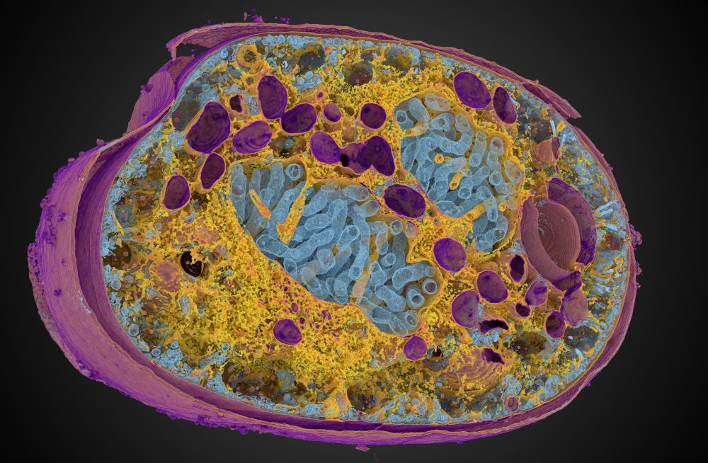
We show that #Expansion #Microscopy is a broad-spectrum modality for Euks, enabling 3D phenotypic maps rooted to phylogeny.
#ProtistsOnSky #SciComm #SciSky
www.cell.com/cell/fulltex...
We show that #Expansion #Microscopy is a broad-spectrum modality for Euks, enabling 3D phenotypic maps rooted to phylogeny.
#ProtistsOnSky #SciComm #SciSky
www.cell.com/cell/fulltex...
October 31, 2025 at 2:42 PM
🚨Our collaboration with @centriolelab.bsky.social & @gautamdey.bsky.social is out today in @cp-cell.bsky.social
We show that #Expansion #Microscopy is a broad-spectrum modality for Euks, enabling 3D phenotypic maps rooted to phylogeny.
#ProtistsOnSky #SciComm #SciSky
www.cell.com/cell/fulltex...
We show that #Expansion #Microscopy is a broad-spectrum modality for Euks, enabling 3D phenotypic maps rooted to phylogeny.
#ProtistsOnSky #SciComm #SciSky
www.cell.com/cell/fulltex...
Reposted by Josiah Passmore
E11 Bio is excited to unveil PRISM technology for mapping brain wiring with simple light microscopes. Today, brain mapping in humans and other mammals is bottlenecked by accurate neuron tracing. PRISM uses molecular ID codes and AI to help neurons trace themselves.
Read more: e11.bio/blog/prism
Read more: e11.bio/blog/prism
October 1, 2025 at 2:16 PM
E11 Bio is excited to unveil PRISM technology for mapping brain wiring with simple light microscopes. Today, brain mapping in humans and other mammals is bottlenecked by accurate neuron tracing. PRISM uses molecular ID codes and AI to help neurons trace themselves.
Read more: e11.bio/blog/prism
Read more: e11.bio/blog/prism
Reposted by Josiah Passmore

Now out on bioRxiv. 🥳My research on #cytokinesis, averaging thousands of #ExM images🔬, creating a dynamic atlas of cytokinesis 🦠⏳. Here's an animated sneak peek of what we found. Better resolution on bioRxiv😄 #PSFoftheGIF
September 28, 2025 at 2:15 PM
Now out on bioRxiv. 🥳My research on #cytokinesis, averaging thousands of #ExM images🔬, creating a dynamic atlas of cytokinesis 🦠⏳. Here's an animated sneak peek of what we found. Better resolution on bioRxiv😄 #PSFoftheGIF
Reposted by Josiah Passmore
Our new preprint is about the airway epithelium microtubule network, cilia, basal body protein composition, averaging of volumetric fluorescence data, and expansion microscopy.
Four years of very hard work from our very talented Emma van Grinsven. www.biorxiv.org/content/10.1... (1/N)
Four years of very hard work from our very talented Emma van Grinsven. www.biorxiv.org/content/10.1... (1/N)
September 5, 2025 at 3:37 PM
Our new preprint is about the airway epithelium microtubule network, cilia, basal body protein composition, averaging of volumetric fluorescence data, and expansion microscopy.
Four years of very hard work from our very talented Emma van Grinsven. www.biorxiv.org/content/10.1... (1/N)
Four years of very hard work from our very talented Emma van Grinsven. www.biorxiv.org/content/10.1... (1/N)
Reposted by Josiah Passmore
Excited to share my postdoc work from the Eggeling lab in Jena (@leibnizipht.bsky.social, #FSUJena, #KTHuniversity) on bringing smart microscopy to super-resolution MINFLUX: event-triggered MINFLUX microscopy.

September 2, 2025 at 8:59 AM
Excited to share my postdoc work from the Eggeling lab in Jena (@leibnizipht.bsky.social, #FSUJena, #KTHuniversity) on bringing smart microscopy to super-resolution MINFLUX: event-triggered MINFLUX microscopy.
Reposted by Josiah Passmore
Collective cell motion has many forms, but rotation is the coolest of them all.
I'm @onenimesa.bsky.social , and in this short🧵, I'll highlight some instances of global tissue rotation like this one from @BauschLab
I'm @onenimesa.bsky.social , and in this short🧵, I'll highlight some instances of global tissue rotation like this one from @BauschLab
August 23, 2025 at 7:00 AM
Collective cell motion has many forms, but rotation is the coolest of them all.
I'm @onenimesa.bsky.social , and in this short🧵, I'll highlight some instances of global tissue rotation like this one from @BauschLab
I'm @onenimesa.bsky.social , and in this short🧵, I'll highlight some instances of global tissue rotation like this one from @BauschLab
Reposted by Josiah Passmore
Automated optogenetic control of hundreds of cells in parallel. Each cell is individually steered, collectively acting as a "tissue printer". Preprint & code out! www.biorxiv.org/content/10.1...
August 21, 2025 at 8:16 PM
Automated optogenetic control of hundreds of cells in parallel. Each cell is individually steered, collectively acting as a "tissue printer". Preprint & code out! www.biorxiv.org/content/10.1...
Reposted by Josiah Passmore
🚨Scaling multiplexed imaging 📈 We are excited to share Pathology-oriented multiPlexing (PathoPlex). Now out in @nature.com: www.nature.com/articles/s41...
🧵Walk-through thread below ⬇️
🧵Walk-through thread below ⬇️
July 18, 2025 at 1:31 PM
🚨Scaling multiplexed imaging 📈 We are excited to share Pathology-oriented multiPlexing (PathoPlex). Now out in @nature.com: www.nature.com/articles/s41...
🧵Walk-through thread below ⬇️
🧵Walk-through thread below ⬇️
Reposted by Josiah Passmore
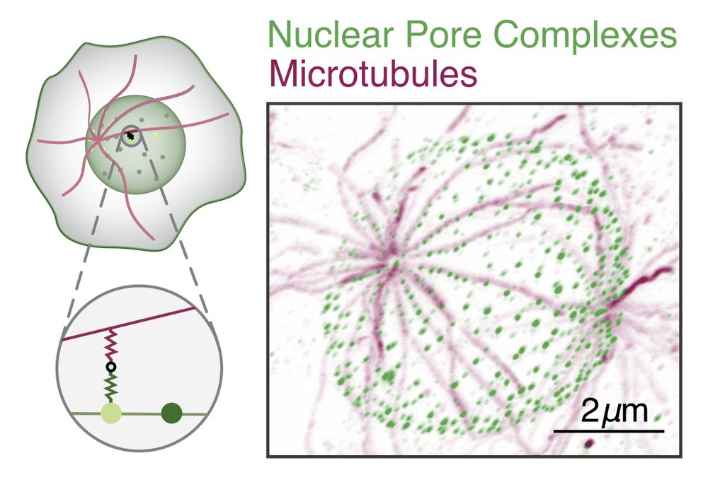
9/ The observed patterns matched our model, and their parameters place these systems near the predicted optimal filtering regime -- These NPCs may act as efficient spatial thresholding filters! #Microscopy #QuantBio #Microtubules #UExM
June 18, 2025 at 5:07 PM
9/ The observed patterns matched our model, and their parameters place these systems near the predicted optimal filtering regime -- These NPCs may act as efficient spatial thresholding filters! #Microscopy #QuantBio #Microtubules #UExM
Reposted by Josiah Passmore

June 6, 2025 at 3:14 PM
Reposted by Josiah Passmore
In the context of our @reviewcommons.org revision process, I'm happy to announce Microscopy Nodes v2.2.0!
This packs lots of new fun features, including new color management 🌈, clearer transparency handling 🫥, custom default settings 🔧 and more!
Preprint at doi.org/10.1101/2025...
This packs lots of new fun features, including new color management 🌈, clearer transparency handling 🫥, custom default settings 🔧 and more!
Preprint at doi.org/10.1101/2025...
May 23, 2025 at 1:13 PM
In the context of our @reviewcommons.org revision process, I'm happy to announce Microscopy Nodes v2.2.0!
This packs lots of new fun features, including new color management 🌈, clearer transparency handling 🫥, custom default settings 🔧 and more!
Preprint at doi.org/10.1101/2025...
This packs lots of new fun features, including new color management 🌈, clearer transparency handling 🫥, custom default settings 🔧 and more!
Preprint at doi.org/10.1101/2025...
Reposted by Josiah Passmore

I’m excited to announce that my paper describing non-canonical mitotic mechanisms in the early mouse embryo is out in @science.org ! (link at end of 🧵)
May 23, 2025 at 7:57 PM
I’m excited to announce that my paper describing non-canonical mitotic mechanisms in the early mouse embryo is out in @science.org ! (link at end of 🧵)
Reposted by Josiah Passmore
Thanks to expansion microscopy, clever labeling, and modern segmentation approaches, doing connectomics with #light #microscopy has become feasible - huge congratulations Mojtaba & the Danzl lab at @istaresearch.bsky.social !
www.nature.com/articles/s41...
www.nature.com/articles/s41...
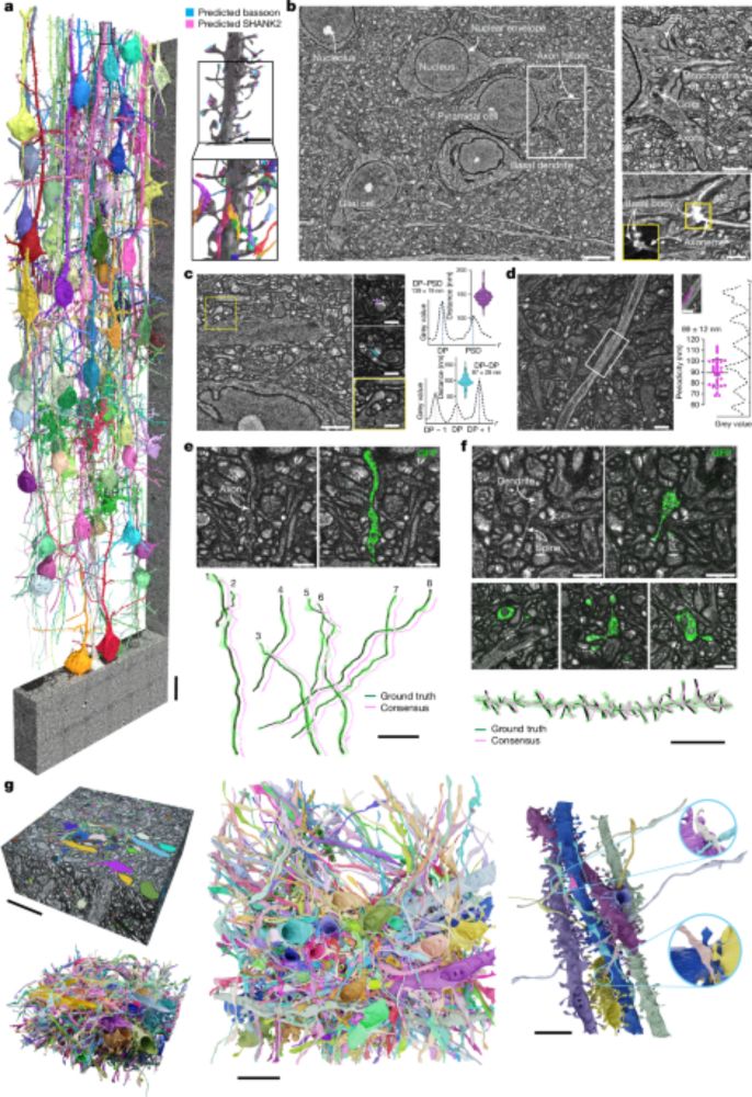
Light-microscopy-based connectomic reconstruction of mammalian brain tissue - Nature
A technique called LICONN (light-microscopy-based connectomics) allows mapping of brain tissue at synapse level and simultaneous measurement of molecular information, thus enabling quantification of c...
www.nature.com
May 8, 2025 at 2:52 AM
Thanks to expansion microscopy, clever labeling, and modern segmentation approaches, doing connectomics with #light #microscopy has become feasible - huge congratulations Mojtaba & the Danzl lab at @istaresearch.bsky.social !
www.nature.com/articles/s41...
www.nature.com/articles/s41...
Reposted by Josiah Passmore

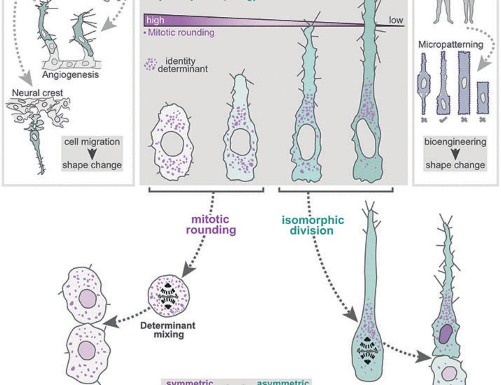
My first post on Bluesky! Very excited to share our work just published in @science.org. We find that “Interphase cell morphology defines the mode, symmetry, and outcome of mitosis” - in angiogenesis and other tissues! www.science.org/doi/abs/10.1... www.science.org/doi/abs/10.1...
Interphase cell morphology defines the mode, symmetry, and outcome of mitosis
During tissue formation, dynamic cell shape changes drive morphogenesis while asymmetric divisions create cellular diversity. We found that the shifts in cell morphology that shape tissues could conco...
www.science.org
May 6, 2025 at 8:39 PM
My first post on Bluesky! Very excited to share our work just published in @science.org. We find that “Interphase cell morphology defines the mode, symmetry, and outcome of mitosis” - in angiogenesis and other tissues! www.science.org/doi/abs/10.1... www.science.org/doi/abs/10.1...
Reposted by Josiah Passmore

If you're curious about how cells make decisions in complex environments, and how mathematical models can capture such behaviour, our preprint is now live!
Check it out here: www.biorxiv.org/content/10.1...
Check it out here: www.biorxiv.org/content/10.1...

Investigating Local Negative Feedback of Rac Activity by Mathematical Models and Cell Motility Simulations
For polarization and directed migration, cells use a combination of local positive feedback and long-range inhibition. We have previously used mathematical models to show the ability of this core…
www.biorxiv.org
May 6, 2025 at 6:09 PM
If you're curious about how cells make decisions in complex environments, and how mathematical models can capture such behaviour, our preprint is now live!
Check it out here: www.biorxiv.org/content/10.1...
Check it out here: www.biorxiv.org/content/10.1...
Reposted by Josiah Passmore
🚀🔬🦠 Releasing 🤖Cellpose-SAM🤖, a cellular segmentation algorithm with superhuman generalization 🦸♀️. Try it now on 🤗 huggingface.co/spaces/mouse...
paper: www.biorxiv.org/content/10.1...
w/ @computingnature.bsky.social 1/n
paper: www.biorxiv.org/content/10.1...
w/ @computingnature.bsky.social 1/n
May 3, 2025 at 7:12 PM
🚀🔬🦠 Releasing 🤖Cellpose-SAM🤖, a cellular segmentation algorithm with superhuman generalization 🦸♀️. Try it now on 🤗 huggingface.co/spaces/mouse...
paper: www.biorxiv.org/content/10.1...
w/ @computingnature.bsky.social 1/n
paper: www.biorxiv.org/content/10.1...
w/ @computingnature.bsky.social 1/n
Reposted by Josiah Passmore
We may not have really squared the circle — but we came close! 😉 In our @NaturePhysics paper w/ @fakhrilab.bsky.social, light-triggered membrane excitability enables programmable shapes — a step toward engineering living matter.
🔗 www.nature.com/articles/s41...
#biophysics #syntheticcell #softmatter
🔗 www.nature.com/articles/s41...
#biophysics #syntheticcell #softmatter
March 31, 2025 at 1:11 PM
We may not have really squared the circle — but we came close! 😉 In our @NaturePhysics paper w/ @fakhrilab.bsky.social, light-triggered membrane excitability enables programmable shapes — a step toward engineering living matter.
🔗 www.nature.com/articles/s41...
#biophysics #syntheticcell #softmatter
🔗 www.nature.com/articles/s41...
#biophysics #syntheticcell #softmatter
Reposted by Josiah Passmore
1/14 “Epithelial” & “Mesenchymal” aren’t binary categories—they form a spectrum, or better yet, a multidimensional space. This becomes especially clear in collective cell migration, which often depends on a finely tuned degree of “mesenchymal-ness”.
#cellbio #devbio #cellmigration
#cellbio #devbio #cellmigration
April 20, 2025 at 7:32 AM
1/14 “Epithelial” & “Mesenchymal” aren’t binary categories—they form a spectrum, or better yet, a multidimensional space. This becomes especially clear in collective cell migration, which often depends on a finely tuned degree of “mesenchymal-ness”.
#cellbio #devbio #cellmigration
#cellbio #devbio #cellmigration
Reposted by Josiah Passmore
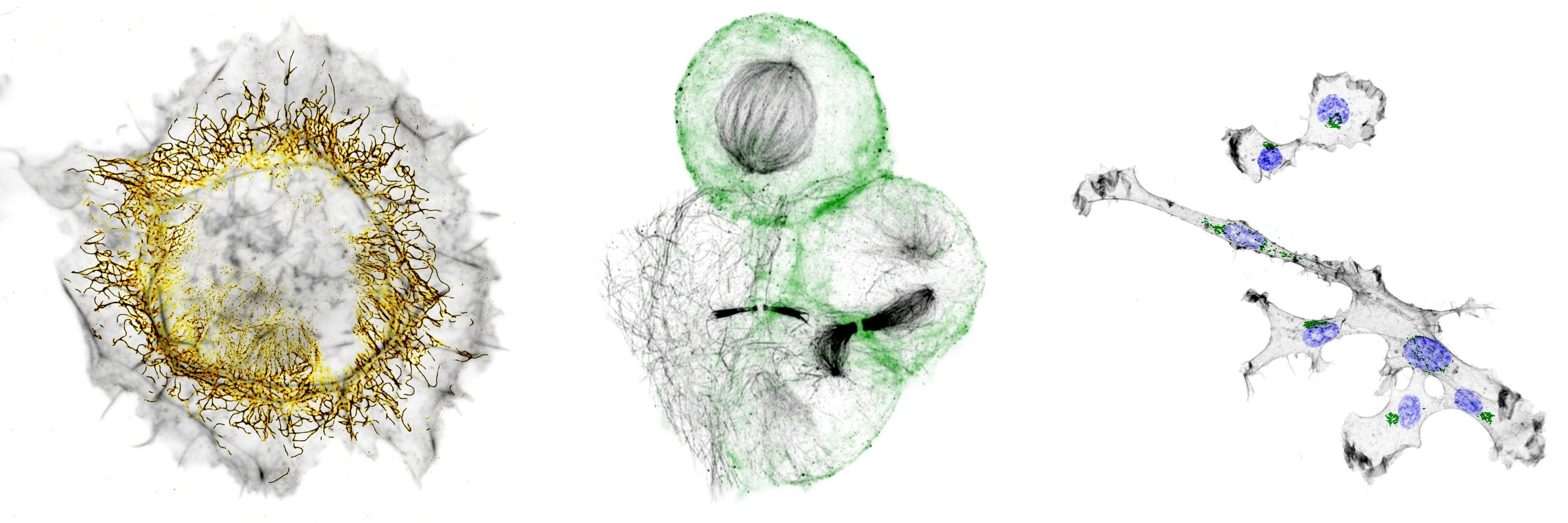
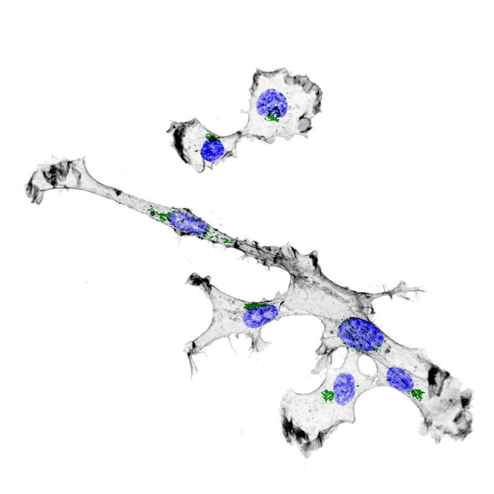
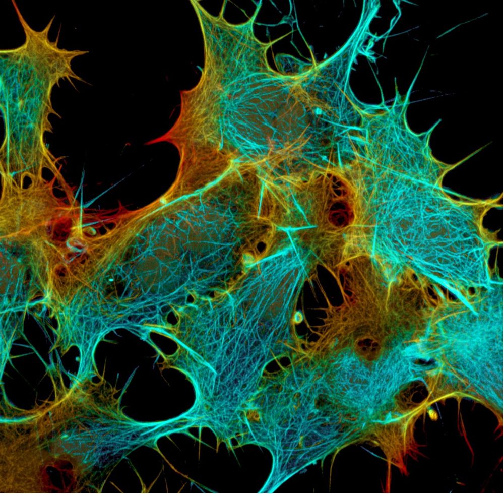
Happy to officially introduce FilaBuster - a strategy for rapid, light-mediated intermediate filament disassembly. Compatible with multiple IF types, modular in design, and precise enough to induce localized filament disassembly in live cells.
www.biorxiv.org/content/10.1...
www.biorxiv.org/content/10.1...
April 22, 2025 at 1:02 AM
Happy to officially introduce FilaBuster - a strategy for rapid, light-mediated intermediate filament disassembly. Compatible with multiple IF types, modular in design, and precise enough to induce localized filament disassembly in live cells.
www.biorxiv.org/content/10.1...
www.biorxiv.org/content/10.1...