


@pierrehener.bsky.social
Reposted
Very glad to be in Beijing at PKU for the Sino-French exchange on biomedical imaging infrastructures. Many thanks to @france-bioimaging.bsky.social, Laurent Bourdieu, Liangyi Chen and many others for the organization!

November 3, 2025 at 1:20 AM
Very glad to be in Beijing at PKU for the Sino-French exchange on biomedical imaging infrastructures. Many thanks to @france-bioimaging.bsky.social, Laurent Bourdieu, Liangyi Chen and many others for the organization!
Reposted
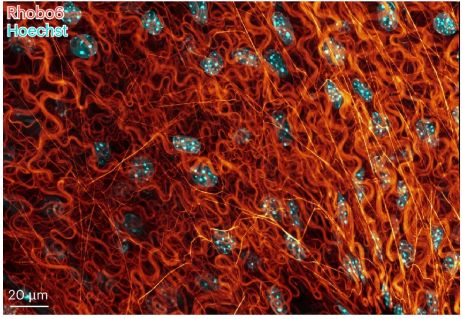
for #FluorescenceFriday : is your ExM sample too large to be imaged with high NA objectives? VIPS it! Very clever way to tackle one of the biggest challenge in large volumetric ExM www.science.org/doi/10.1126/...

Mesoscale volumetric fluorescence imaging at nanoscale resolution by photochemical sectioning
Optical nanoscopy of intact biological specimens has been transformed by recent advancements in hydrogel-based tissue clearing and expansion, enabling the imaging of cellular and subcellular structure...
www.science.org
October 17, 2025 at 12:29 PM
for #FluorescenceFriday : is your ExM sample too large to be imaged with high NA objectives? VIPS it! Very clever way to tackle one of the biggest challenge in large volumetric ExM www.science.org/doi/10.1126/...
Reposted
Great first poster session at #Mifobio2025 — thanks for the inspiring discussions! 🔬
All week long, come (re)discover our poster and explore #OpenCID, making #data #storage, #management & #sharing simpler.
See you soon!
@gdrimabio.bsky.social
@imag-ic.bsky.social
All week long, come (re)discover our poster and explore #OpenCID, making #data #storage, #management & #sharing simpler.
See you soon!
@gdrimabio.bsky.social
@imag-ic.bsky.social

October 11, 2025 at 11:01 AM
Great first poster session at #Mifobio2025 — thanks for the inspiring discussions! 🔬
All week long, come (re)discover our poster and explore #OpenCID, making #data #storage, #management & #sharing simpler.
See you soon!
@gdrimabio.bsky.social
@imag-ic.bsky.social
All week long, come (re)discover our poster and explore #OpenCID, making #data #storage, #management & #sharing simpler.
See you soon!
@gdrimabio.bsky.social
@imag-ic.bsky.social
Reposted

📰 #Mifobio dans la presse
👉 france3-regions.franceinfo.fr/nouvelle-aqu...
Merci @france3-naquitaine.bsky.social pour cet article sur #Mifobio2025
@cnrs.fr @cnrsingenierie.bsky.social @institutfresnel.bsky.social @healthcare.nikon.com @leicamicrosystems.bsky.social @abbelight.bsky.social
👉 france3-regions.franceinfo.fr/nouvelle-aqu...
Merci @france3-naquitaine.bsky.social pour cet article sur #Mifobio2025
@cnrs.fr @cnrsingenierie.bsky.social @institutfresnel.bsky.social @healthcare.nikon.com @leicamicrosystems.bsky.social @abbelight.bsky.social

"On est obligé de refuser des gens !" : 350 chercheurs et scientifiques du monde entier réunis dans les Landes pendant une semaine
350 chercheurs, ingénieurs, mathématiciens, chimistes, biologiques, physiciens... vont passer huit jours ensemble à Seignosse, du 10 au 17 octobre. L'objectif : partager leurs savoirs. Ce rassemblemen...
france3-regions.franceinfo.fr
October 11, 2025 at 7:55 AM
Reposted
🔥 It's Time ! #Mifobio2025
👉 imabio-cnrs.fr
#GDRImabio
@cnrs.fr
@cnrsingenierie.bsky.social
@cnrsbiologie.bsky.social
👉 imabio-cnrs.fr
#GDRImabio
@cnrs.fr
@cnrsingenierie.bsky.social
@cnrsbiologie.bsky.social

October 10, 2025 at 2:05 AM
Reposted
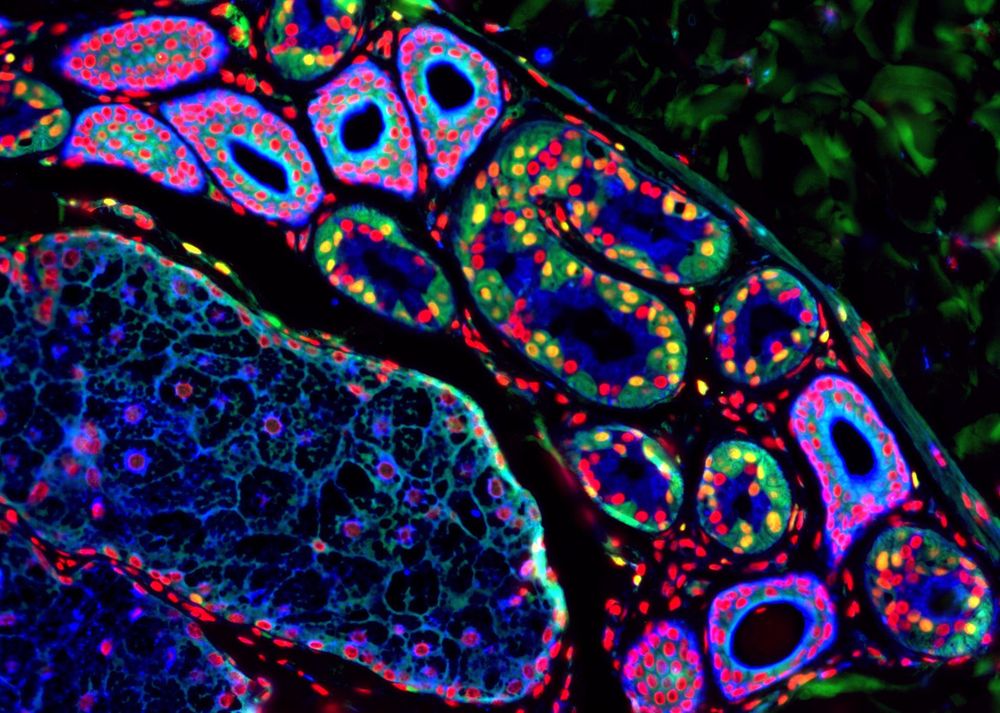
Have a look at this image from the lab, taken by @katkajerabkova.bsky.social, and vote for it as it is competing for the Grand Public Prize of the @sbcf.bsky.social

Cette très jolie image, réalisée par une équipe Inserm dirigée par Jacky Goetz, est en compétition dans la catégorie Grand Public pour l’élection de l’image de l’année 2025, organisée par @sbcf.bsky.social
July 25, 2025 at 3:11 PM
Have a look at this image from the lab, taken by @katkajerabkova.bsky.social, and vote for it as it is competing for the Grand Public Prize of the @sbcf.bsky.social
Reposted

🚨 New EMBO Practical Course!
Ultrastructure Expansion Microscopy: From Cells to Tissue 🔬
📅 20–24 Apr 2026 | 📍 EMBL Heidelberg
Co-organised with @banterlegroup.bsky.social @gautamdey.bsky.social
💡 Register your interest to get notified when registration opens:
🔗 www.embl.org/about/info/c...
Ultrastructure Expansion Microscopy: From Cells to Tissue 🔬
📅 20–24 Apr 2026 | 📍 EMBL Heidelberg
Co-organised with @banterlegroup.bsky.social @gautamdey.bsky.social
💡 Register your interest to get notified when registration opens:
🔗 www.embl.org/about/info/c...
July 21, 2025 at 9:07 AM
🚨 New EMBO Practical Course!
Ultrastructure Expansion Microscopy: From Cells to Tissue 🔬
📅 20–24 Apr 2026 | 📍 EMBL Heidelberg
Co-organised with @banterlegroup.bsky.social @gautamdey.bsky.social
💡 Register your interest to get notified when registration opens:
🔗 www.embl.org/about/info/c...
Ultrastructure Expansion Microscopy: From Cells to Tissue 🔬
📅 20–24 Apr 2026 | 📍 EMBL Heidelberg
Co-organised with @banterlegroup.bsky.social @gautamdey.bsky.social
💡 Register your interest to get notified when registration opens:
🔗 www.embl.org/about/info/c...
Reposted
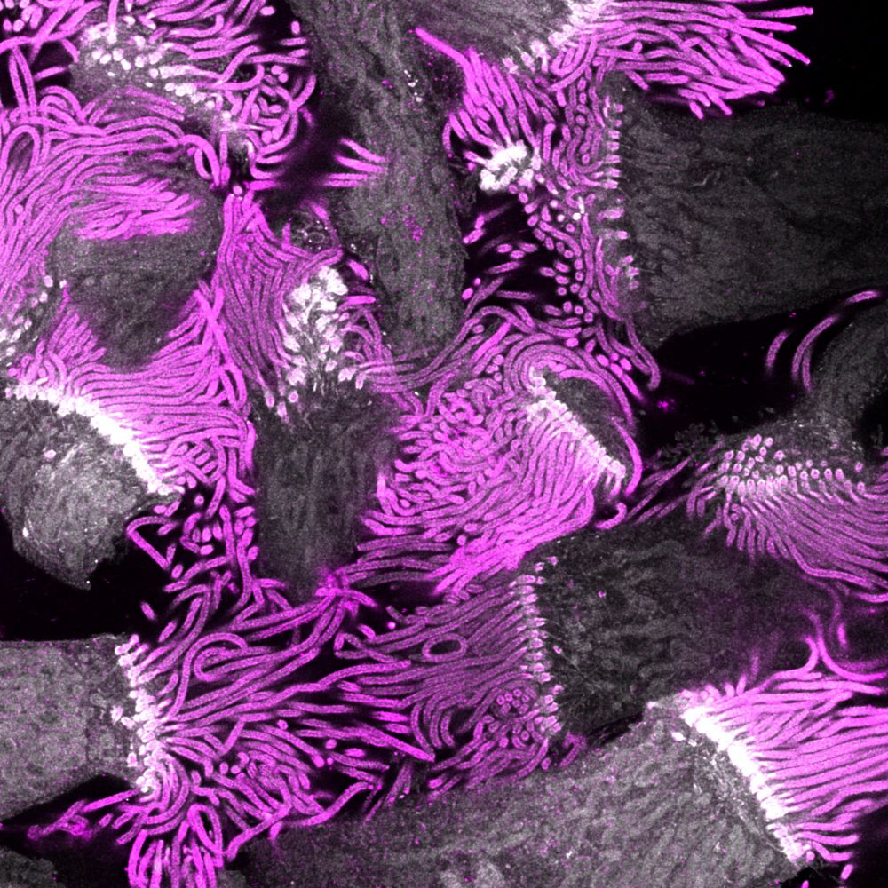
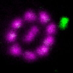
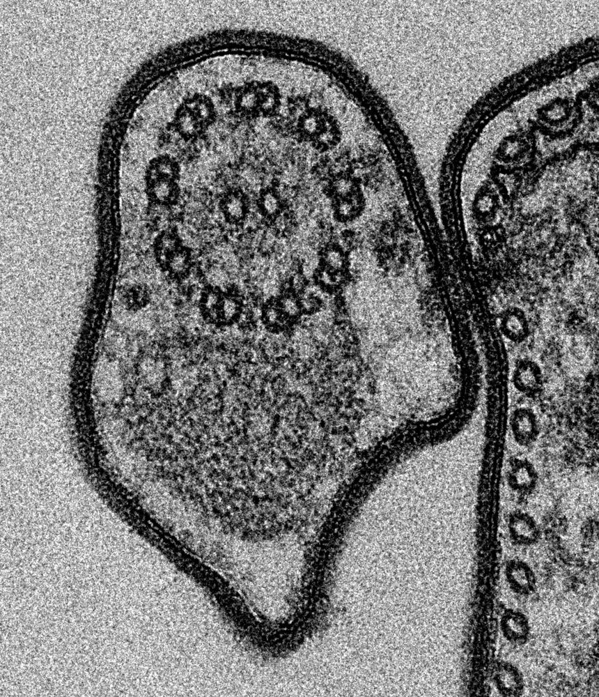
The nucleus is fantastic, but can it beat #cilia and #flagella 😉 here stained for tubulin (magenta) with an intraflagellar transport train (green) after #expansion microscopy and #STED or shown by classic TEM. JCS @jcellsci.bsky.social found the solution and is featuring both! 😍
July 17, 2025 at 9:02 AM
The nucleus is fantastic, but can it beat #cilia and #flagella 😉 here stained for tubulin (magenta) with an intraflagellar transport train (green) after #expansion microscopy and #STED or shown by classic TEM. JCS @jcellsci.bsky.social found the solution and is featuring both! 😍
Reposted

@cnrsecologie.bsky.social regrette profondément l'adoption de cette loi à la vision court-termiste & ses conséquences graves sur l’environnement, qui méprise santé & bien-être de la population & le rôle des espèces sauvages dans la prod. agricole. La communauté scientifique n'a pas été entendue.

Le Parlement a définitivement adopté mardi la proposition de loi agricole Duplomb-Menonville, comprenant entre autres une mesure décriée de réintroduction sous conditions d'un pesticide néonicotinoïde, et présentée comme une réponse à la colère agricole de 2024.

July 9, 2025 at 8:38 AM
@cnrsecologie.bsky.social regrette profondément l'adoption de cette loi à la vision court-termiste & ses conséquences graves sur l’environnement, qui méprise santé & bien-être de la population & le rôle des espèces sauvages dans la prod. agricole. La communauté scientifique n'a pas été entendue.
Reposted
July 3, 2025 at 8:05 AM
Reposted

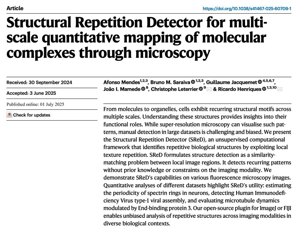
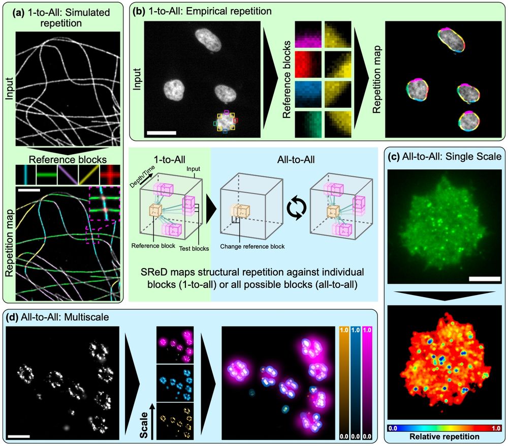
🔬👨💻📰 #SReD is out!
Automated structural detection for #ImageJ & #FIJI, from nano to macro ✨🐘. No training data, no bias - texture analysis with GPU acceleration!⚡️
Brainchild of @afonsomendes92.bsky.social and adventure w @christlet.bsky.social lab + friends.
Check: www.nature.com/articles/s41...
Automated structural detection for #ImageJ & #FIJI, from nano to macro ✨🐘. No training data, no bias - texture analysis with GPU acceleration!⚡️
Brainchild of @afonsomendes92.bsky.social and adventure w @christlet.bsky.social lab + friends.
Check: www.nature.com/articles/s41...
July 2, 2025 at 7:09 AM
🔬👨💻📰 #SReD is out!
Automated structural detection for #ImageJ & #FIJI, from nano to macro ✨🐘. No training data, no bias - texture analysis with GPU acceleration!⚡️
Brainchild of @afonsomendes92.bsky.social and adventure w @christlet.bsky.social lab + friends.
Check: www.nature.com/articles/s41...
Automated structural detection for #ImageJ & #FIJI, from nano to macro ✨🐘. No training data, no bias - texture analysis with GPU acceleration!⚡️
Brainchild of @afonsomendes92.bsky.social and adventure w @christlet.bsky.social lab + friends.
Check: www.nature.com/articles/s41...
Reposted
Finally out!, after a few months of delay, our protocol on ExM in plant roots is now available in The Plant Cell
academic.oup.com/plcell/artic...
Beautiful images in a great collaboration of the @bic-bordeaux.bsky.social with @emmanuellebayer.bsky.social and Magali Grison @lbm-bordeaux.bsky.social
academic.oup.com/plcell/artic...
Beautiful images in a great collaboration of the @bic-bordeaux.bsky.social with @emmanuellebayer.bsky.social and Magali Grison @lbm-bordeaux.bsky.social
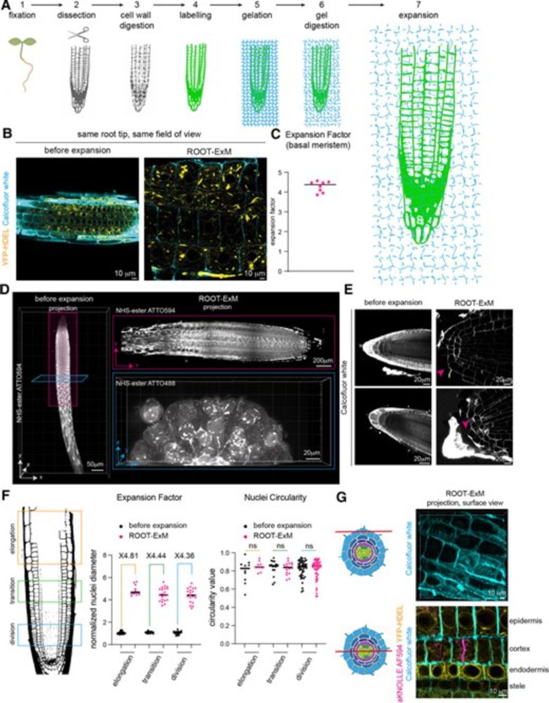
Root expansion microscopy: A robust method for super resolution imaging in Arabidopsis
Root expansion microscopy (ROOT-ExM) achieves super-resolution expansion microscopy in plants.
academic.oup.com
April 14, 2025 at 9:47 AM
Finally out!, after a few months of delay, our protocol on ExM in plant roots is now available in The Plant Cell
academic.oup.com/plcell/artic...
Beautiful images in a great collaboration of the @bic-bordeaux.bsky.social with @emmanuellebayer.bsky.social and Magali Grison @lbm-bordeaux.bsky.social
academic.oup.com/plcell/artic...
Beautiful images in a great collaboration of the @bic-bordeaux.bsky.social with @emmanuellebayer.bsky.social and Magali Grison @lbm-bordeaux.bsky.social
Reposted
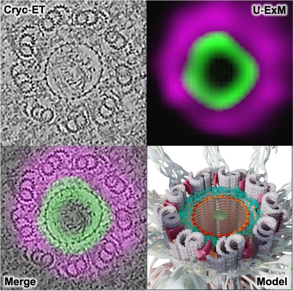
🚨 New preprint!
Using U-ExM + in situ cryo-ET, we show how C2CD3 builds an in-to-out radial architecture connecting the distal centriole lumen to its appendages. Great collab with @cellarchlab.com @chgenoud.bsky.social @stearnslab.bsky.social 🙌. #TeamTomo #UExM
www.biorxiv.org/content/10.1...
Using U-ExM + in situ cryo-ET, we show how C2CD3 builds an in-to-out radial architecture connecting the distal centriole lumen to its appendages. Great collab with @cellarchlab.com @chgenoud.bsky.social @stearnslab.bsky.social 🙌. #TeamTomo #UExM
www.biorxiv.org/content/10.1...
June 19, 2025 at 8:58 AM
🚨 New preprint!
Using U-ExM + in situ cryo-ET, we show how C2CD3 builds an in-to-out radial architecture connecting the distal centriole lumen to its appendages. Great collab with @cellarchlab.com @chgenoud.bsky.social @stearnslab.bsky.social 🙌. #TeamTomo #UExM
www.biorxiv.org/content/10.1...
Using U-ExM + in situ cryo-ET, we show how C2CD3 builds an in-to-out radial architecture connecting the distal centriole lumen to its appendages. Great collab with @cellarchlab.com @chgenoud.bsky.social @stearnslab.bsky.social 🙌. #TeamTomo #UExM
www.biorxiv.org/content/10.1...
Reposted

Proud to share our latest paper. doi.org/10.1016/j.cr...
Through the dedication of @glynnca.bsky.social and @cryingem.bsky.social we report a thorough method to image molecular organisation within hippocampus tissue.
Structural biology in tissue is well and truly here!
@rosfrankinst.bsky.social
Through the dedication of @glynnca.bsky.social and @cryingem.bsky.social we report a thorough method to image molecular organisation within hippocampus tissue.
Structural biology in tissue is well and truly here!
@rosfrankinst.bsky.social

June 17, 2025 at 10:59 AM
Proud to share our latest paper. doi.org/10.1016/j.cr...
Through the dedication of @glynnca.bsky.social and @cryingem.bsky.social we report a thorough method to image molecular organisation within hippocampus tissue.
Structural biology in tissue is well and truly here!
@rosfrankinst.bsky.social
Through the dedication of @glynnca.bsky.social and @cryingem.bsky.social we report a thorough method to image molecular organisation within hippocampus tissue.
Structural biology in tissue is well and truly here!
@rosfrankinst.bsky.social
Reposted
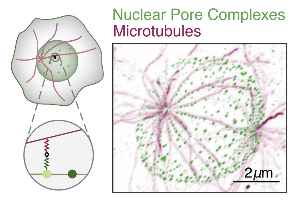
9/ The observed patterns matched our model, and their parameters place these systems near the predicted optimal filtering regime -- These NPCs may act as efficient spatial thresholding filters! #Microscopy #QuantBio #Microtubules #UExM
June 18, 2025 at 5:07 PM
9/ The observed patterns matched our model, and their parameters place these systems near the predicted optimal filtering regime -- These NPCs may act as efficient spatial thresholding filters! #Microscopy #QuantBio #Microtubules #UExM
Reposted
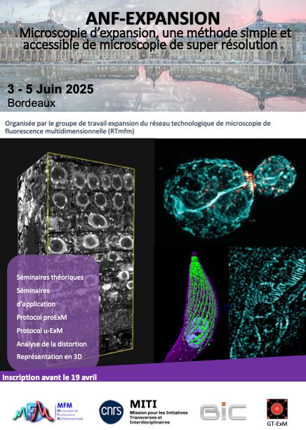
Three full days of #ExM in Bordeaux. Onsite practical course on expansion microscopy. We will be sharing very soon the theoretical and application courses.
June 1, 2025 at 9:18 PM
Three full days of #ExM in Bordeaux. Onsite practical course on expansion microscopy. We will be sharing very soon the theoretical and application courses.
Reposted
Thanks to expansion microscopy, clever labeling, and modern segmentation approaches, doing connectomics with #light #microscopy has become feasible - huge congratulations Mojtaba & the Danzl lab at @istaresearch.bsky.social !
www.nature.com/articles/s41...
www.nature.com/articles/s41...
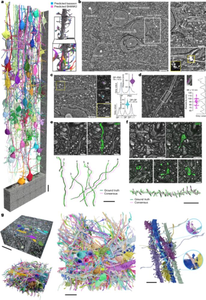
Light-microscopy-based connectomic reconstruction of mammalian brain tissue - Nature
A technique called LICONN (light-microscopy-based connectomics) allows mapping of brain tissue at synapse level and simultaneous measurement of molecular information, thus enabling quantification of c...
www.nature.com
May 8, 2025 at 2:52 AM
Thanks to expansion microscopy, clever labeling, and modern segmentation approaches, doing connectomics with #light #microscopy has become feasible - huge congratulations Mojtaba & the Danzl lab at @istaresearch.bsky.social !
www.nature.com/articles/s41...
www.nature.com/articles/s41...
Reposted
🚨🔬💗Whether investigating cell organelles or mapping proteins, together with Victor Puelles's lab we lay a roadmap for selecting optimal #ExM and #SuperResolution #microscopy combinations. Daria Aristova and Dominik Kylies review with amazing co-authors
pubs.aip.org/aip/apr/arti...
pubs.aip.org/aip/apr/arti...
April 25, 2025 at 5:21 PM
🚨🔬💗Whether investigating cell organelles or mapping proteins, together with Victor Puelles's lab we lay a roadmap for selecting optimal #ExM and #SuperResolution #microscopy combinations. Daria Aristova and Dominik Kylies review with amazing co-authors
pubs.aip.org/aip/apr/arti...
pubs.aip.org/aip/apr/arti...
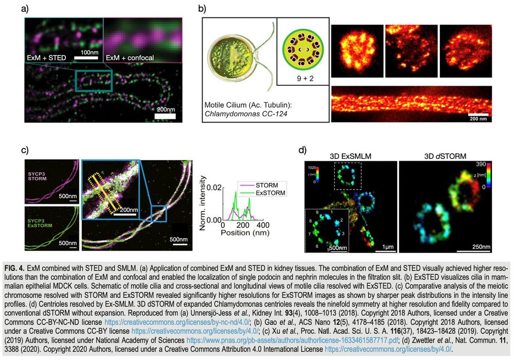
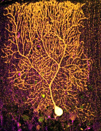
Happy #fluorescentFriday: dendritic spines of Purkinje cell after 10x expansion microscopie #expansionMicroscopie #ExM
April 25, 2025 at 2:25 PM
Happy #fluorescentFriday: dendritic spines of Purkinje cell after 10x expansion microscopie #expansionMicroscopie #ExM
Reposted
I will never tire of looking at these clusters of DRD1🔴/RXFP1🟡/OPRM1🟣 cells - here showing RNAscope in nucleus accumbens of human 🧠. Patterning of them is so neat!
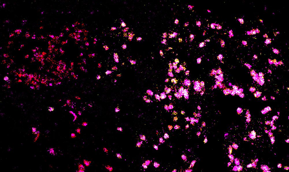
April 23, 2025 at 7:00 PM
I will never tire of looking at these clusters of DRD1🔴/RXFP1🟡/OPRM1🟣 cells - here showing RNAscope in nucleus accumbens of human 🧠. Patterning of them is so neat!
Reposted
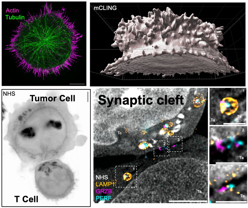
New lab preprint! Unveiling the Molecular Architecture of T Cells and Immune Synapses with Cryo-Expansion Microscopy (Cryo-ExM). A fantastic collaboration with Dr. Benita Wolf. Congratulations to all authors, especially Florent Lemaitre, for leading this amazing work! www.biorxiv.org/content/10.1...
April 23, 2025 at 2:04 PM
New lab preprint! Unveiling the Molecular Architecture of T Cells and Immune Synapses with Cryo-Expansion Microscopy (Cryo-ExM). A fantastic collaboration with Dr. Benita Wolf. Congratulations to all authors, especially Florent Lemaitre, for leading this amazing work! www.biorxiv.org/content/10.1...
Reposted
Incredible 4Pi and pan-expansion imaging of the Golgi 👇

March 29, 2025 at 4:03 PM
Incredible 4Pi and pan-expansion imaging of the Golgi 👇
Reposted
I don't think I'm ever going to get tired of expansion microscopy, so this makes for a great first post here to look back on in the years to come 😄
Mouse brain slices, unexpanded and at two different expansion levels, skillfully prepared by @damstra.bsky.social and Aashir Meeran at E11 Bio! 🧠
Mouse brain slices, unexpanded and at two different expansion levels, skillfully prepared by @damstra.bsky.social and Aashir Meeran at E11 Bio! 🧠
February 15, 2025 at 8:08 AM
I don't think I'm ever going to get tired of expansion microscopy, so this makes for a great first post here to look back on in the years to come 😄
Mouse brain slices, unexpanded and at two different expansion levels, skillfully prepared by @damstra.bsky.social and Aashir Meeran at E11 Bio! 🧠
Mouse brain slices, unexpanded and at two different expansion levels, skillfully prepared by @damstra.bsky.social and Aashir Meeran at E11 Bio! 🧠
Reposted
"Imagine a DAPI-like stain, but for the extracellular matrix." That's basically how this work was pitched to me by Kayvon and Antonio a year or so ago. Now the final product really delivers. Read about their versatile label for ECM in living tissues here: www.nature.com/articles/s41...
February 6, 2025 at 4:02 PM
"Imagine a DAPI-like stain, but for the extracellular matrix." That's basically how this work was pitched to me by Kayvon and Antonio a year or so ago. Now the final product really delivers. Read about their versatile label for ECM in living tissues here: www.nature.com/articles/s41...
Reposted

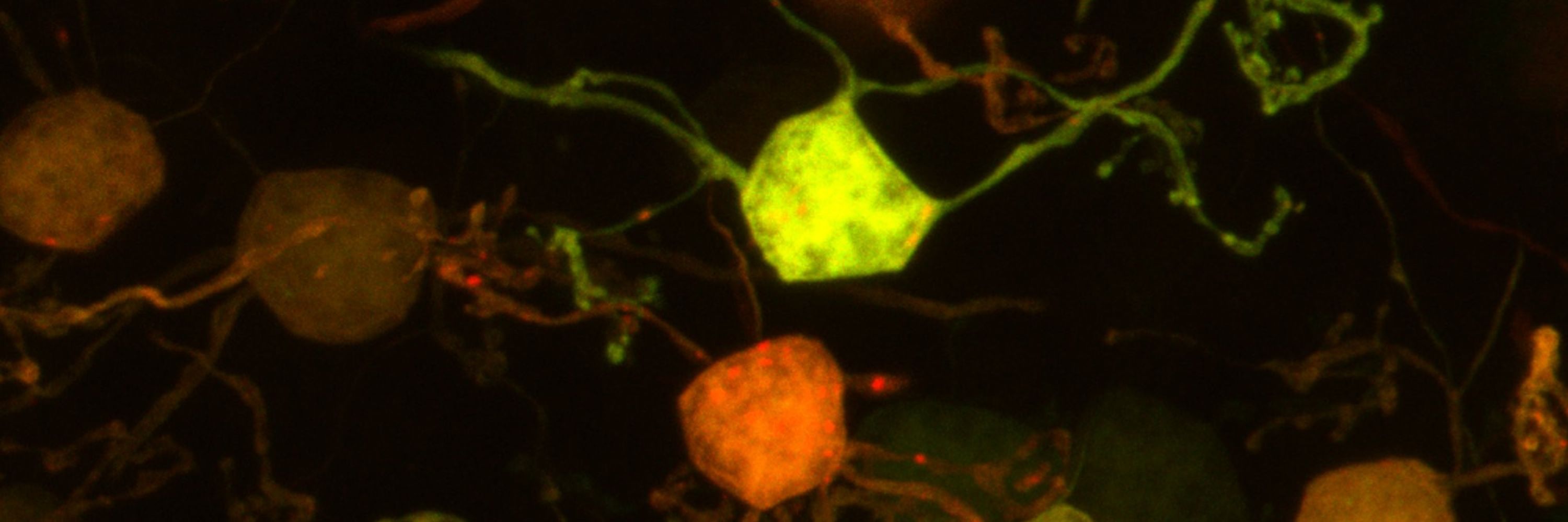
The most beautiful neurons in the brain (fact), and also my villain origin story - #FluorescenceFriday
🧡 biocytin patched and filled Purkinje cell
💚 parvalbumin
💜 AnkG for axon initial segments
🧡 biocytin patched and filled Purkinje cell
💚 parvalbumin
💜 AnkG for axon initial segments
January 24, 2025 at 1:22 PM
The most beautiful neurons in the brain (fact), and also my villain origin story - #FluorescenceFriday
🧡 biocytin patched and filled Purkinje cell
💚 parvalbumin
💜 AnkG for axon initial segments
🧡 biocytin patched and filled Purkinje cell
💚 parvalbumin
💜 AnkG for axon initial segments