


Nat Prunet
@nat-prunet.bsky.social
Microscopic farmer 🔬🌺
Microscopy core director @UNC Chapel Hill
Former plant developmental biologist
Microscopy core director @UNC Chapel Hill
Former plant developmental biologist
I was today years old when I learned that *Drosophila husbandry* was actually a skillset to add to a cell/developmental biology resume. I wonder if I should add Arabidopsis husbandry to mine 😆
#drsophila #cellbio #devbio #arabidopsis #plantscience
#drsophila #cellbio #devbio #arabidopsis #plantscience

November 14, 2025 at 7:41 PM
I was today years old when I learned that *Drosophila husbandry* was actually a skillset to add to a cell/developmental biology resume. I wonder if I should add Arabidopsis husbandry to mine 😆
#drsophila #cellbio #devbio #arabidopsis #plantscience
#drsophila #cellbio #devbio #arabidopsis #plantscience
Reposted by Nat Prunet

If you are looking for #microscopy events, check out our Microscopy events calendar, powered by MicroscopyDB.
🗓️: focalplane.biologists.com/events/
Add an event: microscopydb.io/add-a-commun...
🗓️: focalplane.biologists.com/events/
Add an event: microscopydb.io/add-a-commun...
To help you plan your conference travel for 2026, we’ve been busy updating our #CellBiology events calendar on FocalPlane.
If we are missing your event, feel free to add it to the calendar or reach out to our Community Manager @helenzsci.bsky.social.
focalplane.biologists.com/cell-biology...
If we are missing your event, feel free to add it to the calendar or reach out to our Community Manager @helenzsci.bsky.social.
focalplane.biologists.com/cell-biology...

November 12, 2025 at 2:06 PM
If you are looking for #microscopy events, check out our Microscopy events calendar, powered by MicroscopyDB.
🗓️: focalplane.biologists.com/events/
Add an event: microscopydb.io/add-a-commun...
🗓️: focalplane.biologists.com/events/
Add an event: microscopydb.io/add-a-commun...
Reposted by Nat Prunet
Flashback #FluorescenceFriday to some of the first light-sheet imaging we were doing. Video shows the collecting duct system of the developing kidney labeled with a pan-cytokeratin antibody. Courtesy of former talented technician Deanna Hardesty.
November 7, 2025 at 4:43 PM
Flashback #FluorescenceFriday to some of the first light-sheet imaging we were doing. Video shows the collecting duct system of the developing kidney labeled with a pan-cytokeratin antibody. Courtesy of former talented technician Deanna Hardesty.
Reposted by Nat Prunet
Our #JCSciliaSI is complete
Explore our ToC: journals.biologists.com/jcs/issue/13...
Our cover image shows tracheal epithelial cells from an air-liquid interface culture imaged using U-ExM from Oliver Mercey and Marine Brunet @centriolelab.bsky.social.
journals.biologists.com/jcs/article/...
Explore our ToC: journals.biologists.com/jcs/issue/13...
Our cover image shows tracheal epithelial cells from an air-liquid interface culture imaged using U-ExM from Oliver Mercey and Marine Brunet @centriolelab.bsky.social.
journals.biologists.com/jcs/article/...

November 4, 2025 at 1:36 PM
Our #JCSciliaSI is complete
Explore our ToC: journals.biologists.com/jcs/issue/13...
Our cover image shows tracheal epithelial cells from an air-liquid interface culture imaged using U-ExM from Oliver Mercey and Marine Brunet @centriolelab.bsky.social.
journals.biologists.com/jcs/article/...
Explore our ToC: journals.biologists.com/jcs/issue/13...
Our cover image shows tracheal epithelial cells from an air-liquid interface culture imaged using U-ExM from Oliver Mercey and Marine Brunet @centriolelab.bsky.social.
journals.biologists.com/jcs/article/...

November 6, 2025 at 6:45 PM
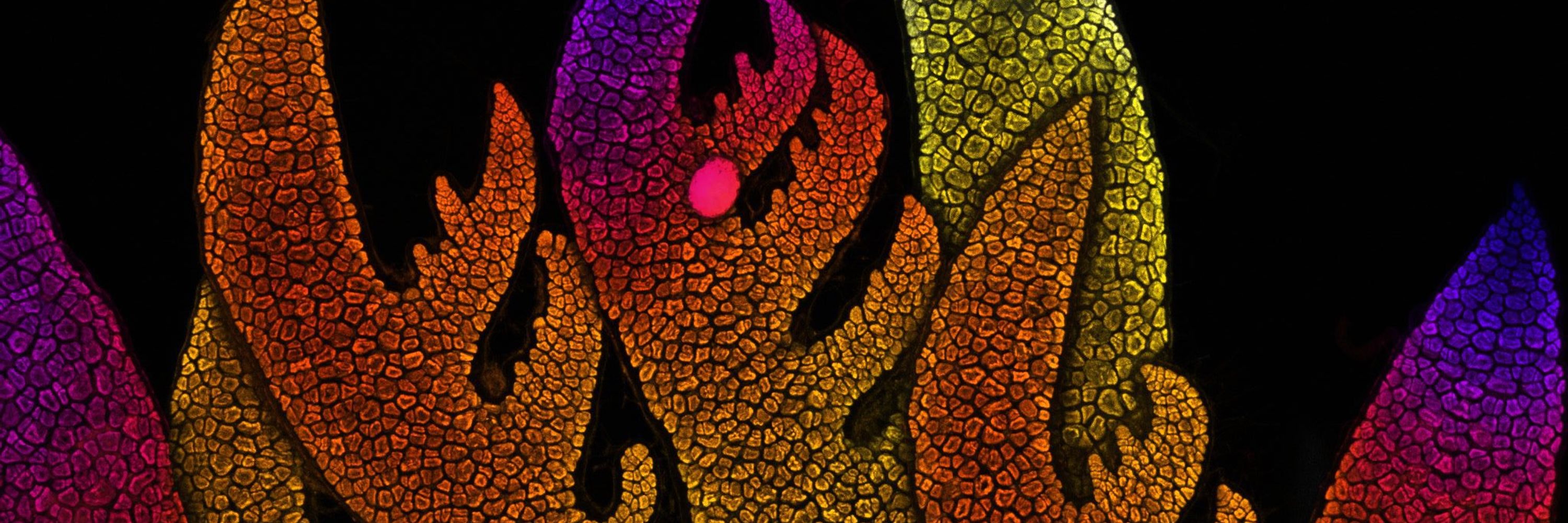
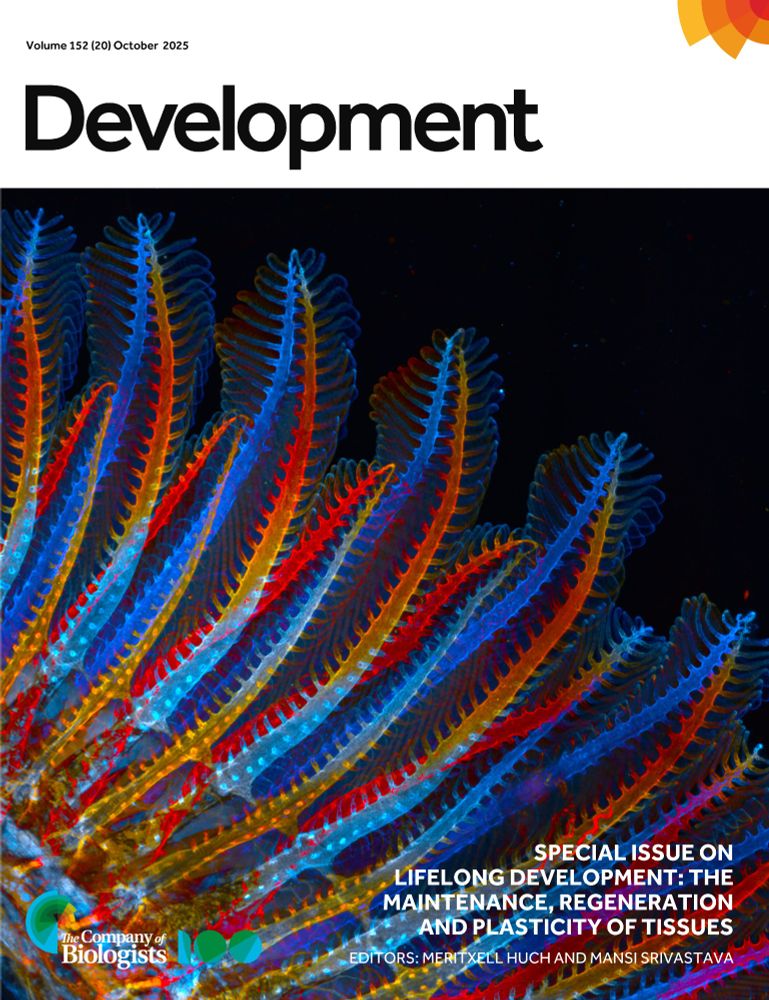
Gorgeous cover by @mathpreu.bsky.social 👏
Our Special Issue on Lifelong Development #LifelongDevSI is now complete!
Guest edited by Meri Huch and Mansi Srivastava, it contains 26 research and review-type articles.
On the cover: the dorsal vasculature of an adult zebrafish's first gill arch. See Preußner et al. doi.org/10.1242/dev....
Guest edited by Meri Huch and Mansi Srivastava, it contains 26 research and review-type articles.
On the cover: the dorsal vasculature of an adult zebrafish's first gill arch. See Preußner et al. doi.org/10.1242/dev....
November 6, 2025 at 11:54 AM
Gorgeous cover by @mathpreu.bsky.social 👏
Reposted by Nat Prunet
Incoming deadline - 7 November 2025
Apply for a @jcellsci.bsky.social @focalplane.bsky.social Training Grant to support your attendance at a #microscopy or #bioimageanalysis training course.
Open to ECRs in #cellbio. For more info: www.biologists.com/grants/jcs-f...
Apply for a @jcellsci.bsky.social @focalplane.bsky.social Training Grant to support your attendance at a #microscopy or #bioimageanalysis training course.
Open to ECRs in #cellbio. For more info: www.biologists.com/grants/jcs-f...

November 5, 2025 at 11:54 AM
Incoming deadline - 7 November 2025
Apply for a @jcellsci.bsky.social @focalplane.bsky.social Training Grant to support your attendance at a #microscopy or #bioimageanalysis training course.
Open to ECRs in #cellbio. For more info: www.biologists.com/grants/jcs-f...
Apply for a @jcellsci.bsky.social @focalplane.bsky.social Training Grant to support your attendance at a #microscopy or #bioimageanalysis training course.
Open to ECRs in #cellbio. For more info: www.biologists.com/grants/jcs-f...
Reposted by Nat Prunet
🔬We are now accepting applications for the 2026 Analytical and Quantitative Light Microscopy course at MBL. Come learn to be an excellent microscopist!
Questions? Ask here or DM me.
@mblscience.bsky.social @aqlm.bsky.social
#microscopy #fluorescence
Questions? Ask here or DM me.
@mblscience.bsky.social @aqlm.bsky.social
#microscopy #fluorescence

October 27, 2025 at 5:37 PM
🔬We are now accepting applications for the 2026 Analytical and Quantitative Light Microscopy course at MBL. Come learn to be an excellent microscopist!
Questions? Ask here or DM me.
@mblscience.bsky.social @aqlm.bsky.social
#microscopy #fluorescence
Questions? Ask here or DM me.
@mblscience.bsky.social @aqlm.bsky.social
#microscopy #fluorescence
Hey plant developmental biologists! Want to do great science with a great guy in a great place? @oconnord.bsky.social is looking for a postdoc to join his lab at Colorado State:
oconnorlab.colostate.edu/wp-content/u...
#devbio #plantscience #botany #bioimaging #microscopy
oconnorlab.colostate.edu/wp-content/u...
#devbio #plantscience #botany #bioimaging #microscopy
October 29, 2025 at 3:00 PM
Hey plant developmental biologists! Want to do great science with a great guy in a great place? @oconnord.bsky.social is looking for a postdoc to join his lab at Colorado State:
oconnorlab.colostate.edu/wp-content/u...
#devbio #plantscience #botany #bioimaging #microscopy
oconnorlab.colostate.edu/wp-content/u...
#devbio #plantscience #botany #bioimaging #microscopy
Love these!
Hello hello world. I’m on here now. I am a microscope addict. I do a lot of plankton chronophotography, see below video of vorticella feeding currents. I love the microworld and consider myself an advocate for these tiny creatures, I like to make them visible. Will post more soon
October 29, 2025 at 2:05 PM
Love these!


Reposted by Nat Prunet

The #mesoSPIM community just celebrated 10 years of open-source light-sheet microscopy in Zürich! For #3DThursday, we're honoring Martina Schaettin & Fabian Voigt’s iconic 3D chick embryo dataset — a luminous benchmark for open science and shared discovery.
#Microscopy #Lightsheet #syGlass
#Microscopy #Lightsheet #syGlass
October 23, 2025 at 3:33 PM
The #mesoSPIM community just celebrated 10 years of open-source light-sheet microscopy in Zürich! For #3DThursday, we're honoring Martina Schaettin & Fabian Voigt’s iconic 3D chick embryo dataset — a luminous benchmark for open science and shared discovery.
#Microscopy #Lightsheet #syGlass
#Microscopy #Lightsheet #syGlass
Reposted by Nat Prunet
The winners for the largest photo microscopy competition were just announced and they do NOT disappoint!!
[Photos shared with permission courtesy of the 2025 Nikon Small World Photo Competition] 🧪🔬
[Photos shared with permission courtesy of the 2025 Nikon Small World Photo Competition] 🧪🔬
October 15, 2025 at 3:49 PM
The winners for the largest photo microscopy competition were just announced and they do NOT disappoint!!
[Photos shared with permission courtesy of the 2025 Nikon Small World Photo Competition] 🧪🔬
[Photos shared with permission courtesy of the 2025 Nikon Small World Photo Competition] 🧪🔬
Reposted by Nat Prunet

Magnifying the minuscule: Nikon Small World photomicrography 2025 – in pictures

Magnifying the minuscule: Nikon Small World photomicrography 2025 – in pictures
Weevils, spores, slime mold and cells in extreme closeup for the 51st anniversary of the Nikon Small World competition
www.theguardian.com
October 15, 2025 at 11:56 PM
Magnifying the minuscule: Nikon Small World photomicrography 2025 – in pictures
Reposted by Nat Prunet

Hello scientists of Bluesky! Since we're new here, what better way to introduce ourselves than to share our collection of light-sheet #microscopy data, featuring a diverse range of species and tissue types 🐠🐁🦎🔬: vimeo.com/lifecanvastech
Stay tuned for more #bioimaging data drops coming soon!
Stay tuned for more #bioimaging data drops coming soon!

October 22, 2025 at 1:43 PM
Hello scientists of Bluesky! Since we're new here, what better way to introduce ourselves than to share our collection of light-sheet #microscopy data, featuring a diverse range of species and tissue types 🐠🐁🦎🔬: vimeo.com/lifecanvastech
Stay tuned for more #bioimaging data drops coming soon!
Stay tuned for more #bioimaging data drops coming soon!
Reposted by Nat Prunet


September 16, 2025 at 11:45 PM
Reposted by Nat Prunet

October 21, 2025 at 10:42 AM
Reposted by Nat Prunet
For this #FluorescenceFriday 🔬
a cryosection of a dissected zebrafish gill, stained with phalloidin (orange) and DAPI (blue-ish).
#Zebrafish #DevBio #Microscopy
a cryosection of a dissected zebrafish gill, stained with phalloidin (orange) and DAPI (blue-ish).
#Zebrafish #DevBio #Microscopy

October 17, 2025 at 3:15 PM
For this #FluorescenceFriday 🔬
a cryosection of a dissected zebrafish gill, stained with phalloidin (orange) and DAPI (blue-ish).
#Zebrafish #DevBio #Microscopy
a cryosection of a dissected zebrafish gill, stained with phalloidin (orange) and DAPI (blue-ish).
#Zebrafish #DevBio #Microscopy
Reposted by Nat Prunet

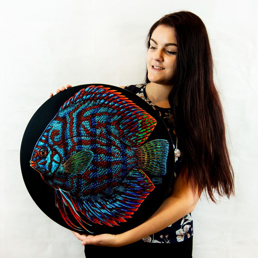
Huge thanks to the incredibly talented Center for Visual Arts students for transforming microscopy images into incredible works of art for Toledo CellulART! Science meets art in the most inspiring way. 🔬❤️🎨 www.toledocellulart.org #SciArt #Microscopy


October 8, 2025 at 12:39 AM
Huge thanks to the incredibly talented Center for Visual Arts students for transforming microscopy images into incredible works of art for Toledo CellulART! Science meets art in the most inspiring way. 🔬❤️🎨 www.toledocellulart.org #SciArt #Microscopy
Reposted by Nat Prunet
October 11, 2025 at 10:52 PM
Reposted by Nat Prunet
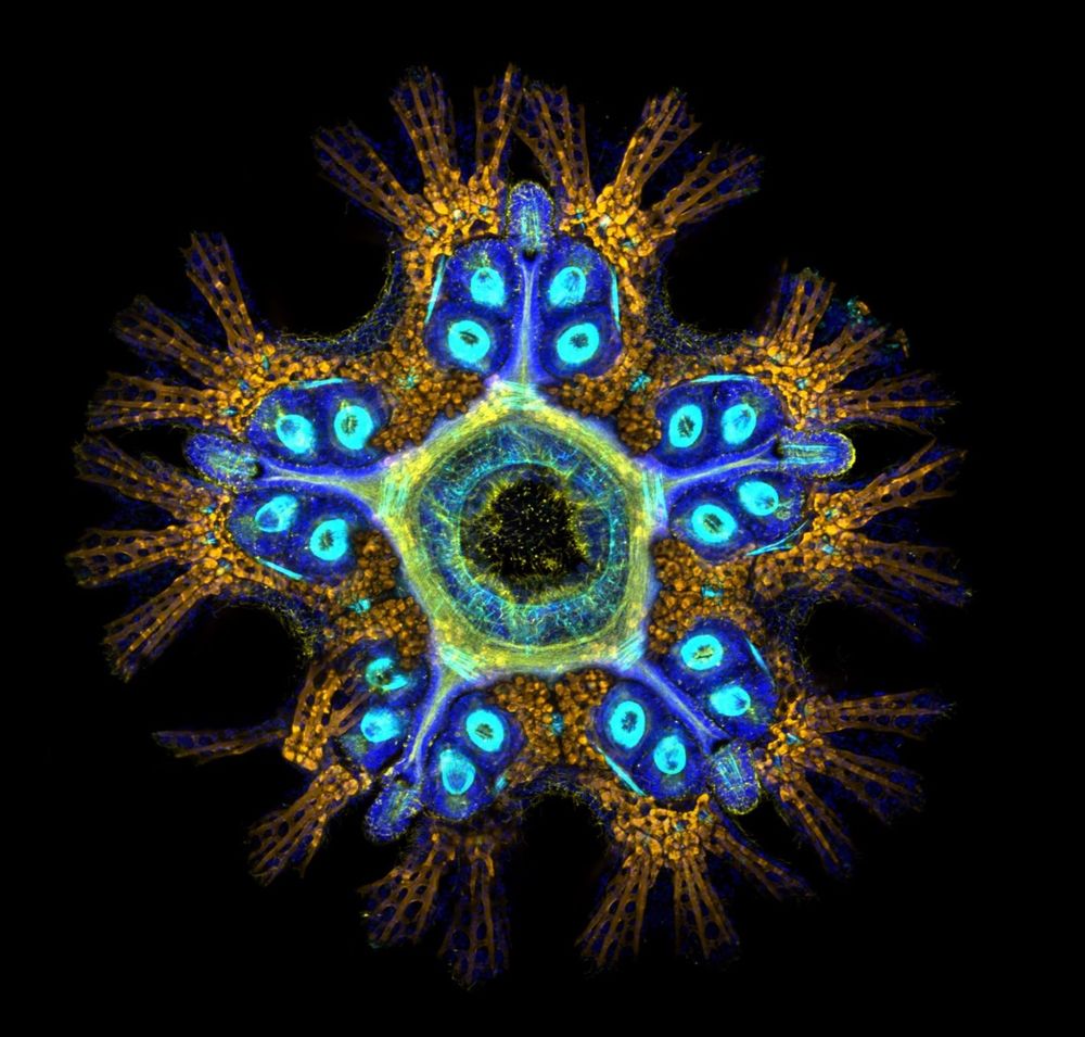
This sea star is only 2 months old 🐣 — but already has a decentralized nervous system. No brain, just a nerve ring + radial cords guiding how it moves, senses & grows.
🌊 Echinoderm family
🧠 Brain-free coordination
🔄 Decentralized = resilient
Credit: Dr. Laurent Formery
#Neurobiology #Science
🌊 Echinoderm family
🧠 Brain-free coordination
🔄 Decentralized = resilient
Credit: Dr. Laurent Formery
#Neurobiology #Science
September 26, 2025 at 7:47 PM
This sea star is only 2 months old 🐣 — but already has a decentralized nervous system. No brain, just a nerve ring + radial cords guiding how it moves, senses & grows.
🌊 Echinoderm family
🧠 Brain-free coordination
🔄 Decentralized = resilient
Credit: Dr. Laurent Formery
#Neurobiology #Science
🌊 Echinoderm family
🧠 Brain-free coordination
🔄 Decentralized = resilient
Credit: Dr. Laurent Formery
#Neurobiology #Science
Reposted by Nat Prunet

🏆 The PPBI Image Contest celebrates the art of science!
🎉 Congratulations to Carina Mónico (CCMAR) for her winning image!
A huge thank-you to everyone who took part.
#PPBI #Microscopy #BioImaging #ScienceArt
🎉 Congratulations to Carina Mónico (CCMAR) for her winning image!
A huge thank-you to everyone who took part.
#PPBI #Microscopy #BioImaging #ScienceArt
October 8, 2025 at 10:20 AM
🏆 The PPBI Image Contest celebrates the art of science!
🎉 Congratulations to Carina Mónico (CCMAR) for her winning image!
A huge thank-you to everyone who took part.
#PPBI #Microscopy #BioImaging #ScienceArt
🎉 Congratulations to Carina Mónico (CCMAR) for her winning image!
A huge thank-you to everyone who took part.
#PPBI #Microscopy #BioImaging #ScienceArt
Reposted by Nat Prunet

Microscopy Image Competition 2025 Finalists
There are two categories:
Category 1: Featuring Proteintech antibodies
Category 2: Featuring any antibodies
The images below are the Category 2 finalists.
Vote before Oct 26 (T&Cs apply): https://ow.ly/qhK350XeWNi
There are two categories:
Category 1: Featuring Proteintech antibodies
Category 2: Featuring any antibodies
The images below are the Category 2 finalists.
Vote before Oct 26 (T&Cs apply): https://ow.ly/qhK350XeWNi


October 20, 2025 at 2:35 PM
Microscopy Image Competition 2025 Finalists
There are two categories:
Category 1: Featuring Proteintech antibodies
Category 2: Featuring any antibodies
The images below are the Category 2 finalists.
Vote before Oct 26 (T&Cs apply): https://ow.ly/qhK350XeWNi
There are two categories:
Category 1: Featuring Proteintech antibodies
Category 2: Featuring any antibodies
The images below are the Category 2 finalists.
Vote before Oct 26 (T&Cs apply): https://ow.ly/qhK350XeWNi
Reposted by Nat Prunet

for #FluorescenceFriday : is your ExM sample too large to be imaged with high NA objectives? VIPS it! Very clever way to tackle one of the biggest challenge in large volumetric ExM www.science.org/doi/10.1126/...

Mesoscale volumetric fluorescence imaging at nanoscale resolution by photochemical sectioning
Optical nanoscopy of intact biological specimens has been transformed by recent advancements in hydrogel-based tissue clearing and expansion, enabling the imaging of cellular and subcellular structure...
www.science.org
October 17, 2025 at 12:29 PM
for #FluorescenceFriday : is your ExM sample too large to be imaged with high NA objectives? VIPS it! Very clever way to tackle one of the biggest challenge in large volumetric ExM www.science.org/doi/10.1126/...
Reposted by Nat Prunet

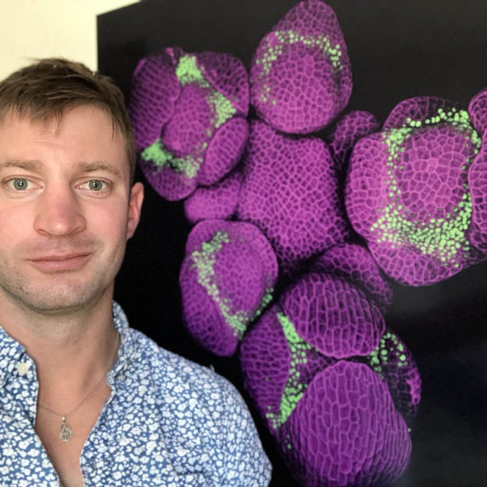
This image of moss Physcomitrium patens beautifully demonstrates what we mean when we say that this cell type is characterized by oblique cell walls.
You can see that the cell walls between cells are at an angle and not perpendicular to the cell axis.
#microscopymonday #moss #plantcells
You can see that the cell walls between cells are at an angle and not perpendicular to the cell axis.
#microscopymonday #moss #plantcells

October 20, 2025 at 1:51 PM
This image of moss Physcomitrium patens beautifully demonstrates what we mean when we say that this cell type is characterized by oblique cell walls.
You can see that the cell walls between cells are at an angle and not perpendicular to the cell axis.
#microscopymonday #moss #plantcells
You can see that the cell walls between cells are at an angle and not perpendicular to the cell axis.
#microscopymonday #moss #plantcells