


Ebrahim Lab
@ebrahim-lab.bsky.social
Lab focused on molecular force-generators and -sensors in living tissues, and all things microscopy @UVA School of Medicine.
https://med.virginia.edu/ebrahim-lab/
https://med.virginia.edu/ebrahim-lab/
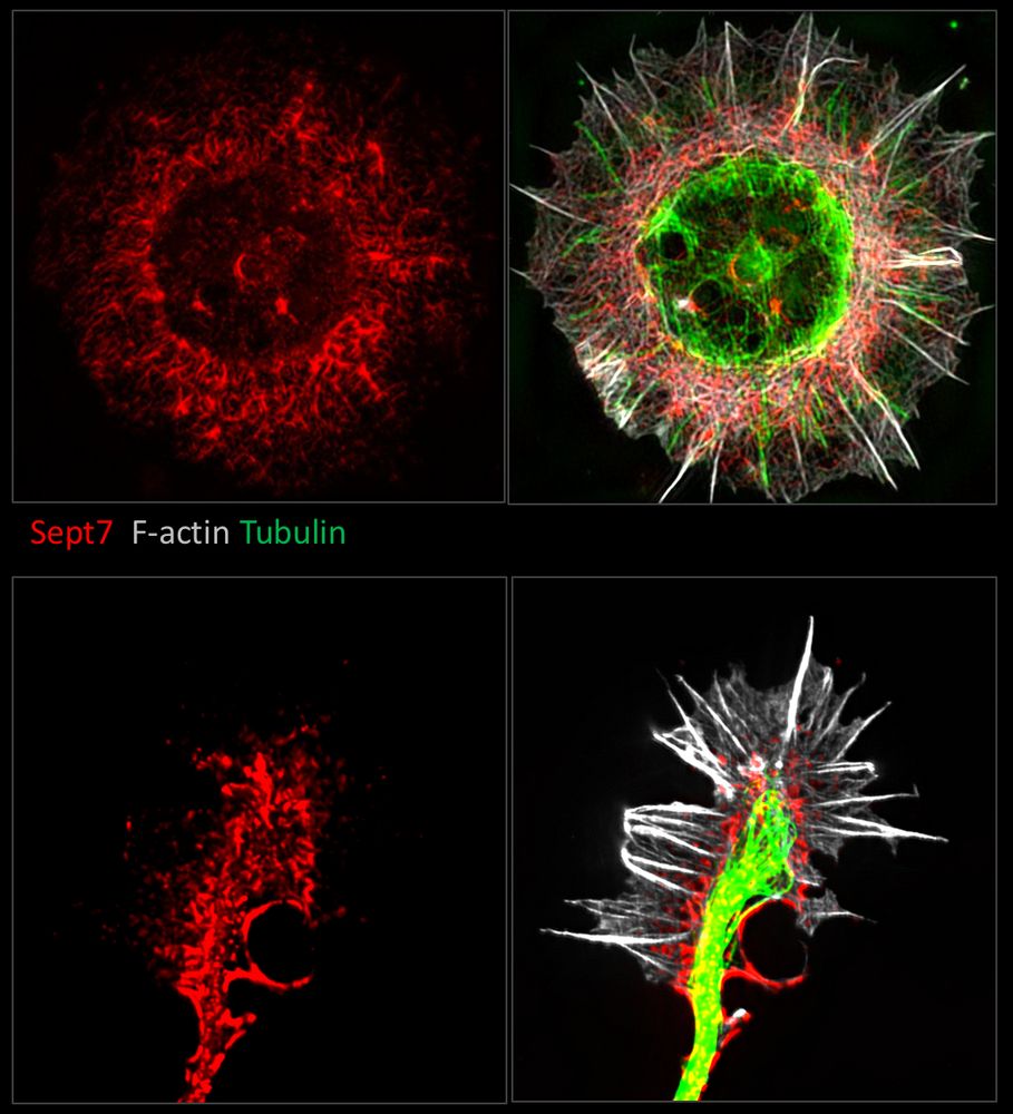
Very excited to share the latest work from our lab in @insight.jci.org, highlighting a role for the #septin #cytoskeleton in protecting against "leaky gut", by recruiting non-muscle myosin II and stabilizing the tight junction.
Credit to @dominikrobak.bsky.social for the stunning cover image!
Credit to @dominikrobak.bsky.social for the stunning cover image!

This issue’s Cover: @ebrahim-lab.bsky.social @dominikrobak.bsky.social& team report on the septin cytoskeleton at apical junctions of intestinal epithelial cells, whereby it maintains barrier integrity & protects from inflammation: insight.jci.org/articles/vie...

November 24, 2025 at 6:01 PM
Very excited to share the latest work from our lab in @insight.jci.org, highlighting a role for the #septin #cytoskeleton in protecting against "leaky gut", by recruiting non-muscle myosin II and stabilizing the tight junction.
Credit to @dominikrobak.bsky.social for the stunning cover image!
Credit to @dominikrobak.bsky.social for the stunning cover image!
Reposted by Ebrahim Lab
Check out our new preprint on the discovery of a molecular switch in NAC that mediates nascent chain sorting on the ribosome and prevents mitochondrial protein mistargeting by SRP. A great collaboration with the Shan Lab @Caltech and the Qi Lab @UVA: www.biorxiv.org/content/10.1...
August 1, 2025 at 3:27 PM
Check out our new preprint on the discovery of a molecular switch in NAC that mediates nascent chain sorting on the ribosome and prevents mitochondrial protein mistargeting by SRP. A great collaboration with the Shan Lab @Caltech and the Qi Lab @UVA: www.biorxiv.org/content/10.1...
Having seen your bingo card, we had to make it happen 😉!
Thank you for highlighting our work @hankgreen.bsky.social ! 🙏🏽
Thank you for highlighting our work @hankgreen.bsky.social ! 🙏🏽

July 26, 2025 at 1:17 AM
Having seen your bingo card, we had to make it happen 😉!
Thank you for highlighting our work @hankgreen.bsky.social ! 🙏🏽
Thank you for highlighting our work @hankgreen.bsky.social ! 🙏🏽
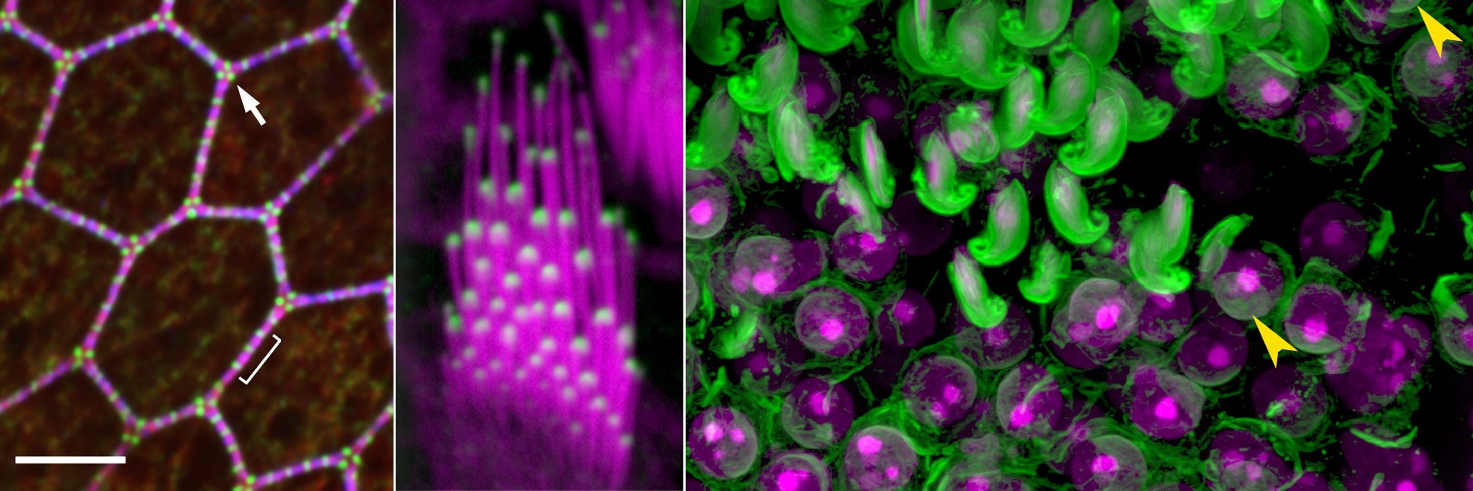
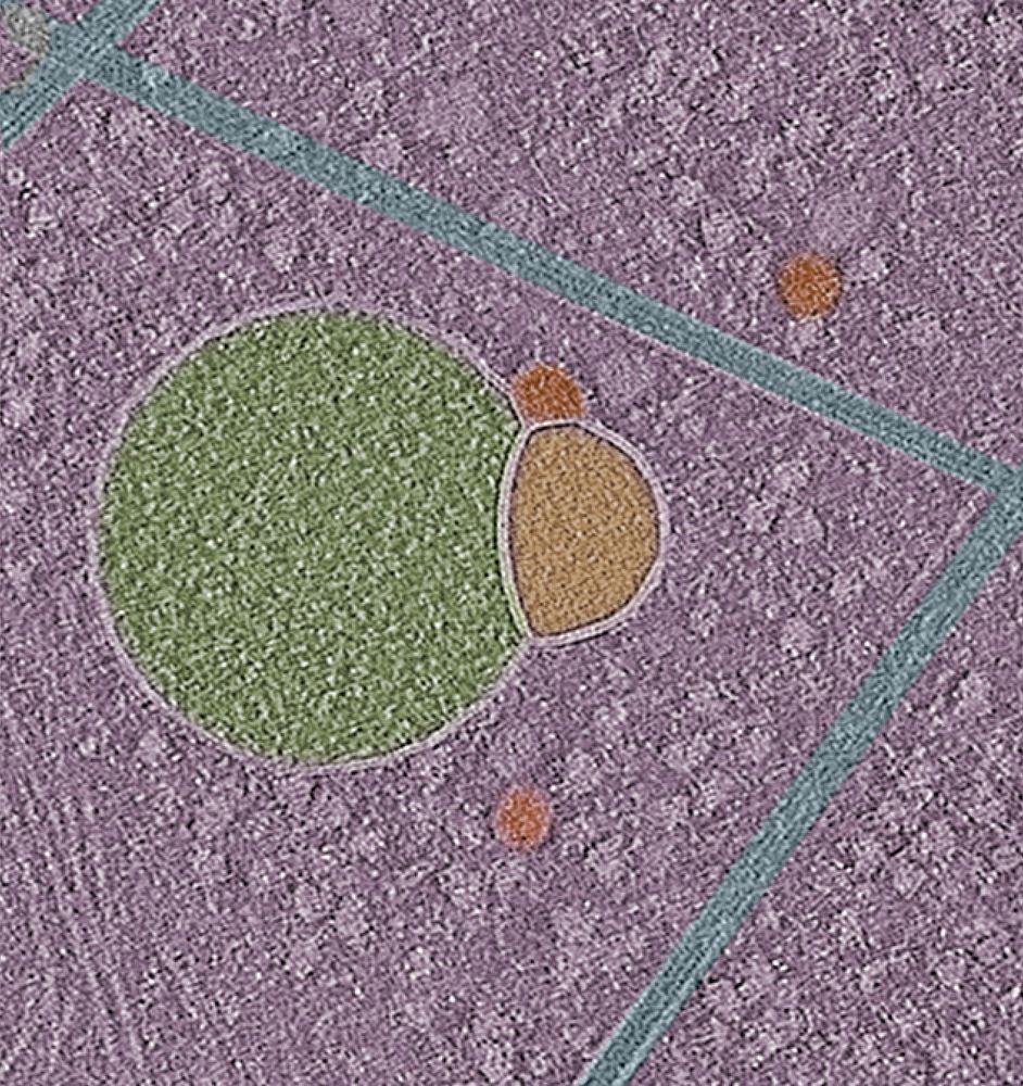
Early for #MicroscopyMonday: #Hemifusomes- Newly discovered ESCRT-independent intermediates of intraluminal vesicle and multivesicular body formation! In collaboration with the Kachar Lab at the NIH and out today in @natcomms.nature.com : rdcu.be/emtQu
#cryoET #trafficking #organelle #hemifusion
#cryoET #trafficking #organelle #hemifusion
May 17, 2025 at 7:37 PM
Early for #MicroscopyMonday: #Hemifusomes- Newly discovered ESCRT-independent intermediates of intraluminal vesicle and multivesicular body formation! In collaboration with the Kachar Lab at the NIH and out today in @natcomms.nature.com : rdcu.be/emtQu
#cryoET #trafficking #organelle #hemifusion
#cryoET #trafficking #organelle #hemifusion
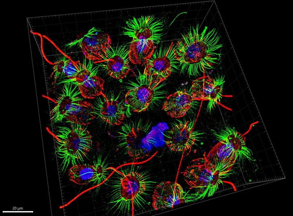
"Stereocilia"- actin-based skyscrapers that adorn sensory cells of our inner ear and covert vibrations into electrical signals—the first step in hearing and balance. #FluorescenceFriday

May 2, 2025 at 1:58 PM
"Stereocilia"- actin-based skyscrapers that adorn sensory cells of our inner ear and covert vibrations into electrical signals—the first step in hearing and balance. #FluorescenceFriday
Reposted by Ebrahim Lab

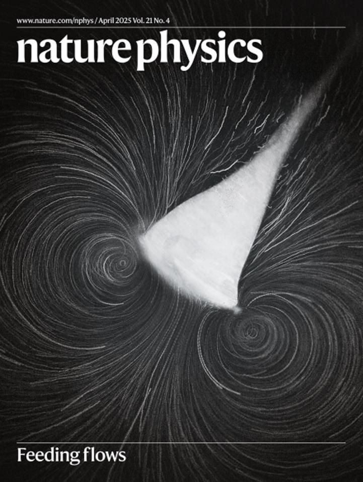
🎉🎉We’re on the cover of Nature Physics! @natphys.nature.com
Here is the original paper www.nature.com/articles/s41...
Here is the original paper www.nature.com/articles/s41...
April 26, 2025 at 2:38 PM
🎉🎉We’re on the cover of Nature Physics! @natphys.nature.com
Here is the original paper www.nature.com/articles/s41...
Here is the original paper www.nature.com/articles/s41...
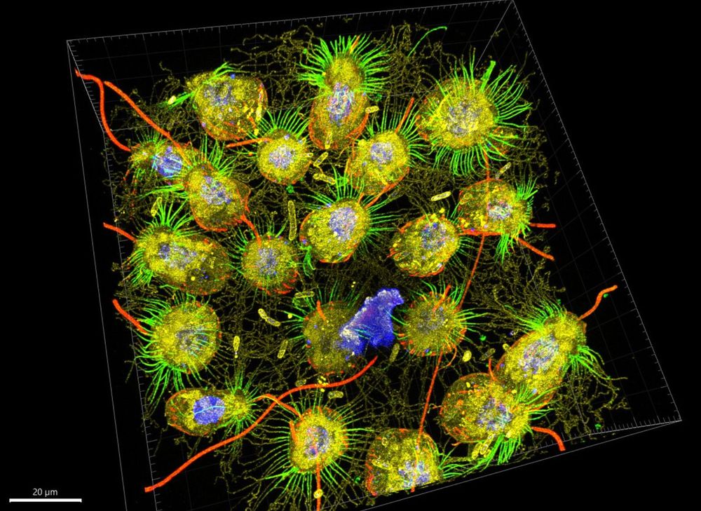
From stem cell to structure-
This human colonoid shows the self-organization of intestinal epithelial cells in 3D—nuclei in magenta, actin in green. A living miniature gut for your #FluorescenceFriday.
This human colonoid shows the self-organization of intestinal epithelial cells in 3D—nuclei in magenta, actin in green. A living miniature gut for your #FluorescenceFriday.

April 25, 2025 at 11:07 AM
From stem cell to structure-
This human colonoid shows the self-organization of intestinal epithelial cells in 3D—nuclei in magenta, actin in green. A living miniature gut for your #FluorescenceFriday.
This human colonoid shows the self-organization of intestinal epithelial cells in 3D—nuclei in magenta, actin in green. A living miniature gut for your #FluorescenceFriday.
Reposted by Ebrahim Lab
A dream come true: the first expansion microscopy images of C. flexa 🤩 Generated by Mylan & Uzuki who learned from the best (@hiralshah.bsky.social @gautamdey.bsky.social @dudinlab.bsky.social). We will learn so much from these!
April 22, 2025 at 4:36 PM
A dream come true: the first expansion microscopy images of C. flexa 🤩 Generated by Mylan & Uzuki who learned from the best (@hiralshah.bsky.social @gautamdey.bsky.social @dudinlab.bsky.social). We will learn so much from these!
Reposted by Ebrahim Lab

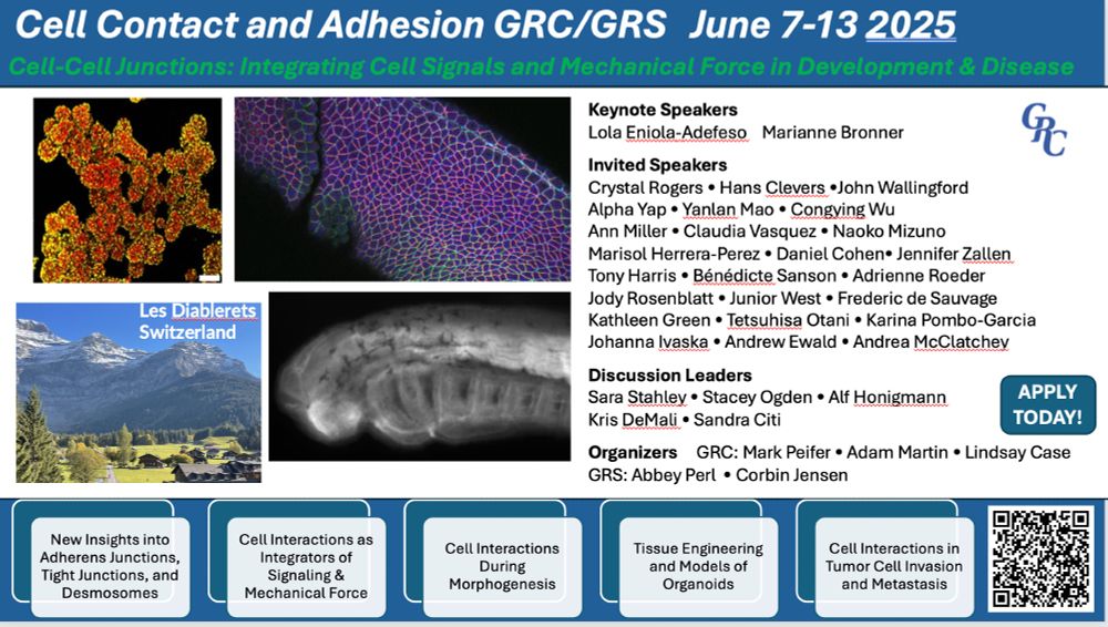
Only 29 slots left out of 200--register now!
Cell and developmental biologists: as you start looking at conferences in 2025, consider joining us at the 2025 Cell Contact & Adhesion GRC. We've assembled a strong and diverse program of speakers at the cutting-edge of science in our field 1/n Please repost
www.grc.org/cell-contact...
www.grc.org/cell-contact...
April 21, 2025 at 2:12 PM
Only 29 slots left out of 200--register now!

April 18, 2025 at 2:33 PM
Reposted by Ebrahim Lab
Two months left for FASEB Conference - Septins: Spatial regulators of cell biology
Register now:
events.faseb.org/event/septin...
Come find out what these filaments do at the interface of the actin and microtubule cytoskeletons and cell membranes. You'll be hooked for life.
Register now:
events.faseb.org/event/septin...
Come find out what these filaments do at the interface of the actin and microtubule cytoskeletons and cell membranes. You'll be hooked for life.
April 16, 2025 at 3:08 PM
Two months left for FASEB Conference - Septins: Spatial regulators of cell biology
Register now:
events.faseb.org/event/septin...
Come find out what these filaments do at the interface of the actin and microtubule cytoskeletons and cell membranes. You'll be hooked for life.
Register now:
events.faseb.org/event/septin...
Come find out what these filaments do at the interface of the actin and microtubule cytoskeletons and cell membranes. You'll be hooked for life.
Reposted by Ebrahim Lab
Is liver zonation conserved from mice to humans? And how does fibrosis affect zonation in patients? Now, these questions are answered using scDVP. Some collaborations are meant to be! @carolineweiss.bsky.social@frosenberger.bsky.social @mannlab.bsky.social@mlsb-borgwardt.bsky.social
Introducing our single-cell DVP framework combining strategic cell selection with protein gradient mapping algorithms. Analyzing hepatocyte proteomes across 18 human livers, revealing cross-species differences, and mapping disruptions in liver fibrosis.
www.biorxiv.org/content/10.1...
www.biorxiv.org/content/10.1...

April 14, 2025 at 2:35 PM
Is liver zonation conserved from mice to humans? And how does fibrosis affect zonation in patients? Now, these questions are answered using scDVP. Some collaborations are meant to be! @carolineweiss.bsky.social@frosenberger.bsky.social @mannlab.bsky.social@mlsb-borgwardt.bsky.social
Reposted by Ebrahim Lab
#MicroscopyMonday | Colon-scoping turned coral reef
Was imaging the colon when I accidentally stumbled into this glowing patch of vascularized white adipose tissue.
🔵 mT/mG mouse (mT-only) → red membranes shown in cyan
Accidental imaging hits different.
#IntravitalImaging #LiveImaging
Was imaging the colon when I accidentally stumbled into this glowing patch of vascularized white adipose tissue.
🔵 mT/mG mouse (mT-only) → red membranes shown in cyan
Accidental imaging hits different.
#IntravitalImaging #LiveImaging


April 14, 2025 at 7:01 PM
#MicroscopyMonday | Colon-scoping turned coral reef
Was imaging the colon when I accidentally stumbled into this glowing patch of vascularized white adipose tissue.
🔵 mT/mG mouse (mT-only) → red membranes shown in cyan
Accidental imaging hits different.
#IntravitalImaging #LiveImaging
Was imaging the colon when I accidentally stumbled into this glowing patch of vascularized white adipose tissue.
🔵 mT/mG mouse (mT-only) → red membranes shown in cyan
Accidental imaging hits different.
#IntravitalImaging #LiveImaging
Reposted by Ebrahim Lab
Excited to share our preprint on the molecular architecture of heterochromatin in human cells 🧬🔬w/ @jpkreysing.bsky.social, @johannesbetz.bsky.social,
@marinalusic.bsky.social, Turoňová lab, @hummerlab.bsky.social @becklab.bsky.social @mpibp.bsky.social
🔗 Preprint here tinyurl.com/3a74uanv
@marinalusic.bsky.social, Turoňová lab, @hummerlab.bsky.social @becklab.bsky.social @mpibp.bsky.social
🔗 Preprint here tinyurl.com/3a74uanv
April 11, 2025 at 8:35 AM
Excited to share our preprint on the molecular architecture of heterochromatin in human cells 🧬🔬w/ @jpkreysing.bsky.social, @johannesbetz.bsky.social,
@marinalusic.bsky.social, Turoňová lab, @hummerlab.bsky.social @becklab.bsky.social @mpibp.bsky.social
🔗 Preprint here tinyurl.com/3a74uanv
@marinalusic.bsky.social, Turoňová lab, @hummerlab.bsky.social @becklab.bsky.social @mpibp.bsky.social
🔗 Preprint here tinyurl.com/3a74uanv
Reposted by Ebrahim Lab
#3DThursday | Intestinal villi in all their folded glory
These self-renewing, nutrient-absorbing structures are beautiful and functional.
🔴 Phalloidin
🔵 DAPI
🟢 Clvd cas-3 (extrusion in action!)
Fixed tissue 3D imaging = big insight, tiny resolution.
#Microscopy #3DImaging #biophysics #intestine
These self-renewing, nutrient-absorbing structures are beautiful and functional.
🔴 Phalloidin
🔵 DAPI
🟢 Clvd cas-3 (extrusion in action!)
Fixed tissue 3D imaging = big insight, tiny resolution.
#Microscopy #3DImaging #biophysics #intestine
April 11, 2025 at 3:08 AM
#3DThursday | Intestinal villi in all their folded glory
These self-renewing, nutrient-absorbing structures are beautiful and functional.
🔴 Phalloidin
🔵 DAPI
🟢 Clvd cas-3 (extrusion in action!)
Fixed tissue 3D imaging = big insight, tiny resolution.
#Microscopy #3DImaging #biophysics #intestine
These self-renewing, nutrient-absorbing structures are beautiful and functional.
🔴 Phalloidin
🔵 DAPI
🟢 Clvd cas-3 (extrusion in action!)
Fixed tissue 3D imaging = big insight, tiny resolution.
#Microscopy #3DImaging #biophysics #intestine
Have cutting-edge cell biology research to share? Submit to Molecular Biology of the Cell (MBoC)- dedicated to advancing discovery and fostering collaboration.
Proud to be part of the editorial team supporting groundbreaking science. molbiolcell.org
#CellBiology #MBoC #Research
Proud to be part of the editorial team supporting groundbreaking science. molbiolcell.org
#CellBiology #MBoC #Research
Molecular Biology of the Cell (MBoC)
molbiolcell.org
April 8, 2025 at 7:15 PM
Have cutting-edge cell biology research to share? Submit to Molecular Biology of the Cell (MBoC)- dedicated to advancing discovery and fostering collaboration.
Proud to be part of the editorial team supporting groundbreaking science. molbiolcell.org
#CellBiology #MBoC #Research
Proud to be part of the editorial team supporting groundbreaking science. molbiolcell.org
#CellBiology #MBoC #Research