


Dr. Caleb Stoltzfus
@cstoltzfus.com
Optics | Microscopy | Physics | Image Analysis | Anything with lasers and data science
Reposted by Dr. Caleb Stoltzfus

A game changer in our hands... www.nature.com/articles/s41...

Universal consensus 3D segmentation of cells from 2D segmented stacks - Nature Methods
u-Segment3D is a universal framework that translates and enhances 2D instance segmentations to a 3D consensus instance segmentation without training data. It performs well across diverse datasets, inc...
www.nature.com
November 11, 2025 at 6:48 PM
A game changer in our hands... www.nature.com/articles/s41...
If you are at #SITC2025 stop by poster #1117 today from 12–2. Chat with Paula Andrea Rotger Gonzalez about Exploring Ovarian Cancer Subtypes with AlpenInsight 3D: A Scalable Workflow for 3D Tissue Classification Using Texture-Based Features and Light-Sheet Microscopy.
November 7, 2025 at 4:35 PM
If you are at #SITC2025 stop by poster #1117 today from 12–2. Chat with Paula Andrea Rotger Gonzalez about Exploring Ovarian Cancer Subtypes with AlpenInsight 3D: A Scalable Workflow for 3D Tissue Classification Using Texture-Based Features and Light-Sheet Microscopy.
Reposted by Dr. Caleb Stoltzfus

3D spatial multi-omic assessment of tumors is not a luxury, it is necessary.
2D assessments of tumors are simply insufficient.
The whole field of spatial omics needs to move to 3D.
Read about this here: www.cell.com/action/showP...
2D assessments of tumors are simply insufficient.
The whole field of spatial omics needs to move to 3D.
Read about this here: www.cell.com/action/showP...

June 9, 2025 at 5:39 PM
3D spatial multi-omic assessment of tumors is not a luxury, it is necessary.
2D assessments of tumors are simply insufficient.
The whole field of spatial omics needs to move to 3D.
Read about this here: www.cell.com/action/showP...
2D assessments of tumors are simply insufficient.
The whole field of spatial omics needs to move to 3D.
Read about this here: www.cell.com/action/showP...
Reposted by Dr. Caleb Stoltzfus
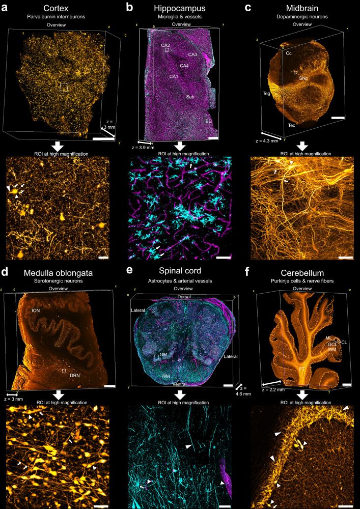
The culmination of years of hard work by Anna Maria Reuss & Co: aDISCO - a protocol for clearing & antibody-staining human formalin-fixed paraffin-embedded (FFPE) tissue! With lots of @mesospim.bsky.social #lightsheet #Microscopy @ptrrupprecht.bsky.social & many more!
www.biorxiv.org/content/10.1...
www.biorxiv.org/content/10.1...
May 27, 2025 at 7:30 AM
The culmination of years of hard work by Anna Maria Reuss & Co: aDISCO - a protocol for clearing & antibody-staining human formalin-fixed paraffin-embedded (FFPE) tissue! With lots of @mesospim.bsky.social #lightsheet #Microscopy @ptrrupprecht.bsky.social & many more!
www.biorxiv.org/content/10.1...
www.biorxiv.org/content/10.1...
Reposted by Dr. Caleb Stoltzfus

We are excited to share our development of a new 10X lens with NA=0.50 over a 7.2 mm FOV and a 35 mm working distance (water), fully corrected from 400-850 nm. This lens will power our upcoming ExA-SPIM "2" and is available for dissemination - please get in touch if interested!
May 19, 2025 at 1:10 PM
We are excited to share our development of a new 10X lens with NA=0.50 over a 7.2 mm FOV and a 35 mm working distance (water), fully corrected from 400-850 nm. This lens will power our upcoming ExA-SPIM "2" and is available for dissemination - please get in touch if interested!
Reposted by Dr. Caleb Stoltzfus

Are you a visionary leader in quantitative cellular imaging?
We're looking for a Director of Quantitative Cellular Imaging. This role is pivotal in our mission to demystify multicellular morphogenesis through innovative imaging assays and data analysis.
🔗 alleninstitute.org/careers/jobs...
We're looking for a Director of Quantitative Cellular Imaging. This role is pivotal in our mission to demystify multicellular morphogenesis through innovative imaging assays and data analysis.
🔗 alleninstitute.org/careers/jobs...

May 5, 2025 at 6:15 PM
Are you a visionary leader in quantitative cellular imaging?
We're looking for a Director of Quantitative Cellular Imaging. This role is pivotal in our mission to demystify multicellular morphogenesis through innovative imaging assays and data analysis.
🔗 alleninstitute.org/careers/jobs...
We're looking for a Director of Quantitative Cellular Imaging. This role is pivotal in our mission to demystify multicellular morphogenesis through innovative imaging assays and data analysis.
🔗 alleninstitute.org/careers/jobs...
Reposted by Dr. Caleb Stoltzfus
To look into: a Python alternative to ZEMAX... github.com/HarrisonKram...

GitHub - HarrisonKramer/optiland: Comprehensive optical design, optimization, and analysis in Python, including GPU-accelerated and differentiable ray tracing via PyTorch.
Comprehensive optical design, optimization, and analysis in Python, including GPU-accelerated and differentiable ray tracing via PyTorch. - HarrisonKramer/optiland
github.com
May 2, 2025 at 10:56 PM
To look into: a Python alternative to ZEMAX... github.com/HarrisonKram...
The team published LUMI v2.0! The best new feature defines ROIs on low-res 3D scout scans!
Scan your whole tissue at low resolution
Define a bounding box using something like BigStitcher
Import and scan the ROI
Take your time finding and defining an ROI without worrying about photobleaching!
Scan your whole tissue at low resolution
Define a bounding box using something like BigStitcher
Import and scan the ROI
Take your time finding and defining an ROI without worrying about photobleaching!

March 21, 2025 at 3:56 PM
The team published LUMI v2.0! The best new feature defines ROIs on low-res 3D scout scans!
Scan your whole tissue at low resolution
Define a bounding box using something like BigStitcher
Import and scan the ROI
Take your time finding and defining an ROI without worrying about photobleaching!
Scan your whole tissue at low resolution
Define a bounding box using something like BigStitcher
Import and scan the ROI
Take your time finding and defining an ROI without worrying about photobleaching!
Reposted by Dr. Caleb Stoltzfus

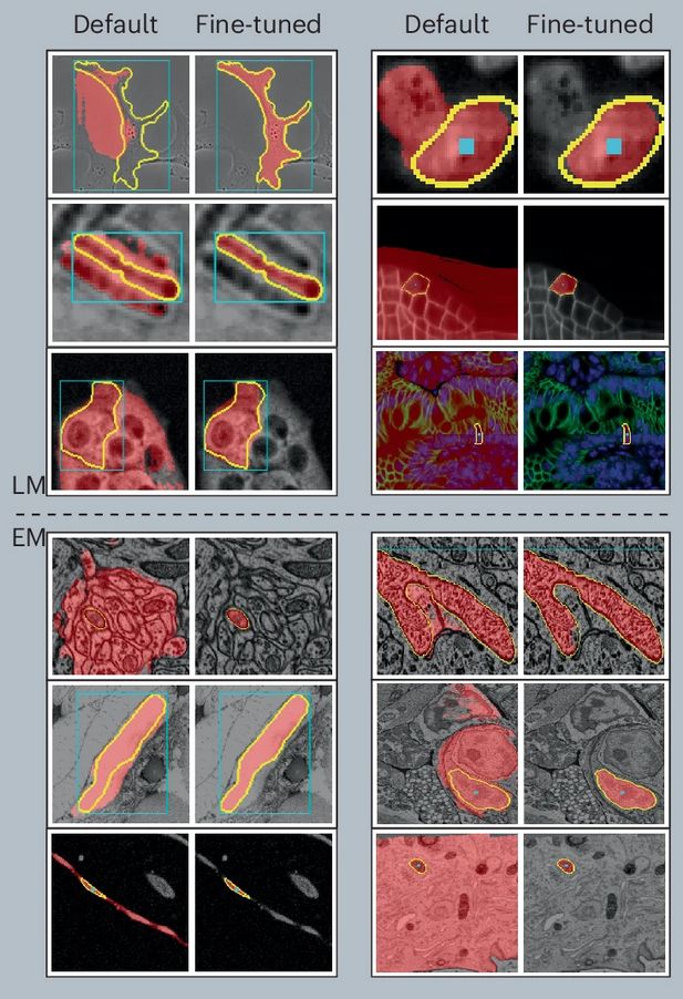
After a long journey, Segment Anything for Microscopy is now published in Nature Methods! We significantly improve SAM for interactive and automatic segmentation in light and electron microscopy and build a user-friendly tool.
www.nature.com/articles/s41...
www.nature.com/articles/s41...
February 12, 2025 at 11:41 AM
After a long journey, Segment Anything for Microscopy is now published in Nature Methods! We significantly improve SAM for interactive and automatic segmentation in light and electron microscopy and build a user-friendly tool.
www.nature.com/articles/s41...
www.nature.com/articles/s41...
Reposted by Dr. Caleb Stoltzfus
I used VVD Viewer for this #FluorescenceFriday video: 141 volumes from the #HHMIJanelia FlyLight Split-GAL4 #Drosophila driver collection. The animation comes from a high-level description processed by neuVid. Such a rapid tour of the data has questionable scientific value, but it's fun to watch!
December 20, 2024 at 9:57 AM
I used VVD Viewer for this #FluorescenceFriday video: 141 volumes from the #HHMIJanelia FlyLight Split-GAL4 #Drosophila driver collection. The animation comes from a high-level description processed by neuVid. Such a rapid tour of the data has questionable scientific value, but it's fun to watch!
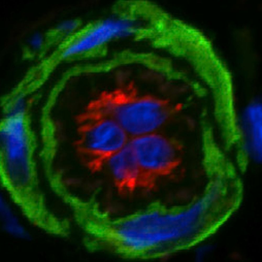
I hope I never get tired of how amazing cells look in 3D. Happy Holidays and Happy #FluorescenceFriday! Here are CD45+ cells in a skin biopsy. The segmented cells and nerves at the end of the video kind of look like strings of lights
December 20, 2024 at 7:44 PM
I hope I never get tired of how amazing cells look in 3D. Happy Holidays and Happy #FluorescenceFriday! Here are CD45+ cells in a skin biopsy. The segmented cells and nerves at the end of the video kind of look like strings of lights
The Washington Research Foundation announced the next cohort of 12 postdoctoral fellowships! Congratulations to the fellows! I can't wait to see what they accomplish. www.wrfseattle.org/news/washing...
Washington Research Foundation awards 12 three-year postdoctoral fellowships - Washington Research Foundation
www.wrfseattle.org
December 18, 2024 at 7:14 PM
The Washington Research Foundation announced the next cohort of 12 postdoctoral fellowships! Congratulations to the fellows! I can't wait to see what they accomplish. www.wrfseattle.org/news/washing...
For #fluorescencefriday here is a mouse brain stained with Eosin and To-PRO-3 showing a segmented Glioblastoma in yellow. While this whole brain computational H&E staining is cool, I think I prefer TH staining for the dendrites. Imaged on Alpenglow Bioscience's 3Di #light-sheet #microscopy
December 6, 2024 at 5:05 PM
For #fluorescencefriday here is a mouse brain stained with Eosin and To-PRO-3 showing a segmented Glioblastoma in yellow. While this whole brain computational H&E staining is cool, I think I prefer TH staining for the dendrites. Imaged on Alpenglow Bioscience's 3Di #light-sheet #microscopy
Reposted by Dr. Caleb Stoltzfus
Microscopy/live imaging people! Need your help 😅
I’ve only just recently started working with large time series files (1,000+ slices) and don’t know where to start when it comes to rendering and exporting them. What software(s) do you use?
I’ve only just recently started working with large time series files (1,000+ slices) and don’t know where to start when it comes to rendering and exporting them. What software(s) do you use?
November 27, 2024 at 4:54 PM
Microscopy/live imaging people! Need your help 😅
I’ve only just recently started working with large time series files (1,000+ slices) and don’t know where to start when it comes to rendering and exporting them. What software(s) do you use?
I’ve only just recently started working with large time series files (1,000+ slices) and don’t know where to start when it comes to rendering and exporting them. What software(s) do you use?
Come check out the work we have been doing on 3D morphometrics using #light-sheet #microscopy!
🔬 Sneak peek! I'm presenting at Multi-Omics Brisbane 2024 on our automated 3D cell classification & segmentation system!
📽️ Check out our 3D melanoma tissue imaging: neutrophils (red) & lymphocytes (green) and nuclei (blue). Credit: Dr Alexandra Alvarsson
📽️ Check out our 3D melanoma tissue imaging: neutrophils (red) & lymphocytes (green) and nuclei (blue). Credit: Dr Alexandra Alvarsson
November 27, 2024 at 4:23 PM
Come check out the work we have been doing on 3D morphometrics using #light-sheet #microscopy!
Reposted by Dr. Caleb Stoltzfus
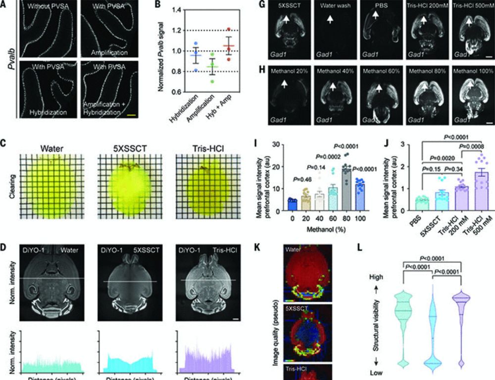
TRISCO
A solid step closer to 3D #SpatialTranscriptomics 😎
Optimized probe-based 3D RNA imaging in whole organs
isHCR
Tris 500 mM
4'C
Validated probe list in Table S1
Brain Heart Lung Kidney
@peruhlen.bsky.social @science.org 2024
www.science.org/doi/10.1126/...
A solid step closer to 3D #SpatialTranscriptomics 😎
Optimized probe-based 3D RNA imaging in whole organs
isHCR
Tris 500 mM
4'C
Validated probe list in Table S1
Brain Heart Lung Kidney
@peruhlen.bsky.social @science.org 2024
www.science.org/doi/10.1126/...

November 26, 2024 at 1:10 PM
TRISCO
A solid step closer to 3D #SpatialTranscriptomics 😎
Optimized probe-based 3D RNA imaging in whole organs
isHCR
Tris 500 mM
4'C
Validated probe list in Table S1
Brain Heart Lung Kidney
@peruhlen.bsky.social @science.org 2024
www.science.org/doi/10.1126/...
A solid step closer to 3D #SpatialTranscriptomics 😎
Optimized probe-based 3D RNA imaging in whole organs
isHCR
Tris 500 mM
4'C
Validated probe list in Table S1
Brain Heart Lung Kidney
@peruhlen.bsky.social @science.org 2024
www.science.org/doi/10.1126/...
Here is a murine heart video we imaged with the Red-Horse lab for #FluorescenceFriday.
November 22, 2024 at 6:34 PM
Here is a murine heart video we imaged with the Red-Horse lab for #FluorescenceFriday.
Reposted by Dr. Caleb Stoltzfus
Let’s see how well bsky does with external media. 🧪 #Vanderbilt Neurovisualization Lab is among the first to clear, label and image an intact NHP hemisphere. Please set your player to 4K! #lightsheet #microscopy #science #brain. If your lab wants to add lightsheet to your work we can help!

Marmoset brain hemisphere imaged with lightsheet
YouTube video by Neurovisualization Lab
youtu.be
November 20, 2024 at 2:20 PM
Let’s see how well bsky does with external media. 🧪 #Vanderbilt Neurovisualization Lab is among the first to clear, label and image an intact NHP hemisphere. Please set your player to 4K! #lightsheet #microscopy #science #brain. If your lab wants to add lightsheet to your work we can help!
Whoa! Visualization of RNA gene expression in intact rodent brains using TRISCO clearing and light-sheet #microscopy. www.science.org/doi/10.1126/...
Whole-brain spatial transcriptional analysis at cellular resolution
Recent advances in RNA analysis have deepened our understanding of cellular states in biological tissues. However, a substantial gap remains in integrating RNA expression data with spatial context acr...
www.science.org
November 21, 2024 at 8:12 PM
Whoa! Visualization of RNA gene expression in intact rodent brains using TRISCO clearing and light-sheet #microscopy. www.science.org/doi/10.1126/...
Reposted by Dr. Caleb Stoltzfus
Axially Swept Light-Sheet Microscopy (#ASLM) synchronizes the camera’s rolling shutter with axial remote focusing (sweeping) of the light sheet. This enables 3D imaging with a thin optical section and isotropic resolution across large FOVs. Ideal for imaging cleared tissue!
doi.org/10.1016/j.bp...
doi.org/10.1016/j.bp...
November 16, 2024 at 8:12 PM
Axially Swept Light-Sheet Microscopy (#ASLM) synchronizes the camera’s rolling shutter with axial remote focusing (sweeping) of the light sheet. This enables 3D imaging with a thin optical section and isotropic resolution across large FOVs. Ideal for imaging cleared tissue!
doi.org/10.1016/j.bp...
doi.org/10.1016/j.bp...
Lets start this off with some 3D imaging of human placenta. Samples from Mira Moufarrej and the lab of Kristy Red Horse at Stanford. Imaged at Alpenglow Biosciences in Seattle by our amazing team of scientists and engineers.
November 14, 2024 at 6:02 PM
Lets start this off with some 3D imaging of human placenta. Samples from Mira Moufarrej and the lab of Kristy Red Horse at Stanford. Imaged at Alpenglow Biosciences in Seattle by our amazing team of scientists and engineers.