


Tanner Fadero
@tanner-fadero.bsky.social
(he/him) Light microscopy scientist interested in developing and deploying better live-cell fluorescence microscopy technology. Tweets about microscopy🔬, cell biology, and optics. Conflicts of interest: http://tinyurl.com/y25ctubo
Motorized slit! Off-the-shelf part from ScienceTech. Thorlabs stepper motor driven by K-cube controller.
www.sciencetech-inc.com/shop/product...
@amsikking.bsky.social @retof.bsky.social @kevin-dean.bsky.social
www.sciencetech-inc.com/shop/product...
@amsikking.bsky.social @retof.bsky.social @kevin-dean.bsky.social

November 19, 2025 at 6:06 PM
Motorized slit! Off-the-shelf part from ScienceTech. Thorlabs stepper motor driven by K-cube controller.
www.sciencetech-inc.com/shop/product...
@amsikking.bsky.social @retof.bsky.social @kevin-dean.bsky.social
www.sciencetech-inc.com/shop/product...
@amsikking.bsky.social @retof.bsky.social @kevin-dean.bsky.social

But yeah, a modest tilt angle between projections looks similar!


August 30, 2025 at 11:25 PM
But yeah, a modest tilt angle between projections looks similar!
Optics folks: does anyone know a manufacturer of a dichroic beamsplitter *cube*?
Essentially, a dichroic version of this (for fluorescence microscopy):
Essentially, a dichroic version of this (for fluorescence microscopy):

August 28, 2025 at 12:15 AM
Optics folks: does anyone know a manufacturer of a dichroic beamsplitter *cube*?
Essentially, a dichroic version of this (for fluorescence microscopy):
Essentially, a dichroic version of this (for fluorescence microscopy):
Here's a six-month time-lapse of me building the single objective light sheet fluorescence microscope at the UCSF Center for Advanced Light Microscopy!
Also pictured is @nicostuurman.bsky.social and @kahsage.bsky.social.
#snouty
Also pictured is @nicostuurman.bsky.social and @kahsage.bsky.social.
#snouty
August 20, 2025 at 6:57 PM
Here's a six-month time-lapse of me building the single objective light sheet fluorescence microscope at the UCSF Center for Advanced Light Microscopy!
Also pictured is @nicostuurman.bsky.social and @kahsage.bsky.social.
#snouty
Also pictured is @nicostuurman.bsky.social and @kahsage.bsky.social.
#snouty
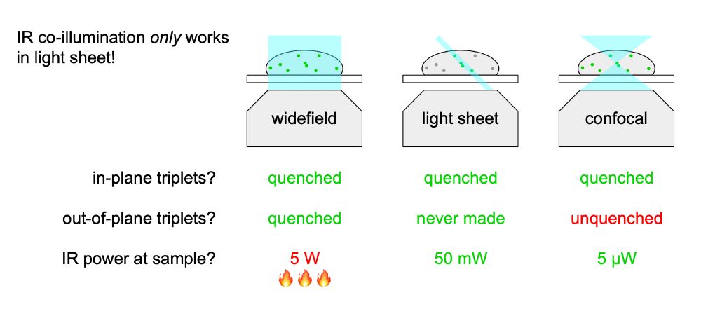
Unlike confocal, light sheet also doesn't need to deplete triplets outside of the focal plane, because light sheet doesn't *generate* out-of-plane triplets.
August 19, 2025 at 7:40 PM
Unlike confocal, light sheet also doesn't need to deplete triplets outside of the focal plane, because light sheet doesn't *generate* out-of-plane triplets.
How many photons are in a GFP? — more than last year, and more than you thought. Here's a simple, cheap, and practical method to break a fundamental limit in fluorescence microscopy. But it only works in light sheet!
August 19, 2025 at 7:31 PM
How many photons are in a GFP? — more than last year, and more than you thought. Here's a simple, cheap, and practical method to break a fundamental limit in fluorescence microscopy. But it only works in light sheet!
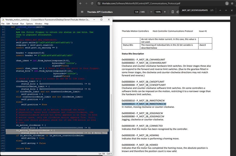
Hey @thorlabs.bsky.social! Thanks for such a well-documented APT Controller Communication Protocol! It was fun coding against it.
@andrewgyork.bsky.social and I did find a small bug, though. When talking to a MFF102 flipper, it only ever reports clockwise motion (never counterclockwise).
@andrewgyork.bsky.social and I did find a small bug, though. When talking to a MFF102 flipper, it only ever reports clockwise motion (never counterclockwise).
August 4, 2025 at 11:48 PM
Hey @thorlabs.bsky.social! Thanks for such a well-documented APT Controller Communication Protocol! It was fun coding against it.
@andrewgyork.bsky.social and I did find a small bug, though. When talking to a MFF102 flipper, it only ever reports clockwise motion (never counterclockwise).
@andrewgyork.bsky.social and I did find a small bug, though. When talking to a MFF102 flipper, it only ever reports clockwise motion (never counterclockwise).
A @bob-goldstein-art.bsky.social original!
August 4, 2025 at 4:31 PM
A @bob-goldstein-art.bsky.social original!
If you've ever wanted to know what the actin filaments on the scales in a developing moth wing look like in 3D, here you go!
Kyle DeMarr from @mullinslab.bsky.social acquired these beautiful data on the single objective light sheet at the UCSF Center for Advanced Light Microscopy.
#snouty
Kyle DeMarr from @mullinslab.bsky.social acquired these beautiful data on the single objective light sheet at the UCSF Center for Advanced Light Microscopy.
#snouty
July 26, 2025 at 7:18 PM
If you've ever wanted to know what the actin filaments on the scales in a developing moth wing look like in 3D, here you go!
Kyle DeMarr from @mullinslab.bsky.social acquired these beautiful data on the single objective light sheet at the UCSF Center for Advanced Light Microscopy.
#snouty
Kyle DeMarr from @mullinslab.bsky.social acquired these beautiful data on the single objective light sheet at the UCSF Center for Advanced Light Microscopy.
#snouty
Interestingly, it already is! I used plane waves to calculate this. Plane waves don't experience a focal plane, they're "collimated." Despite this, they can form 3D patterns (e.g. 3D SIM).
In fact, you can't assess axial magnification without considering focal planes other than the native one!
In fact, you can't assess axial magnification without considering focal planes other than the native one!

July 1, 2025 at 8:11 PM
Interestingly, it already is! I used plane waves to calculate this. Plane waves don't experience a focal plane, they're "collimated." Despite this, they can form 3D patterns (e.g. 3D SIM).
In fact, you can't assess axial magnification without considering focal planes other than the native one!
In fact, you can't assess axial magnification without considering focal planes other than the native one!
If you zoom into the region where M_z = M_xy, you'll find the region where remote refocus works! When M_z = M_xy = n_s at all wave angles. This is where you can form a non-aberrated remote 3D image.
This is why @amsikking.bsky.social's any immersion remote refocus works!
This is why @amsikking.bsky.social's any immersion remote refocus works!

July 1, 2025 at 4:17 PM
If you zoom into the region where M_z = M_xy, you'll find the region where remote refocus works! When M_z = M_xy = n_s at all wave angles. This is where you can form a non-aberrated remote 3D image.
This is why @amsikking.bsky.social's any immersion remote refocus works!
This is why @amsikking.bsky.social's any immersion remote refocus works!
I spent some time finally deriving the relationship between lateral image magnification and axial magnification! It's an interesting equation, with dependencies on sample refractive index and the wave propogation angle w.r.t. the optical axis.
Play with it yourself! www.desmos.com/calculator/q...
Play with it yourself! www.desmos.com/calculator/q...


July 1, 2025 at 3:59 PM
I spent some time finally deriving the relationship between lateral image magnification and axial magnification! It's an interesting equation, with dependencies on sample refractive index and the wave propogation angle w.r.t. the optical axis.
Play with it yourself! www.desmos.com/calculator/q...
Play with it yourself! www.desmos.com/calculator/q...
Always good advice from @kahsage.bsky.social. Don't make your imaging experiment take longer than it needs to!

June 26, 2025 at 6:59 PM
Always good advice from @kahsage.bsky.social. Don't make your imaging experiment take longer than it needs to!
@kahsage.bsky.social is killing it with these teaching tools for #snoutscope training at the UCSF Center for Advanced Light Microscopy. Fantastic illustrations and a whiteboard in the room. Brilliant!


June 26, 2025 at 6:57 PM
@kahsage.bsky.social is killing it with these teaching tools for #snoutscope training at the UCSF Center for Advanced Light Microscopy. Fantastic illustrations and a whiteboard in the room. Brilliant!
Another #snoutscope imaging session at the UCSF Center for Advanced Light Microscopy! This time, with neutrophils! Courtesy of Patrick Zager in the Weiner lab.
Neutrophil plasma membranes are labeled to show 3D filopodia and lamellipodia dynamics at 1 volume/second.
Neutrophil plasma membranes are labeled to show 3D filopodia and lamellipodia dynamics at 1 volume/second.
June 24, 2025 at 1:31 AM
Another #snoutscope imaging session at the UCSF Center for Advanced Light Microscopy! This time, with neutrophils! Courtesy of Patrick Zager in the Weiner lab.
Neutrophil plasma membranes are labeled to show 3D filopodia and lamellipodia dynamics at 1 volume/second.
Neutrophil plasma membranes are labeled to show 3D filopodia and lamellipodia dynamics at 1 volume/second.
I couldn't be happier to wake up to this text at 1 am. 1 volume/second for tens of minutes of single molecule imaging!
Thanks to @kahsage.bsky.social for training Yovan, and thanks to Hernan Garcia's lab @ucberkeleyofficial.bsky.social for bringing their biology to the scope!
Thanks to @kahsage.bsky.social for training Yovan, and thanks to Hernan Garcia's lab @ucberkeleyofficial.bsky.social for bringing their biology to the scope!

June 21, 2025 at 2:04 PM
I couldn't be happier to wake up to this text at 1 am. 1 volume/second for tens of minutes of single molecule imaging!
Thanks to @kahsage.bsky.social for training Yovan, and thanks to Hernan Garcia's lab @ucberkeleyofficial.bsky.social for bringing their biology to the scope!
Thanks to @kahsage.bsky.social for training Yovan, and thanks to Hernan Garcia's lab @ucberkeleyofficial.bsky.social for bringing their biology to the scope!
I got a text from Yovan Bodal, who just used our #snoutscope @UCSF to image fly embryos. His quote says it all!
"This was the best results we've gotten on any instrument, possibly for the least amount of work on my end. Here's a GIF of individual mRNAs coming off the gene as they get transcribed:"
"This was the best results we've gotten on any instrument, possibly for the least amount of work on my end. Here's a GIF of individual mRNAs coming off the gene as they get transcribed:"
June 21, 2025 at 2:00 PM
I got a text from Yovan Bodal, who just used our #snoutscope @UCSF to image fly embryos. His quote says it all!
"This was the best results we've gotten on any instrument, possibly for the least amount of work on my end. Here's a GIF of individual mRNAs coming off the gene as they get transcribed:"
"This was the best results we've gotten on any instrument, possibly for the least amount of work on my end. Here's a GIF of individual mRNAs coming off the gene as they get transcribed:"
Here's the reflectivity at 33.5° and 56.5°. Seems like 808 nm is lost after some critical angle. Maybe I'll just build the two-galvo system at <45° to compensate?

May 13, 2025 at 1:35 PM
Here's the reflectivity at 33.5° and 56.5°. Seems like 808 nm is lost after some critical angle. Maybe I'll just build the two-galvo system at <45° to compensate?
@jennysachweh.bsky.social inspired me to make a quick and dirty model of "Two Mirror Beam Scanning" this morning. It's a nice visual demonstration of how two galvo mirrors can translate a beam with no angular motion.
Try it for yourself! www.geogebra.org/calculator/s...
Try it for yourself! www.geogebra.org/calculator/s...
May 8, 2025 at 4:37 PM
@jennysachweh.bsky.social inspired me to make a quick and dirty model of "Two Mirror Beam Scanning" this morning. It's a nice visual demonstration of how two galvo mirrors can translate a beam with no angular motion.
Try it for yourself! www.geogebra.org/calculator/s...
Try it for yourself! www.geogebra.org/calculator/s...
It's the last #KingSnout ever! 😭
I'm looking forward to what's next in the #snouty objective lens line from ASI and Special Optics. Onwards to bigger and better things!
#snoutclub
@jsdaniel02.bsky.social @amsikking.bsky.social
I'm looking forward to what's next in the #snouty objective lens line from ASI and Special Optics. Onwards to bigger and better things!
#snoutclub
@jsdaniel02.bsky.social @amsikking.bsky.social

April 18, 2025 at 4:37 PM
It's the last #KingSnout ever! 😭
I'm looking forward to what's next in the #snouty objective lens line from ASI and Special Optics. Onwards to bigger and better things!
#snoutclub
@jsdaniel02.bsky.social @amsikking.bsky.social
I'm looking forward to what's next in the #snouty objective lens line from ASI and Special Optics. Onwards to bigger and better things!
#snoutclub
@jsdaniel02.bsky.social @amsikking.bsky.social
Awh yeah, got my own hardware cart:

April 16, 2025 at 1:30 AM
Awh yeah, got my own hardware cart:
best microscope I ever built, who agrees?

March 7, 2025 at 6:57 PM
best microscope I ever built, who agrees?
I saw that ThorLabs part. Good idea about the c-shaped insert.
There are two set screws (or at least M3 tapped holes for set screws) in the Edmund 18291 19.1 mm ID adapter:
There are two set screws (or at least M3 tapped holes for set screws) in the Edmund 18291 19.1 mm ID adapter:

March 5, 2025 at 7:57 PM
I saw that ThorLabs part. Good idea about the c-shaped insert.
There are two set screws (or at least M3 tapped holes for set screws) in the Edmund 18291 19.1 mm ID adapter:
There are two set screws (or at least M3 tapped holes for set screws) in the Edmund 18291 19.1 mm ID adapter:
Upon further review, it seems Edmund suggests this part for this exact purpose: productimages.edmundoptics.com/20168.jpg?qu...

March 5, 2025 at 7:51 PM
Upon further review, it seems Edmund suggests this part for this exact purpose: productimages.edmundoptics.com/20168.jpg?qu...