Max Schelski
banner
maxschelski.bsky.social
Max Schelski
@maxschelski.bsky.social
Neuroscientist. Developing mathematical models of cell biology underlying synaptic plasticity. Coding Python.

PhD done @ Bradke lab #NeuroDev #Microtubules

PostDoc in Tchumatchenko group. #SynapticPlasticity #Dendrites
https://github.com/maxschelski/
What slows down microtubule retrograde flow (MT-RF) in the axon is an even more open question. It could be the change in MT orientation in the axon - too few microtubules with their plus-ends to the soma. However, I also saw that Taxol slows down MT-RF before changing MT orientation (right side).
November 25, 2024 at 6:07 AM
To make matters even more interesting, I also see that inhibiting Myosin II slows down MT-RF (in the middle). This could indicate that Myosin might also somehow be involved in driving MT-RF.
November 25, 2024 at 5:55 AM
However, I also recruited two different Kinesin motor domains to the plasma membrane. For both motor domains I saw something like faster MT-RF - with a delay after recruiting them to the membrane. I would say, the jury is still out on whether Dynein or Kinesins drive MT-RF physiologically.
November 25, 2024 at 5:55 AM
This would suggest that Dynein could speed up MT-RF through microtubules that have their plus-end towards the soma. One result supporting this, is that recruiting Dynein to the plasma membrane speeds up MT-RF in dendrites (50% plus-ends to soma) but not in axons (2% plus-ends to soma).
November 25, 2024 at 5:55 AM
I also found that recruiting endogous Dynein or an overexpressed Dynein motor domain is sufficient to speed up MT-RF. Recruting a motor deficient point mutant had no effect (see video). I saw the same also for the overexpressed minus-end directed motor domain of KIFC1.
November 25, 2024 at 5:55 AM
#Microtubules flow retrogradely in all neurites before axon specification - labeled CAMSAP3 on the right. After axon outgrowth, this retrograde flow slows down - to enable stable axon growth. Could this retrograde flow also exist in #microglia, #astrocytes & #cancer cells? #FluorescenceFriday 🧪 🧠📈
November 22, 2024 at 10:26 AM
Thanks a lot for the post. :) Just as a site note - I also saw the same retrograde movement, when I photoconverted small patches of microtubules (on the right).
November 20, 2024 at 7:59 AM
I also built a simple-to-use #Python toolbox to generate publication-grade movies and figures easily: github.com/maxschelski/.... I optimized it particularly for #microscopy data. Scripts for 25 example figures and 12 movies are available. Documentation under figureflow.readthedocs.io/en/latest/ 🔬🧪📈
November 20, 2024 at 7:46 AM
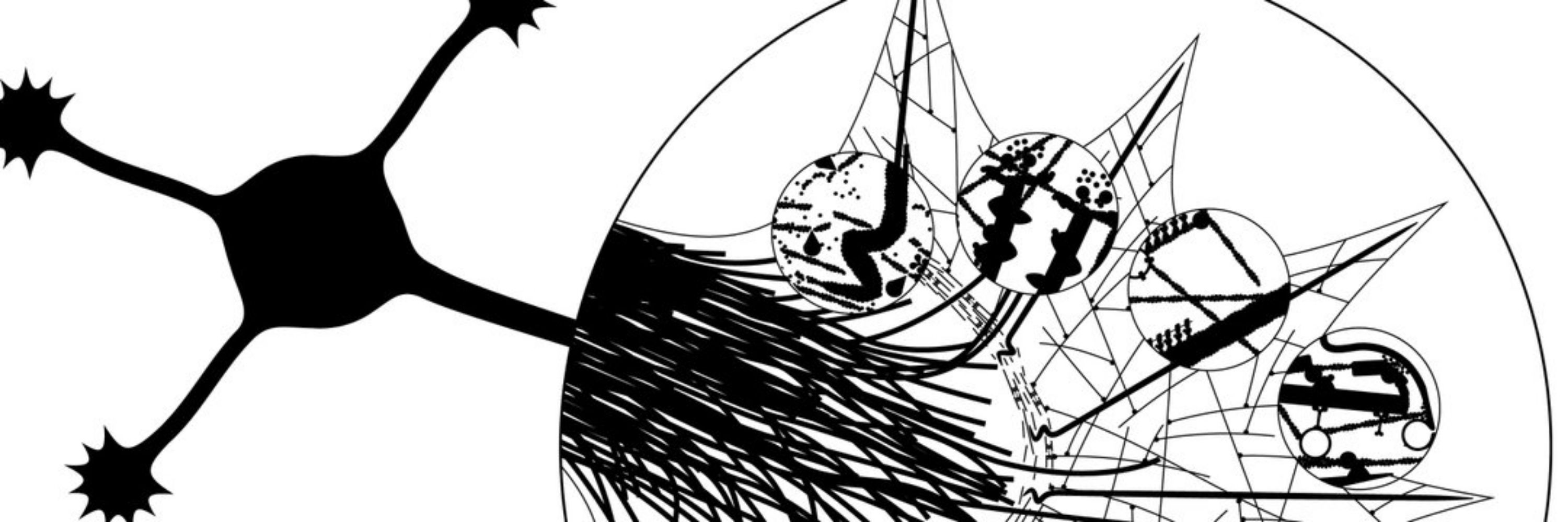
Hi! I'm Max. I'm developing a #biophysical model of #microtubules in #neurons during #development. The model is based on my surprising experimental finding (lots of live-cell imaging) that the entire microtubule array in neurites flows retrogradely into the soma: www.science.org/doi/10.1126/... 🧪 🧠📈
November 20, 2024 at 7:37 AM