


Electron Microscopy Facility, University of Oslo
@emlaboslo.bsky.social
Established in 1966, today equipped with 4 electron microscopes (two transmission electron microscopes (TEM) / two scanning electron microscopes (SEM)) and a wide range of preparation equipment covering most of the current preparation methods.
Pinned

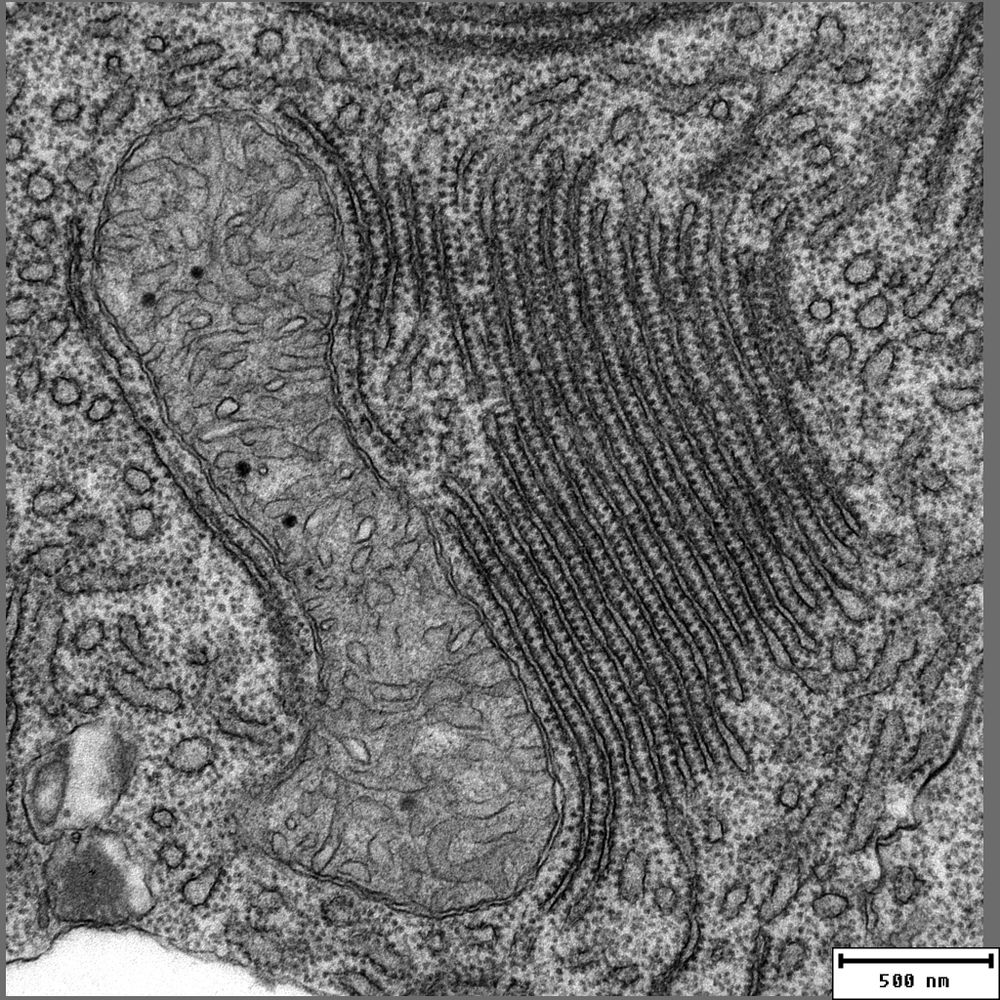
Expanding the field of view – a simple approach for interactive visualisation of electron microscopy data
Summary: A simple and easy to implement interactive visualisation for EM data, inspired by digital maps, which compensates for the loss of field of view at high magnifications and improves data commun...
doi.org
A simple and easy to implement interactive visualisation approach for EM data. doi.org/10.1242/jcs....
Try it: wohlmann.github.io/2024_EMMA_F0...
(zebrafish [fin] tissue, 6pdf)
Try it: wohlmann.github.io/2024_EMMA_F0...
(zebrafish [fin] tissue, 6pdf)
Reposted by Electron Microscopy Facility, University of Oslo
(1/n) DNA-PAINT imaging inside the nucleus at single antibody resolution using TIRF? Ultrathin sectioning makes it happen!
Grateful to share my postdoctoral work introducing “tomographic & kinetically-enhanced DNA-PAINT” or in brief: tkPAINT. Out in @pnas.org!
doi.org/10.1073/pnas...
👇🧵
Grateful to share my postdoctoral work introducing “tomographic & kinetically-enhanced DNA-PAINT” or in brief: tkPAINT. Out in @pnas.org!
doi.org/10.1073/pnas...
👇🧵

August 13, 2025 at 2:19 PM
(1/n) DNA-PAINT imaging inside the nucleus at single antibody resolution using TIRF? Ultrathin sectioning makes it happen!
Grateful to share my postdoctoral work introducing “tomographic & kinetically-enhanced DNA-PAINT” or in brief: tkPAINT. Out in @pnas.org!
doi.org/10.1073/pnas...
👇🧵
Grateful to share my postdoctoral work introducing “tomographic & kinetically-enhanced DNA-PAINT” or in brief: tkPAINT. Out in @pnas.org!
doi.org/10.1073/pnas...
👇🧵
Reposted by Electron Microscopy Facility, University of Oslo
Astronomy Picture from 21/04/2008
Bacteriophages: The Most Common Life-Like Form on Earth
Source: https://apod.nasa.gov/apod/ap080421.htm<a href="/hashtag/Bacteriophages" class="hover:underline text-blue-600 dark:text-sky-400 no-card-link">#Bacteriophagesi#Viruses##ElectronMicroscopyc#Microbiologyo#Astronomys#Spacey#Science##BacteriaB#Phages #TinyWorldsnyWorlds

March 10, 2025 at 4:15 PM
Astronomy Picture from 21/04/2008
Bacteriophages: The Most Common Life-Like Form on Earth
Source: https://apod.nasa.gov/apod/ap080421.htm<a href="/hashtag/Bacteriophages" class="hover:underline text-blue-600 dark:text-sky-400 no-card-link">#Bacteriophagesi#Viruses##ElectronMicroscopyc#Microbiologyo#Astronomys#Spacey#Science##BacteriaB#Phages #TinyWorldsnyWorlds
Bacteriophages: The Most Common Life-Like Form on Earth
Source: https://apod.nasa.gov/apod/ap080421.htm<a href="/hashtag/Bacteriophages" class="hover:underline text-blue-600 dark:text-sky-400 no-card-link">#Bacteriophagesi#Viruses##ElectronMicroscopyc#Microbiologyo#Astronomys#Spacey#Science##BacteriaB#Phages #TinyWorldsnyWorlds
Reposted by Electron Microscopy Facility, University of Oslo

Apply for a position as Postdoctoral Research Fellow in Fish Immunology/Microscopy @biovitenskap.bsky.social
This role contributes to the ERC-funded project “Fish-S.H.I.E.L.D.”, which investigate the fish immune surveillance system and its importance for the fish defense against infectious diseases
This role contributes to the ERC-funded project “Fish-S.H.I.E.L.D.”, which investigate the fish immune surveillance system and its importance for the fish defense against infectious diseases

Postdoctoral Research Fellow in Fish Immunology/Microscopy (278595) | University of Oslo
Job title: Postdoctoral Research Fellow in Fish Immunology/Microscopy (278595), Employer: University of Oslo, Deadline: Wednesday, April 30, 2025
www.jobbnorge.no
April 4, 2025 at 11:21 AM
Apply for a position as Postdoctoral Research Fellow in Fish Immunology/Microscopy @biovitenskap.bsky.social
This role contributes to the ERC-funded project “Fish-S.H.I.E.L.D.”, which investigate the fish immune surveillance system and its importance for the fish defense against infectious diseases
This role contributes to the ERC-funded project “Fish-S.H.I.E.L.D.”, which investigate the fish immune surveillance system and its importance for the fish defense against infectious diseases
Reposted by Electron Microscopy Facility, University of Oslo
We are proud to announce the launch of FAIR Image Analysis Across Sciences, supported by the OSCARS project, in collaboration with Simula Research Laboratory, #EPFL & University of Bergen (UiB)! 🎉

March 31, 2025 at 3:55 PM
We are proud to announce the launch of FAIR Image Analysis Across Sciences, supported by the OSCARS project, in collaboration with Simula Research Laboratory, #EPFL & University of Bergen (UiB)! 🎉
Reposted by Electron Microscopy Facility, University of Oslo
Vi har mange ledige #PhD-stillinger ledige ved @biovitenskap.bsky.social med søknadsfrist 28. februar 2025 Kanskje noen av stillingene er aktuelle for deg eller noen du kjenner? Ta en titt og del gjerne!
www.mn.uio.no/ibv/om/ledig...
www.mn.uio.no/ibv/om/ledig...

Ledige stillingar frå instituttet - Institutt for biovitenskap
Les denne saken på UiOs nettsider.
www.mn.uio.no
February 21, 2025 at 11:27 AM
Vi har mange ledige #PhD-stillinger ledige ved @biovitenskap.bsky.social med søknadsfrist 28. februar 2025 Kanskje noen av stillingene er aktuelle for deg eller noen du kjenner? Ta en titt og del gjerne!
www.mn.uio.no/ibv/om/ledig...
www.mn.uio.no/ibv/om/ledig...
Reposted by Electron Microscopy Facility, University of Oslo
Hyperparasitisme : un virus qui parasite un virus qui infecte une amibe

Hyperparasitisme : un virus qui parasite un virus qui infecte une amibe
Les virus aussi peuvent être infectés ! Un virus géant infectant une amibe est lui-même attaqué par un virophage, à son tour influencé par un élément génétique mobile, un transpoviron. Une cascade d’interactions étonnante révélée par des chercheurs marseillais.
www.pourlascience.fr
February 12, 2025 at 7:19 AM
Hyperparasitisme : un virus qui parasite un virus qui infecte une amibe
Reposted by Electron Microscopy Facility, University of Oslo
We have a PhD position available at the Department of Biosciences, University of Oslo to work on peptide signaling in plants and insects. Please share.
www.jobbnorge.no/en/available...
www.jobbnorge.no/en/available...
PhD Research Fellow in functional genomics (274518) | University of Oslo
Job title: PhD Research Fellow in functional genomics (274518), Employer: University of Oslo, Deadline: Friday, February 28, 2025
www.jobbnorge.no
February 10, 2025 at 2:05 PM
We have a PhD position available at the Department of Biosciences, University of Oslo to work on peptide signaling in plants and insects. Please share.
www.jobbnorge.no/en/available...
www.jobbnorge.no/en/available...
Reposted by Electron Microscopy Facility, University of Oslo

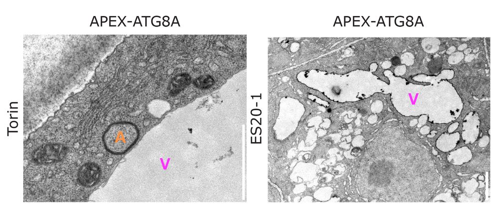
For the first time in plants, we used APEX-based electron microscopy to map the precise localization of ATG8 at the vacuolar membrane after stress! 🚀
Pushing the boundaries of plant cell biology—one EM image at a time. ⚡👀
#PlantScience #ElectronMicroscopy #Autophagy #Vacuole #NaturePlants
Pushing the boundaries of plant cell biology—one EM image at a time. ⚡👀
#PlantScience #ElectronMicroscopy #Autophagy #Vacuole #NaturePlants
February 7, 2025 at 11:11 AM
For the first time in plants, we used APEX-based electron microscopy to map the precise localization of ATG8 at the vacuolar membrane after stress! 🚀
Pushing the boundaries of plant cell biology—one EM image at a time. ⚡👀
#PlantScience #ElectronMicroscopy #Autophagy #Vacuole #NaturePlants
Pushing the boundaries of plant cell biology—one EM image at a time. ⚡👀
#PlantScience #ElectronMicroscopy #Autophagy #Vacuole #NaturePlants
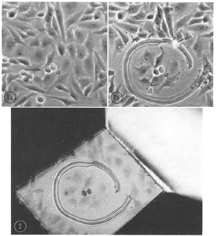
#Correlative Light and Electron Microscopy, #CLEM, is a modern #method combining light and #electronmicroscopy data. Although it is considered “new”, it was already used in the early 1960s, for instance to study the #ultrastructure of mitotic cells in this beautiful example: doi.org/10.1083/jcb....
January 13, 2025 at 7:47 AM
#Correlative Light and Electron Microscopy, #CLEM, is a modern #method combining light and #electronmicroscopy data. Although it is considered “new”, it was already used in the early 1960s, for instance to study the #ultrastructure of mitotic cells in this beautiful example: doi.org/10.1083/jcb....
Reposted by Electron Microscopy Facility, University of Oslo
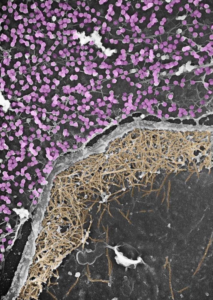
sauerkraut with a variety of bacteria magnified 8,000x

December 17, 2024 at 6:36 PM
sauerkraut with a variety of bacteria magnified 8,000x
Reposted by Electron Microscopy Facility, University of Oslo
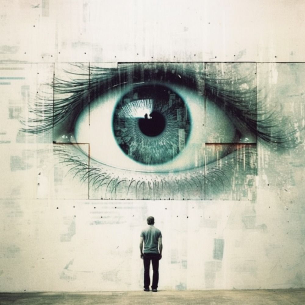
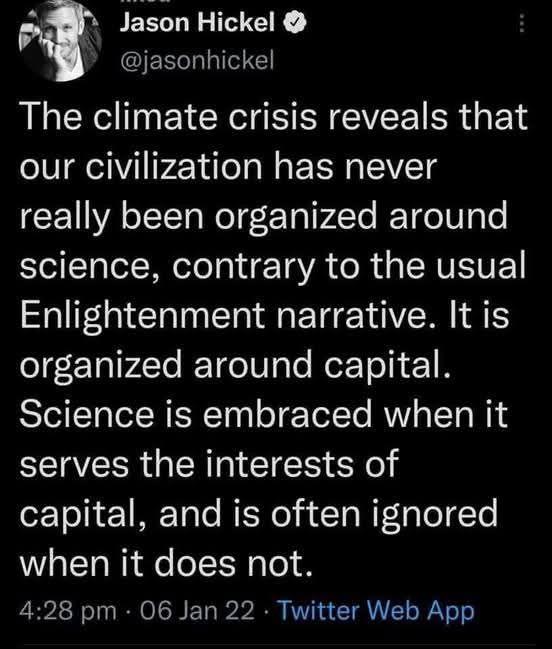
I fear there is deep truth here.
January 11, 2025 at 8:47 PM
I fear there is deep truth here.
Reposted by Electron Microscopy Facility, University of Oslo
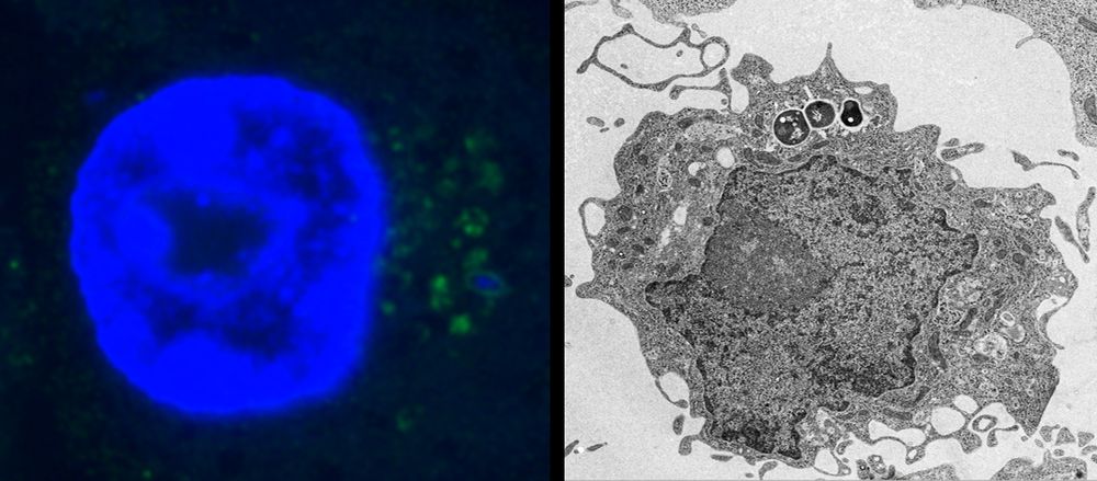
“One of the challenges in science journalism is the oversimplification of research findings to attract attention. Headlines such as “scientists find cure for cancer” or “ozone layer is healing” are designed to attract readers’ attention but often misrepresent the complexity of scientific research.”
Beyond misalignment of science in the news and in schools
Almost 40 years ago, the American astronomer, planetary scientist, and science communicator, Carl Sagan, reflected on the role of mass media in science communication. “How much science and technology ...
www.science.org
January 9, 2025 at 3:56 PM
“One of the challenges in science journalism is the oversimplification of research findings to attract attention. Headlines such as “scientists find cure for cancer” or “ozone layer is healing” are designed to attract readers’ attention but often misrepresent the complexity of scientific research.”
This is probably what a #rotifer gets to see when it encounters a (seemingly bad-tempered) 6dpf #zebrafish larva. #Creepy! But maybe the fluffy #nostrils or the funny #neuromast beard make the situation a little better...
Scanning #electronmicroscopy / #SEM
Scanning #electronmicroscopy / #SEM

January 9, 2025 at 7:38 AM
This is probably what a #rotifer gets to see when it encounters a (seemingly bad-tempered) 6dpf #zebrafish larva. #Creepy! But maybe the fluffy #nostrils or the funny #neuromast beard make the situation a little better...
Scanning #electronmicroscopy / #SEM
Scanning #electronmicroscopy / #SEM
Still our favorite #review! Unfortunately, as relevant today as when it was published, it is an excellent illustration of the #problems arising from the neglect of #ultrastructure and #electronmicroscopy in #cell-biology and the over-reliance on #fluorescence #microscopy.
doi.org/10.1016/0962...
doi.org/10.1016/0962...
January 8, 2025 at 8:18 AM
Still our favorite #review! Unfortunately, as relevant today as when it was published, it is an excellent illustration of the #problems arising from the neglect of #ultrastructure and #electronmicroscopy in #cell-biology and the over-reliance on #fluorescence #microscopy.
doi.org/10.1016/0962...
doi.org/10.1016/0962...
Reposted by Electron Microscopy Facility, University of Oslo

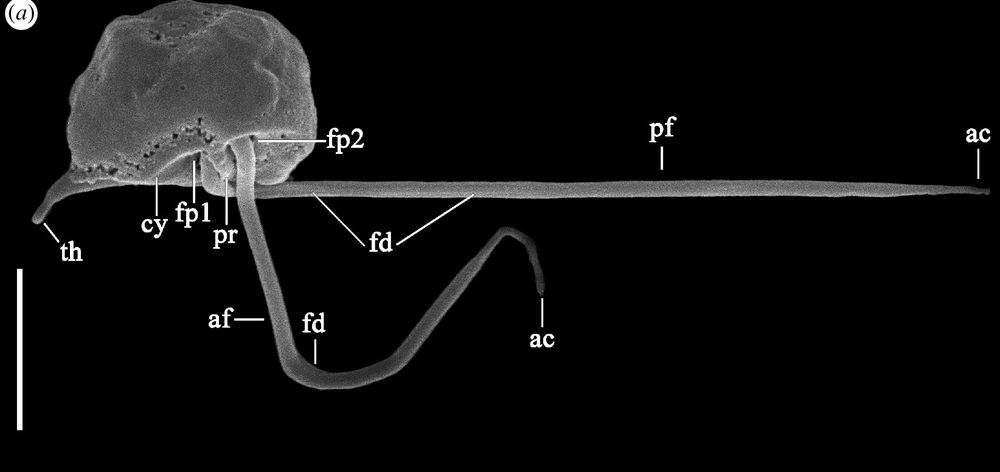
New #ISEPpapers! The nature of ‘jaws’: a new predatory representative of #Provora and the ultrastructure of nibbling protists royalsocietypublishing.org/doi/10.1098/...
#Protists #Microbes #Biology #TreeOfLife #Microscopy
#Protists #Microbes #Biology #TreeOfLife #Microscopy
December 21, 2024 at 6:51 PM
New #ISEPpapers! The nature of ‘jaws’: a new predatory representative of #Provora and the ultrastructure of nibbling protists royalsocietypublishing.org/doi/10.1098/...
#Protists #Microbes #Biology #TreeOfLife #Microscopy
#Protists #Microbes #Biology #TreeOfLife #Microscopy
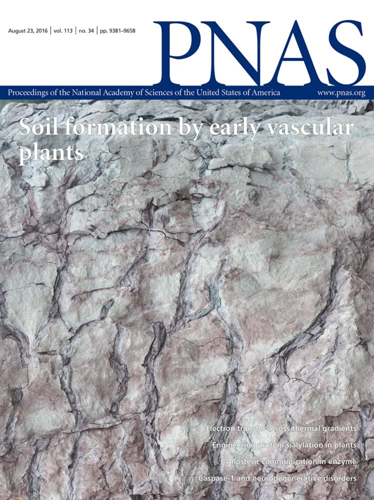
#Science in the age of #selfies
A short #opinion article that we think hits a very important point!
Albert #Einstein: “an academic career, in which a person is forced to produce #scientific writings in great amounts, creates a #danger of intellectual superficiality”
www.pnas.org/doi/10.1073/...
A short #opinion article that we think hits a very important point!
Albert #Einstein: “an academic career, in which a person is forced to produce #scientific writings in great amounts, creates a #danger of intellectual superficiality”
www.pnas.org/doi/10.1073/...
Science in the age of selfies | PNAS
Science in the age of selfies
www.pnas.org
January 6, 2025 at 7:35 AM
#Science in the age of #selfies
A short #opinion article that we think hits a very important point!
Albert #Einstein: “an academic career, in which a person is forced to produce #scientific writings in great amounts, creates a #danger of intellectual superficiality”
www.pnas.org/doi/10.1073/...
A short #opinion article that we think hits a very important point!
Albert #Einstein: “an academic career, in which a person is forced to produce #scientific writings in great amounts, creates a #danger of intellectual superficiality”
www.pnas.org/doi/10.1073/...
Reposted by Electron Microscopy Facility, University of Oslo
Was gifted this one for xmas: www.goodreads.com/book/show/26..., looking forward to reading 👀
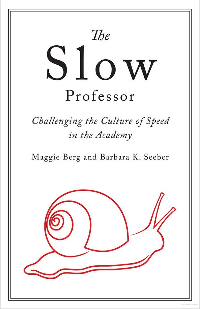
Slow Professor: Challenging the Culture of Speed in the…
If there is one sector of society that should be cultiv…
www.goodreads.com
December 26, 2024 at 3:41 AM
Was gifted this one for xmas: www.goodreads.com/book/show/26..., looking forward to reading 👀
Reposted by Electron Microscopy Facility, University of Oslo
Dynamic duo at the cell's edge: Caveolae and the actin cytoskeleton work together to sense and respond to mechanical stress. Caveolae buffer tension, while actin provides structure and force. Ultimate biomechanics team. #CellBiology #Mechanotransduction
December 21, 2024 at 5:38 PM
Dynamic duo at the cell's edge: Caveolae and the actin cytoskeleton work together to sense and respond to mechanical stress. Caveolae buffer tension, while actin provides structure and force. Ultimate biomechanics team. #CellBiology #Mechanotransduction
Reposted by Electron Microscopy Facility, University of Oslo
Instead of listing my publications, as the year draws to an end, I want to shine the spotlight on the commonplace assumption that productivity must always increase. Good research is disruptive and thinking time is central to high quality scholarship and necessary for disruptive research.
December 20, 2024 at 11:18 AM
Instead of listing my publications, as the year draws to an end, I want to shine the spotlight on the commonplace assumption that productivity must always increase. Good research is disruptive and thinking time is central to high quality scholarship and necessary for disruptive research.
Reposted by Electron Microscopy Facility, University of Oslo
At my institution, they want to install profiles on our personal phones that will give IT root level access to our personal phones.
And allow them to remotely wipe our phones. Our personal phones.
Damn straight, take your work email off your phone. And remove your work profiles from your phones.
And allow them to remotely wipe our phones. Our personal phones.
Damn straight, take your work email off your phone. And remove your work profiles from your phones.
Take your work email off your phone.
You’re welcome
You’re welcome
December 21, 2024 at 3:42 PM
At my institution, they want to install profiles on our personal phones that will give IT root level access to our personal phones.
And allow them to remotely wipe our phones. Our personal phones.
Damn straight, take your work email off your phone. And remove your work profiles from your phones.
And allow them to remotely wipe our phones. Our personal phones.
Damn straight, take your work email off your phone. And remove your work profiles from your phones.
Reposted by Electron Microscopy Facility, University of Oslo
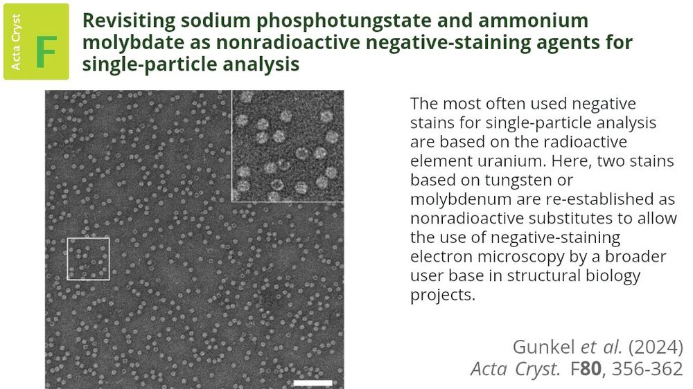
Safer, nonradioactive staining alternatives for electron microscopy are introduced, simplifying sample preparation, reducing costs and making structural biology more accessible to researchers globally #NegativeStaining #ElectronMicroscopy doi.org/10.1107/S205...
December 17, 2024 at 5:13 PM
Safer, nonradioactive staining alternatives for electron microscopy are introduced, simplifying sample preparation, reducing costs and making structural biology more accessible to researchers globally #NegativeStaining #ElectronMicroscopy doi.org/10.1107/S205...
Reposted by Electron Microscopy Facility, University of Oslo
This is our IT department telling us that we cannot copy confocal microscopy data to our Imaris workstation so that we can... analyze the confocal data.

December 17, 2024 at 11:36 PM
This is our IT department telling us that we cannot copy confocal microscopy data to our Imaris workstation so that we can... analyze the confocal data.
Did you know that just 100-200 gold particles in your #immuno-EM data can give you an incredibly good idea of the #labelling distribution and reveal positive signals that you may not immediately recognise? It's amazing how reproducible this " #100-gold-method" is. #stereology
doi.org/10.1369/jhc....
doi.org/10.1369/jhc....

December 17, 2024 at 7:20 AM
Did you know that just 100-200 gold particles in your #immuno-EM data can give you an incredibly good idea of the #labelling distribution and reveal positive signals that you may not immediately recognise? It's amazing how reproducible this " #100-gold-method" is. #stereology
doi.org/10.1369/jhc....
doi.org/10.1369/jhc....
Reposted by Electron Microscopy Facility, University of Oslo
Join us for the last Pub before the Winter Break! ☃️
We'll learn about #VolumeEM approaches from the experts at #EuroBioImaging’s Advanced Light & Electron Prague Node, part of @volumeem1.bsky.social community.
🗓️Fri, Dec 20 @ 13:00 CET
All are welcome 🔽
www.eurobioimaging.eu/events/diver...
We'll learn about #VolumeEM approaches from the experts at #EuroBioImaging’s Advanced Light & Electron Prague Node, part of @volumeem1.bsky.social community.
🗓️Fri, Dec 20 @ 13:00 CET
All are welcome 🔽
www.eurobioimaging.eu/events/diver...

December 16, 2024 at 5:17 PM
Join us for the last Pub before the Winter Break! ☃️
We'll learn about #VolumeEM approaches from the experts at #EuroBioImaging’s Advanced Light & Electron Prague Node, part of @volumeem1.bsky.social community.
🗓️Fri, Dec 20 @ 13:00 CET
All are welcome 🔽
www.eurobioimaging.eu/events/diver...
We'll learn about #VolumeEM approaches from the experts at #EuroBioImaging’s Advanced Light & Electron Prague Node, part of @volumeem1.bsky.social community.
🗓️Fri, Dec 20 @ 13:00 CET
All are welcome 🔽
www.eurobioimaging.eu/events/diver...
Reposted by Electron Microscopy Facility, University of Oslo
I wrote some thoughts about why peer review matters
It shapes scientific standards, maintains field coherence & trains new researchers
Yes, it needs improvement—but it's the glue that holds scientific progress together
briscoelab.org/2024/12/11/i...
It shapes scientific standards, maintains field coherence & trains new researchers
Yes, it needs improvement—but it's the glue that holds scientific progress together
briscoelab.org/2024/12/11/i...

In Praise of Peer Review
As an Editor of the journal Development, I often hear complaints about peer review. We all know the problems – from lengthy review times to requests for seemingly unnecessary experiments. I a…
briscoelab.org
December 11, 2024 at 10:09 AM
I wrote some thoughts about why peer review matters
It shapes scientific standards, maintains field coherence & trains new researchers
Yes, it needs improvement—but it's the glue that holds scientific progress together
briscoelab.org/2024/12/11/i...
It shapes scientific standards, maintains field coherence & trains new researchers
Yes, it needs improvement—but it's the glue that holds scientific progress together
briscoelab.org/2024/12/11/i...