


Houart Lab
@houartlab.bsky.social
Our lab studies fundamental mechanisms in early neurodevelopment and neurodegenerative diseases | Centre for Developmental Neurobiology @ King's College London | Satellite lab @ The Francis Crick Institute
We're so proud of our very own Richard for being awarded this fellowship! Onwards and upwards!
🚨Meet the latest early career researchers awarded Race Against Dementia Fellowships🚨
These rising stars are challenging old ideas and driving bold new approaches to speed up progress @RichardTaylor @sorayam.bsky.social @HelenRowland
These rising stars are challenging old ideas and driving bold new approaches to speed up progress @RichardTaylor @sorayam.bsky.social @HelenRowland


November 20, 2025 at 9:22 PM
We're so proud of our very own Richard for being awarded this fellowship! Onwards and upwards!
Reposted by Houart Lab
Can’t be happier seeing Richard starting this. His original angle to an old problem ( 😉) has great potential for success.
Fascinating to see how findings in dev. neuro. leads to insights in age-related degenerative disorders!
@houartlab.bsky.social @devneuro.bsky.social @kingsioppn.bsky.social
Fascinating to see how findings in dev. neuro. leads to insights in age-related degenerative disorders!
@houartlab.bsky.social @devneuro.bsky.social @kingsioppn.bsky.social
🚨Meet the latest early career researchers awarded Race Against Dementia Fellowships🚨
These rising stars are challenging old ideas and driving bold new approaches to speed up progress @RichardTaylor @sorayam.bsky.social @HelenRowland
These rising stars are challenging old ideas and driving bold new approaches to speed up progress @RichardTaylor @sorayam.bsky.social @HelenRowland
November 20, 2025 at 5:00 PM
Can’t be happier seeing Richard starting this. His original angle to an old problem ( 😉) has great potential for success.
Fascinating to see how findings in dev. neuro. leads to insights in age-related degenerative disorders!
@houartlab.bsky.social @devneuro.bsky.social @kingsioppn.bsky.social
Fascinating to see how findings in dev. neuro. leads to insights in age-related degenerative disorders!
@houartlab.bsky.social @devneuro.bsky.social @kingsioppn.bsky.social
Reposted by Houart Lab

🚨Meet the latest early career researchers awarded Race Against Dementia Fellowships🚨
These rising stars are challenging old ideas and driving bold new approaches to speed up progress @RichardTaylor @sorayam.bsky.social @HelenRowland
These rising stars are challenging old ideas and driving bold new approaches to speed up progress @RichardTaylor @sorayam.bsky.social @HelenRowland
November 20, 2025 at 9:52 AM
🚨Meet the latest early career researchers awarded Race Against Dementia Fellowships🚨
These rising stars are challenging old ideas and driving bold new approaches to speed up progress @RichardTaylor @sorayam.bsky.social @HelenRowland
These rising stars are challenging old ideas and driving bold new approaches to speed up progress @RichardTaylor @sorayam.bsky.social @HelenRowland
Reposted by Houart Lab

Axonal pathfinding of zebrafish retinal ganglion cells forms the optic nerve. Credit to Dr. Matthew Bostock @houartlab.bsky.social. #ZebrafishZunday 🧪
November 16, 2025 at 8:30 PM
Axonal pathfinding of zebrafish retinal ganglion cells forms the optic nerve. Credit to Dr. Matthew Bostock @houartlab.bsky.social. #ZebrafishZunday 🧪
Reposted by Houart Lab
What a day! Congrats to @danafd.bsky.social for great achievement. Big thanks to Sylvie Retaux & @jamesasharpe.bsky.social for very useful examination of thesis.
Absolutely loved joint supervision w. Zena! Perfect @crick.ac.uk & @kingsioppn.bsky.social combo
Dana’s day had a stiff competition 😉😉😉
Absolutely loved joint supervision w. Zena! Perfect @crick.ac.uk & @kingsioppn.bsky.social combo
Dana’s day had a stiff competition 😉😉😉

October 24, 2025 at 5:45 PM
What a day! Congrats to @danafd.bsky.social for great achievement. Big thanks to Sylvie Retaux & @jamesasharpe.bsky.social for very useful examination of thesis.
Absolutely loved joint supervision w. Zena! Perfect @crick.ac.uk & @kingsioppn.bsky.social combo
Dana’s day had a stiff competition 😉😉😉
Absolutely loved joint supervision w. Zena! Perfect @crick.ac.uk & @kingsioppn.bsky.social combo
Dana’s day had a stiff competition 😉😉😉
Reposted by Houart Lab

Couldn’t be prouder of the first PhD graduate of the lab, super congrats @danafd.bsky.social 🙌🎓🌟. Such a pleasure to supervise you together with @cohouart.bsky.social @houartlab.bsky.social, the learning journey never ends 😌. Huge thanks to @jamesasharpe.bsky.social and Sylvie Retaux for examining 💥


October 24, 2025 at 8:15 AM
Couldn’t be prouder of the first PhD graduate of the lab, super congrats @danafd.bsky.social 🙌🎓🌟. Such a pleasure to supervise you together with @cohouart.bsky.social @houartlab.bsky.social, the learning journey never ends 😌. Huge thanks to @jamesasharpe.bsky.social and Sylvie Retaux for examining 💥
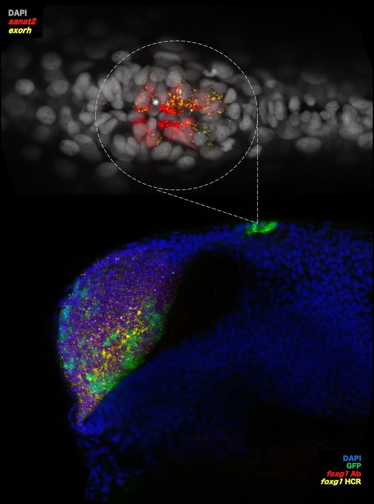
How does foxg1 impact pineal gland development? What are the implications for FOXG1 syndrome?
Lewis Hill, masters student, investigates this in #zebrafish. His image shows in-situ HCR + immuno in foxg1,Gal4;UAS,GFP zebrafish at 24hpf revealing foxg1 mRNA/protein with pineal markers aanat2 & exorh.
Lewis Hill, masters student, investigates this in #zebrafish. His image shows in-situ HCR + immuno in foxg1,Gal4;UAS,GFP zebrafish at 24hpf revealing foxg1 mRNA/protein with pineal markers aanat2 & exorh.
July 29, 2025 at 4:23 PM
How does foxg1 impact pineal gland development? What are the implications for FOXG1 syndrome?
Lewis Hill, masters student, investigates this in #zebrafish. His image shows in-situ HCR + immuno in foxg1,Gal4;UAS,GFP zebrafish at 24hpf revealing foxg1 mRNA/protein with pineal markers aanat2 & exorh.
Lewis Hill, masters student, investigates this in #zebrafish. His image shows in-situ HCR + immuno in foxg1,Gal4;UAS,GFP zebrafish at 24hpf revealing foxg1 mRNA/protein with pineal markers aanat2 & exorh.
Reposted by Houart Lab
Human cortical neurons growing in a dish, with their signal-receiving structures - dendrites - coloured in green by Richard Taylor.
Richard explores how translation of mRNAs into proteins is regulated in dendrites and whether disruptions in this process in dementia contribute to their degeneration.
Richard explores how translation of mRNAs into proteins is regulated in dendrites and whether disruptions in this process in dementia contribute to their degeneration.
July 18, 2025 at 2:59 PM
Human cortical neurons growing in a dish, with their signal-receiving structures - dendrites - coloured in green by Richard Taylor.
Richard explores how translation of mRNAs into proteins is regulated in dendrites and whether disruptions in this process in dementia contribute to their degeneration.
Richard explores how translation of mRNAs into proteins is regulated in dendrites and whether disruptions in this process in dementia contribute to their degeneration.
July 18, 2025 at 2:59 PM
Human cortical neurons growing in a dish, with their signal-receiving structures - dendrites - coloured in green by Richard Taylor.
Richard explores how translation of mRNAs into proteins is regulated in dendrites and whether disruptions in this process in dementia contribute to their degeneration.
Richard explores how translation of mRNAs into proteins is regulated in dendrites and whether disruptions in this process in dementia contribute to their degeneration.
Reposted by Houart Lab
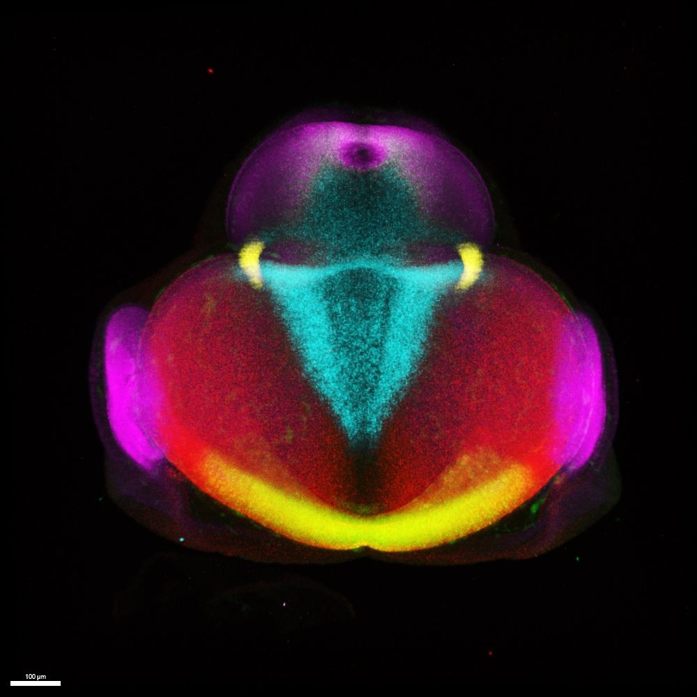
This week, post-doc Katie Adamson shares an incredible image of a #zebrafish embryo forebrain at 24hpf with membrane labelled tbr2+ excitatory neurons.
Just 24 hours post fertilisation, neuronal cell types are already being born and will come to form the forebrain of this magnificent beast.
Just 24 hours post fertilisation, neuronal cell types are already being born and will come to form the forebrain of this magnificent beast.
June 24, 2025 at 4:54 PM
This week, post-doc Katie Adamson shares an incredible image of a #zebrafish embryo forebrain at 24hpf with membrane labelled tbr2+ excitatory neurons.
Just 24 hours post fertilisation, neuronal cell types are already being born and will come to form the forebrain of this magnificent beast.
Just 24 hours post fertilisation, neuronal cell types are already being born and will come to form the forebrain of this magnificent beast.
June 24, 2025 at 4:54 PM
This week, post-doc Katie Adamson shares an incredible image of a #zebrafish embryo forebrain at 24hpf with membrane labelled tbr2+ excitatory neurons.
Just 24 hours post fertilisation, neuronal cell types are already being born and will come to form the forebrain of this magnificent beast.
Just 24 hours post fertilisation, neuronal cell types are already being born and will come to form the forebrain of this magnificent beast.
Reposted by Houart Lab
Movie of the week - joined collab of @houartlab.bsky.social & Long lab @kingsioppn.bsky.social @devneuro.bsky.social
@crick.ac.uk
Joining cell identity to cell behaviour in human embryonic brain.
Fascinating >10 days timelapse imaging by @gessicagoncalves.bsky.social
A technical tour de force!
@crick.ac.uk
Joining cell identity to cell behaviour in human embryonic brain.
Fascinating >10 days timelapse imaging by @gessicagoncalves.bsky.social
A technical tour de force!
A 3D view of apical radial glia finding their way in the developing human cortex.
A stunning video shared by @gessicagoncalves.bsky.social who cultures organotypic cortical slices to study cell dynamics during human cortex development.
A stunning video shared by @gessicagoncalves.bsky.social who cultures organotypic cortical slices to study cell dynamics during human cortex development.
June 20, 2025 at 4:40 PM
Movie of the week - joined collab of @houartlab.bsky.social & Long lab @kingsioppn.bsky.social @devneuro.bsky.social
@crick.ac.uk
Joining cell identity to cell behaviour in human embryonic brain.
Fascinating >10 days timelapse imaging by @gessicagoncalves.bsky.social
A technical tour de force!
@crick.ac.uk
Joining cell identity to cell behaviour in human embryonic brain.
Fascinating >10 days timelapse imaging by @gessicagoncalves.bsky.social
A technical tour de force!
A 3D view of apical radial glia finding their way in the developing human cortex.
A stunning video shared by @gessicagoncalves.bsky.social who cultures organotypic cortical slices to study cell dynamics during human cortex development.
A stunning video shared by @gessicagoncalves.bsky.social who cultures organotypic cortical slices to study cell dynamics during human cortex development.
June 20, 2025 at 1:59 PM
A 3D view of apical radial glia finding their way in the developing human cortex.
A stunning video shared by @gessicagoncalves.bsky.social who cultures organotypic cortical slices to study cell dynamics during human cortex development.
A stunning video shared by @gessicagoncalves.bsky.social who cultures organotypic cortical slices to study cell dynamics during human cortex development.
Reposted by Houart Lab
Do tissue material properties impact morphogen transport? Yes! And morphogens know it. Thanks to @nicolettapetridou.bsky.social who got us involved and to Bernat Corominas-Murtra, and kudos to @cami-autorino.bsky.social @dianakhorom.bsky.social and all the authors! 🧵 below if you are curious 👇
🔔 Proud to share the preprint of my PhD work in the Petridou group @nicolettapetridou.bsky.social @embl.org ✨
“A closed feedback between tissue phase transitions and morphogen gradients drives patterning dynamics” 🐟 🔁 📶
🔗 www.biorxiv.org/content/10.1...
#devbio #biophysics
🧵⤵️
“A closed feedback between tissue phase transitions and morphogen gradients drives patterning dynamics” 🐟 🔁 📶
🔗 www.biorxiv.org/content/10.1...
#devbio #biophysics
🧵⤵️
June 12, 2025 at 4:23 PM
Do tissue material properties impact morphogen transport? Yes! And morphogens know it. Thanks to @nicolettapetridou.bsky.social who got us involved and to Bernat Corominas-Murtra, and kudos to @cami-autorino.bsky.social @dianakhorom.bsky.social and all the authors! 🧵 below if you are curious 👇
Reposted by Houart Lab
Amazing work by Afnan @stochastalive.bsky.social and our lab satellite colleagues @crick.ac.uk, comparing human vs mouse forebrain signalling centres & progenitor diversity in nascent embryonic telencephalon. Outcome of exploration soon on bioRxiv.
@kingsioppn.bsky.social @devneuro.bsky.social
@kingsioppn.bsky.social @devneuro.bsky.social
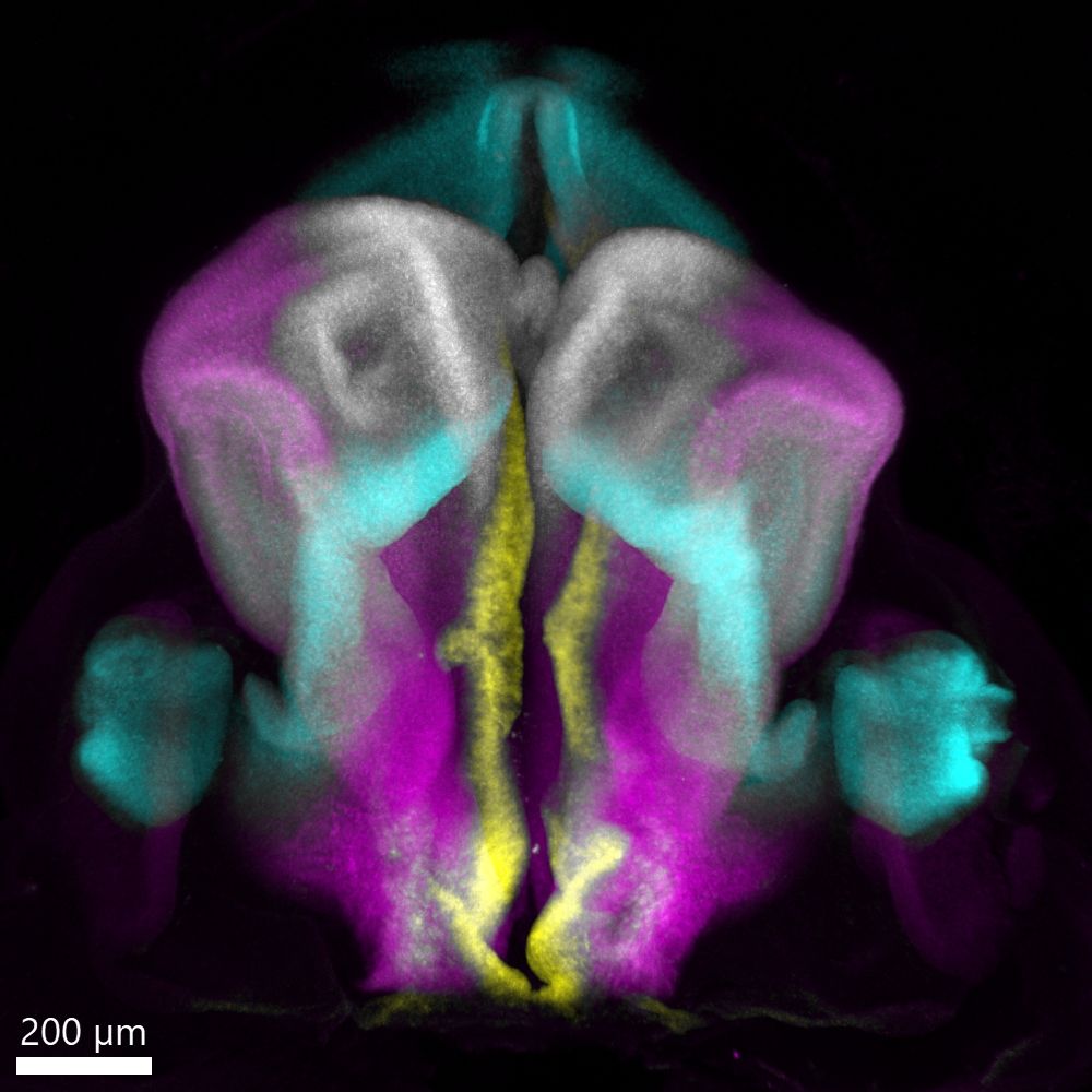
A look at the developing human embryonic forebrain in 3D!
Post-doc @stochastalive.bsky.social shares this week's image showing a dorsal view of the human embryonic forebrain at Carnegie Stage 16 (~6 weeks post conception). Major regions marked by FOXG1, WNT8B, PAX6, and SHH are displayed.
Post-doc @stochastalive.bsky.social shares this week's image showing a dorsal view of the human embryonic forebrain at Carnegie Stage 16 (~6 weeks post conception). Major regions marked by FOXG1, WNT8B, PAX6, and SHH are displayed.
June 11, 2025 at 7:28 PM
Amazing work by Afnan @stochastalive.bsky.social and our lab satellite colleagues @crick.ac.uk, comparing human vs mouse forebrain signalling centres & progenitor diversity in nascent embryonic telencephalon. Outcome of exploration soon on bioRxiv.
@kingsioppn.bsky.social @devneuro.bsky.social
@kingsioppn.bsky.social @devneuro.bsky.social
A look at the developing human embryonic forebrain in 3D!
Post-doc @stochastalive.bsky.social shares this week's image showing a dorsal view of the human embryonic forebrain at Carnegie Stage 16 (~6 weeks post conception). Major regions marked by FOXG1, WNT8B, PAX6, and SHH are displayed.
Post-doc @stochastalive.bsky.social shares this week's image showing a dorsal view of the human embryonic forebrain at Carnegie Stage 16 (~6 weeks post conception). Major regions marked by FOXG1, WNT8B, PAX6, and SHH are displayed.
June 11, 2025 at 4:54 PM
A look at the developing human embryonic forebrain in 3D!
Post-doc @stochastalive.bsky.social shares this week's image showing a dorsal view of the human embryonic forebrain at Carnegie Stage 16 (~6 weeks post conception). Major regions marked by FOXG1, WNT8B, PAX6, and SHH are displayed.
Post-doc @stochastalive.bsky.social shares this week's image showing a dorsal view of the human embryonic forebrain at Carnegie Stage 16 (~6 weeks post conception). Major regions marked by FOXG1, WNT8B, PAX6, and SHH are displayed.
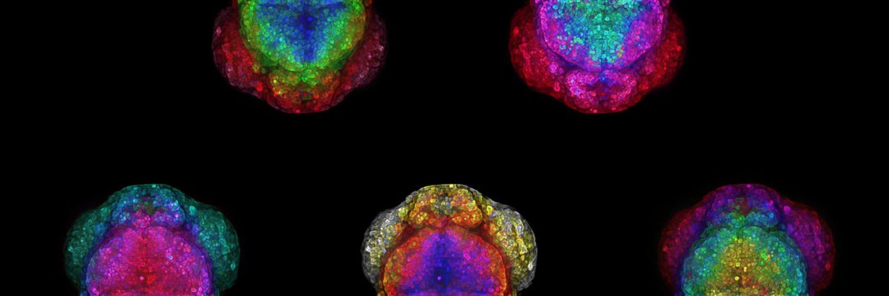
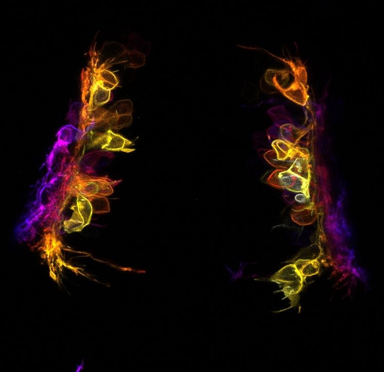
Another week, another Houart lab image🔬
This week, it's a 3D image of a multiplex, in-situ of a chick forebrain by @danafd.bsky.social who studies early forebrain development in vertebrates & uses HCR to show the expression patterns of gene products instrumental in pallial/subpallial specification.
This week, it's a 3D image of a multiplex, in-situ of a chick forebrain by @danafd.bsky.social who studies early forebrain development in vertebrates & uses HCR to show the expression patterns of gene products instrumental in pallial/subpallial specification.
June 3, 2025 at 1:28 PM
Another week, another Houart lab image🔬
This week, it's a 3D image of a multiplex, in-situ of a chick forebrain by @danafd.bsky.social who studies early forebrain development in vertebrates & uses HCR to show the expression patterns of gene products instrumental in pallial/subpallial specification.
This week, it's a 3D image of a multiplex, in-situ of a chick forebrain by @danafd.bsky.social who studies early forebrain development in vertebrates & uses HCR to show the expression patterns of gene products instrumental in pallial/subpallial specification.
Reposted by Houart Lab
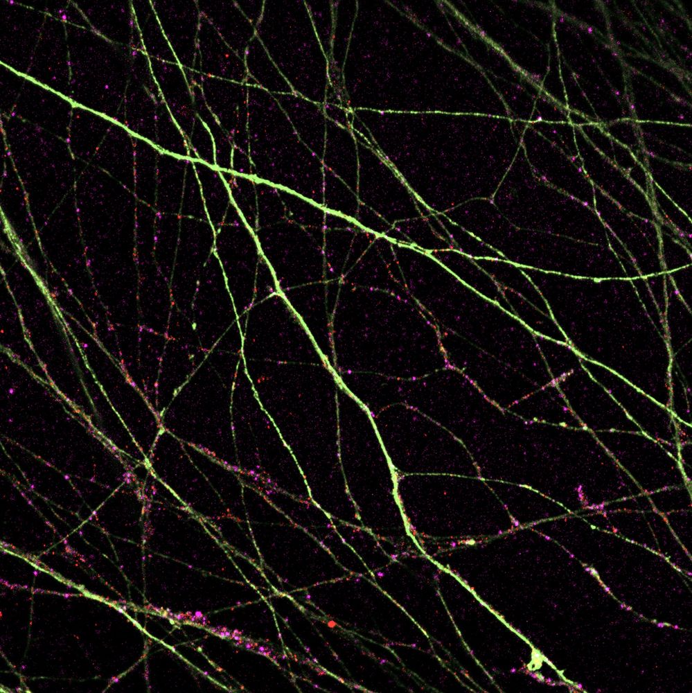
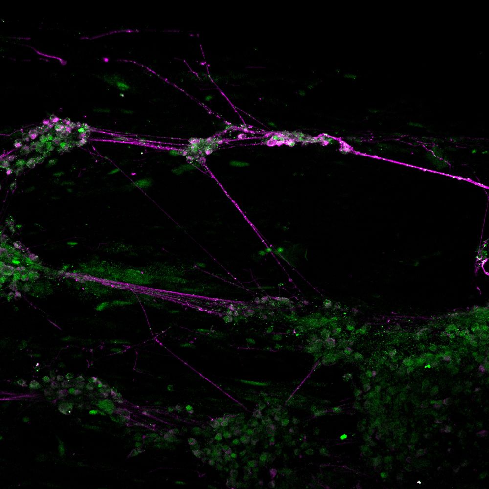
This week, Rachel Moore shares a gorgeous image from her #zebrafish neuron culture. In a dish, these neurons can grow long axons (magenta). If you look closely, you might be able to see some RNA splicing proteins (green) localised as small puncta along the axons.
May 28, 2025 at 8:41 AM
This week, Rachel Moore shares a gorgeous image from her #zebrafish neuron culture. In a dish, these neurons can grow long axons (magenta). If you look closely, you might be able to see some RNA splicing proteins (green) localised as small puncta along the axons.
This week, Rachel Moore shares a gorgeous image from her #zebrafish neuron culture. In a dish, these neurons can grow long axons (magenta). If you look closely, you might be able to see some RNA splicing proteins (green) localised as small puncta along the axons.
May 28, 2025 at 8:41 AM
This week, Rachel Moore shares a gorgeous image from her #zebrafish neuron culture. In a dish, these neurons can grow long axons (magenta). If you look closely, you might be able to see some RNA splicing proteins (green) localised as small puncta along the axons.
Reposted by Houart Lab
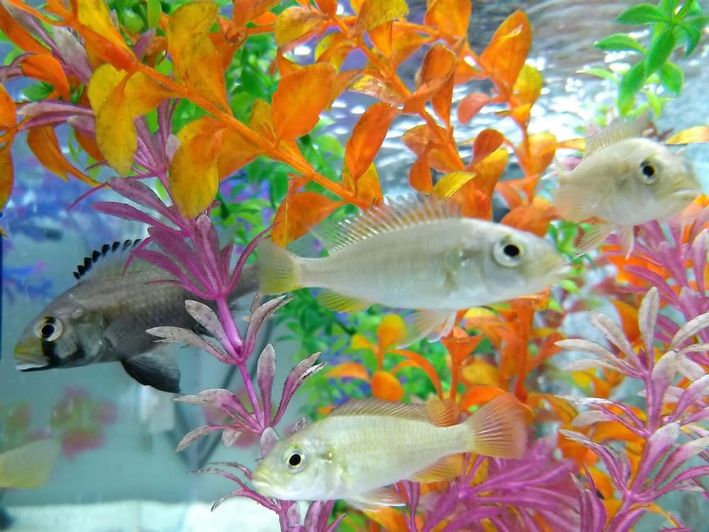
Experimental embryology postdoc available in my lab at the @biology.ox.ac.uk @ox.ac.uk working on the evolution of vertebral counts. Reach out if you’re passionate about EvoDevo, enjoy lab work and microscopy and are into or could get into cichlid fishes. Deadline on the 16th June. Please share!
May 19, 2025 at 4:10 PM
Experimental embryology postdoc available in my lab at the @biology.ox.ac.uk @ox.ac.uk working on the evolution of vertebral counts. Reach out if you’re passionate about EvoDevo, enjoy lab work and microscopy and are into or could get into cichlid fishes. Deadline on the 16th June. Please share!
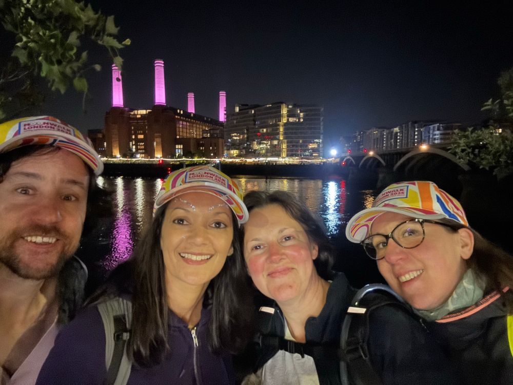
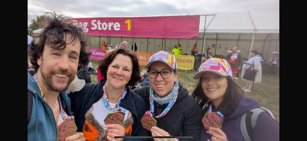
We are so proud of Triona, Stephania, Joao & Vicky for finishing the London Moonwalk for Marie Johansson, a dear member of our lab and the CDN, who lost her battle against breast cancer last June. Marie was a talented scientist and an exceptional colleague whom we miss terribly.
Donate link below⬇️
Donate link below⬇️

May 20, 2025 at 9:10 AM
We are so proud of Triona, Stephania, Joao & Vicky for finishing the London Moonwalk for Marie Johansson, a dear member of our lab and the CDN, who lost her battle against breast cancer last June. Marie was a talented scientist and an exceptional colleague whom we miss terribly.
Donate link below⬇️
Donate link below⬇️
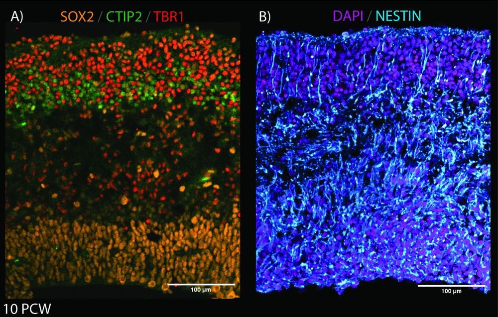
This week's image is by @joseriveraz.bsky.social who's tracing the lineage of progenitors in the human developing cortex.
Here, José shows the building blocks of the developing cortex at 10pcw with SOX2 (apical progenitors), CTIP2 & TBR1(neuronal markers), NESTIN (radial glial fibers), and DAPI.
Here, José shows the building blocks of the developing cortex at 10pcw with SOX2 (apical progenitors), CTIP2 & TBR1(neuronal markers), NESTIN (radial glial fibers), and DAPI.
May 17, 2025 at 1:15 PM
This week's image is by @joseriveraz.bsky.social who's tracing the lineage of progenitors in the human developing cortex.
Here, José shows the building blocks of the developing cortex at 10pcw with SOX2 (apical progenitors), CTIP2 & TBR1(neuronal markers), NESTIN (radial glial fibers), and DAPI.
Here, José shows the building blocks of the developing cortex at 10pcw with SOX2 (apical progenitors), CTIP2 & TBR1(neuronal markers), NESTIN (radial glial fibers), and DAPI.
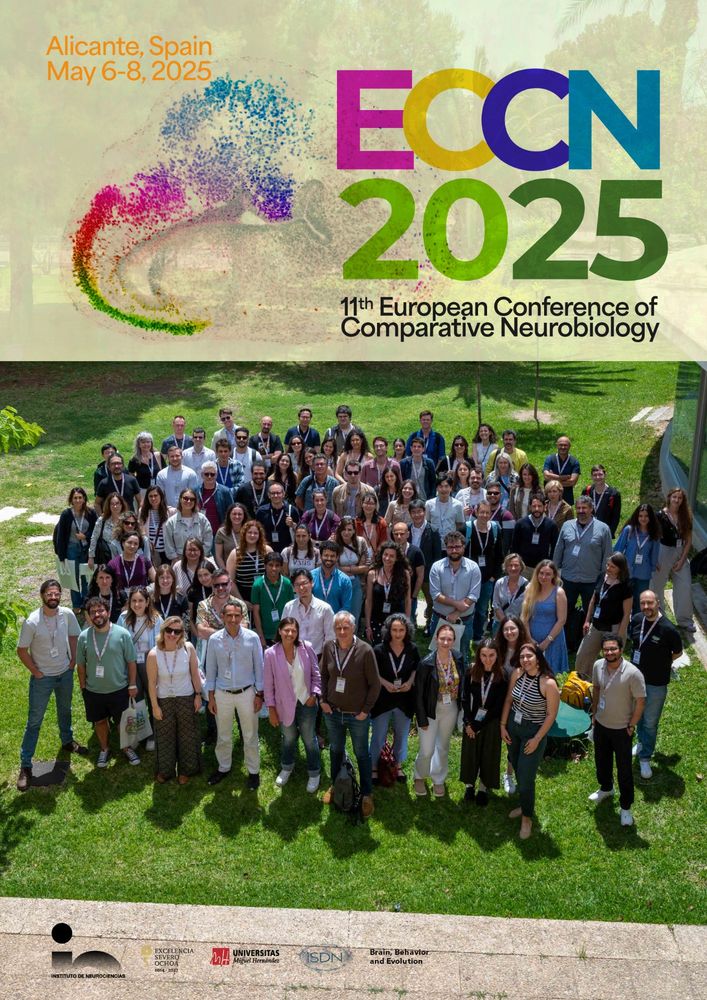
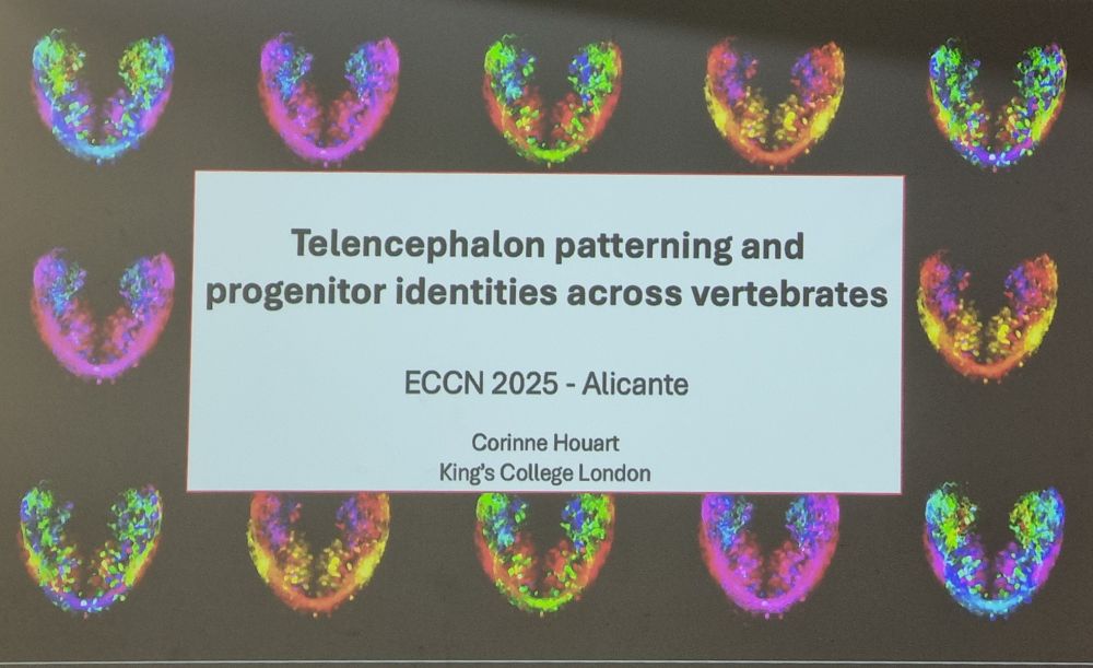
It has been an absolute pleasure for our lab to attend #ECCN2025 for the first time this year.
We had meaningful discussions and learned a lot from a vibrant community tackling tough questions on brain evolution and development! Super excited for what lies ahead!
We had meaningful discussions and learned a lot from a vibrant community tackling tough questions on brain evolution and development! Super excited for what lies ahead!
May 9, 2025 at 11:44 AM
It has been an absolute pleasure for our lab to attend #ECCN2025 for the first time this year.
We had meaningful discussions and learned a lot from a vibrant community tackling tough questions on brain evolution and development! Super excited for what lies ahead!
We had meaningful discussions and learned a lot from a vibrant community tackling tough questions on brain evolution and development! Super excited for what lies ahead!
Reposted by Houart Lab
Dkk genes dynamically expressed in the zebrafish nervous system, some in very discreet brain populations (watch this space for brain images).
Malik Missaoui is using genetics to follow behaviour of endogenous proteins and crack their local functions.
@mrc-cndd.bsky.social @kingsioppn.bsky.social
Malik Missaoui is using genetics to follow behaviour of endogenous proteins and crack their local functions.
@mrc-cndd.bsky.social @kingsioppn.bsky.social
🎣 Fishing for Dkk in #zebrafish 🎣
In the lab, PhD student Malik Misssaoui looks for the expression patterns of the dkk genes in zebrafish. Dkk proteins are modulators of wnt singalling, and changes in their expression levels have been linked to neurodegenerative diseases like Alzheimers.
In the lab, PhD student Malik Misssaoui looks for the expression patterns of the dkk genes in zebrafish. Dkk proteins are modulators of wnt singalling, and changes in their expression levels have been linked to neurodegenerative diseases like Alzheimers.
May 8, 2025 at 1:08 PM
Dkk genes dynamically expressed in the zebrafish nervous system, some in very discreet brain populations (watch this space for brain images).
Malik Missaoui is using genetics to follow behaviour of endogenous proteins and crack their local functions.
@mrc-cndd.bsky.social @kingsioppn.bsky.social
Malik Missaoui is using genetics to follow behaviour of endogenous proteins and crack their local functions.
@mrc-cndd.bsky.social @kingsioppn.bsky.social
Reposted by Houart Lab
@houartlab.bsky.social first time at the comparative neuro conference. Fantastic #ECCN2025 meeting! The mix of ‘old’ and new technologies brings the field a new edge and the potential for new understanding of brain evolutionary mechanisms. Exciting!

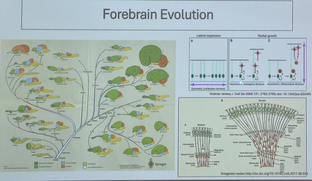
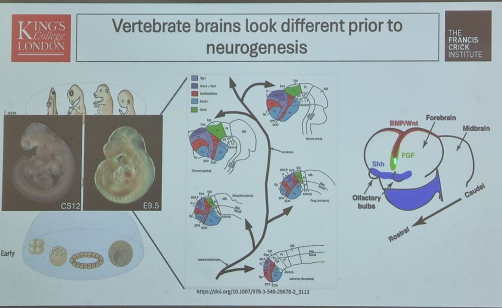
Differences in neurogenesis across species appear since the earliest stages of development, as shows @cohouart.bsky.social
#ECCN2025 @borrell-lab.bsky.social @k4tj4.bsky.social
#ECCN2025 @borrell-lab.bsky.social @k4tj4.bsky.social
May 8, 2025 at 1:43 PM
@houartlab.bsky.social first time at the comparative neuro conference. Fantastic #ECCN2025 meeting! The mix of ‘old’ and new technologies brings the field a new edge and the potential for new understanding of brain evolutionary mechanisms. Exciting!