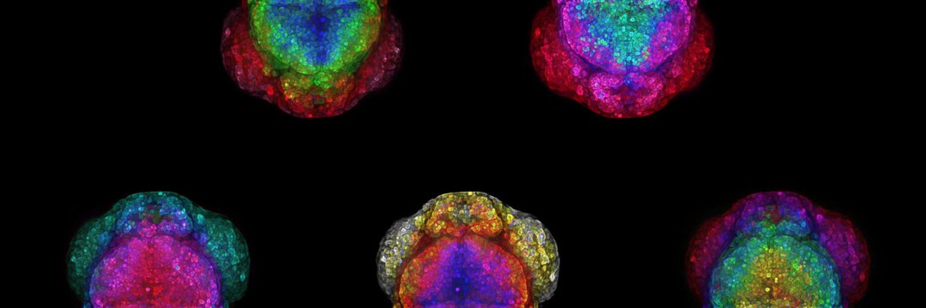
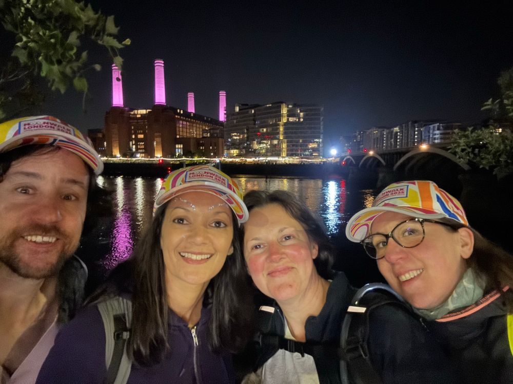
Absolutely loved joint supervision w. Zena! Perfect @crick.ac.uk & @kingsioppn.bsky.social combo
Dana’s day had a stiff competition 😉😉😉

Absolutely loved joint supervision w. Zena! Perfect @crick.ac.uk & @kingsioppn.bsky.social combo
Dana’s day had a stiff competition 😉😉😉


Lewis Hill, masters student, investigates this in #zebrafish. His image shows in-situ HCR + immuno in foxg1,Gal4;UAS,GFP zebrafish at 24hpf revealing foxg1 mRNA/protein with pineal markers aanat2 & exorh.
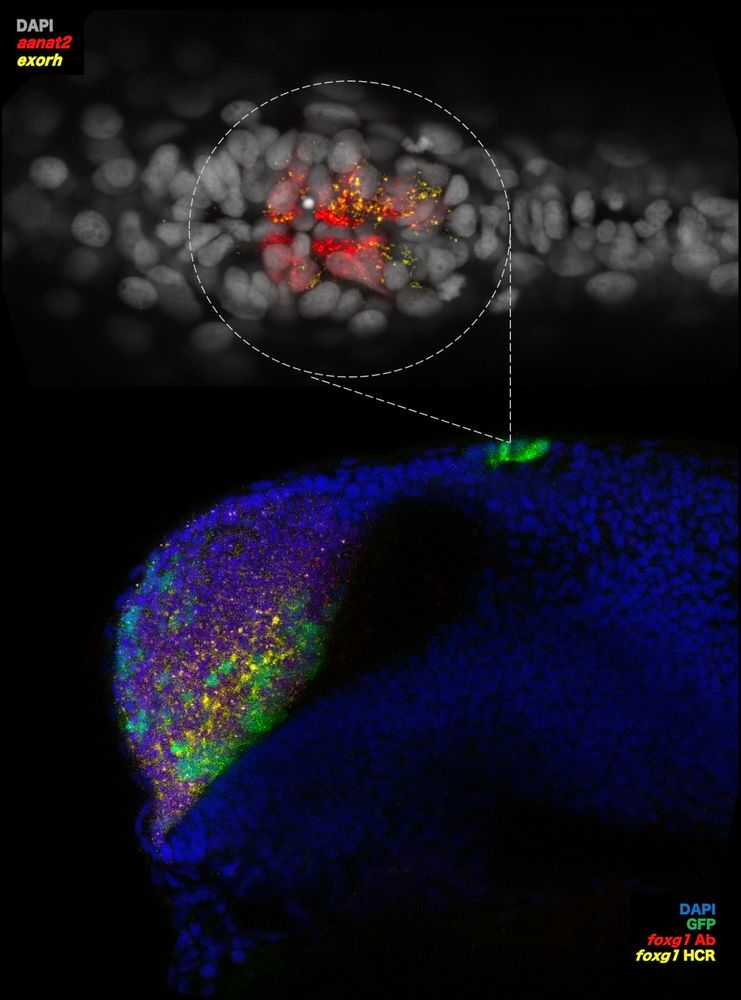
Lewis Hill, masters student, investigates this in #zebrafish. His image shows in-situ HCR + immuno in foxg1,Gal4;UAS,GFP zebrafish at 24hpf revealing foxg1 mRNA/protein with pineal markers aanat2 & exorh.
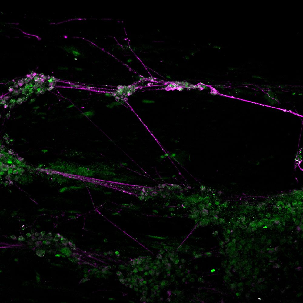
Richard explores how translation of mRNAs into proteins is regulated in dendrites and whether disruptions in this process in dementia contribute to their degeneration.
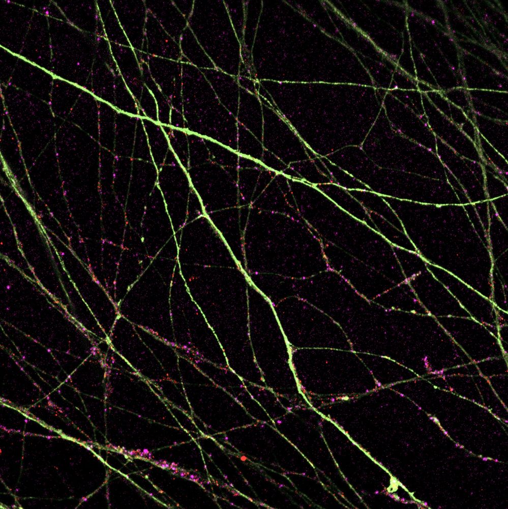
Richard explores how translation of mRNAs into proteins is regulated in dendrites and whether disruptions in this process in dementia contribute to their degeneration.
Richard explores how translation of mRNAs into proteins is regulated in dendrites and whether disruptions in this process in dementia contribute to their degeneration.

Richard explores how translation of mRNAs into proteins is regulated in dendrites and whether disruptions in this process in dementia contribute to their degeneration.
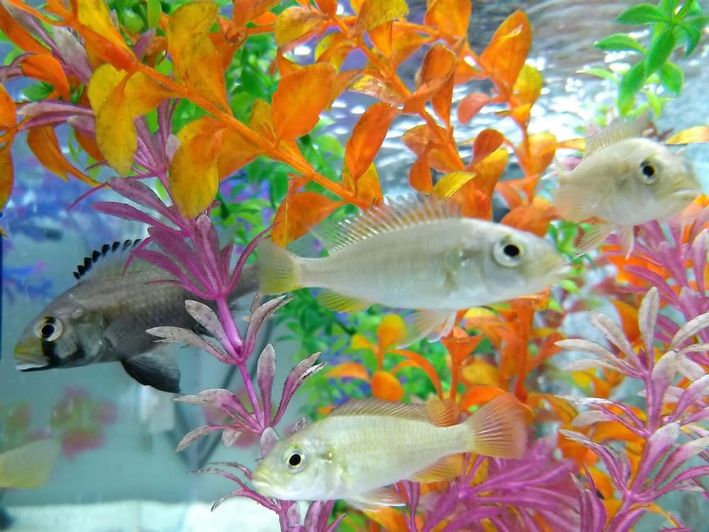
Just 24 hours post fertilisation, neuronal cell types are already being born and will come to form the forebrain of this magnificent beast.
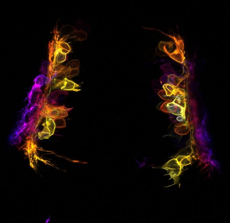
Just 24 hours post fertilisation, neuronal cell types are already being born and will come to form the forebrain of this magnificent beast.
Just 24 hours post fertilisation, neuronal cell types are already being born and will come to form the forebrain of this magnificent beast.

Just 24 hours post fertilisation, neuronal cell types are already being born and will come to form the forebrain of this magnificent beast.
@crick.ac.uk
Joining cell identity to cell behaviour in human embryonic brain.
Fascinating >10 days timelapse imaging by @gessicagoncalves.bsky.social
A technical tour de force!
A stunning video shared by @gessicagoncalves.bsky.social who cultures organotypic cortical slices to study cell dynamics during human cortex development.
A stunning video shared by @gessicagoncalves.bsky.social who cultures organotypic cortical slices to study cell dynamics during human cortex development.
“A closed feedback between tissue phase transitions and morphogen gradients drives patterning dynamics” 🐟 🔁 📶
🔗 www.biorxiv.org/content/10.1...
#devbio #biophysics
🧵⤵️
@kingsioppn.bsky.social @devneuro.bsky.social
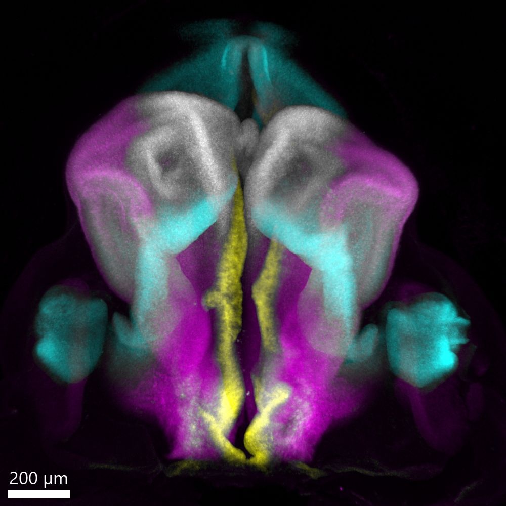
Post-doc @stochastalive.bsky.social shares this week's image showing a dorsal view of the human embryonic forebrain at Carnegie Stage 16 (~6 weeks post conception). Major regions marked by FOXG1, WNT8B, PAX6, and SHH are displayed.
@kingsioppn.bsky.social @devneuro.bsky.social
Post-doc @stochastalive.bsky.social shares this week's image showing a dorsal view of the human embryonic forebrain at Carnegie Stage 16 (~6 weeks post conception). Major regions marked by FOXG1, WNT8B, PAX6, and SHH are displayed.

Post-doc @stochastalive.bsky.social shares this week's image showing a dorsal view of the human embryonic forebrain at Carnegie Stage 16 (~6 weeks post conception). Major regions marked by FOXG1, WNT8B, PAX6, and SHH are displayed.
This week, it's a 3D image of a multiplex, in-situ of a chick forebrain by @danafd.bsky.social who studies early forebrain development in vertebrates & uses HCR to show the expression patterns of gene products instrumental in pallial/subpallial specification.
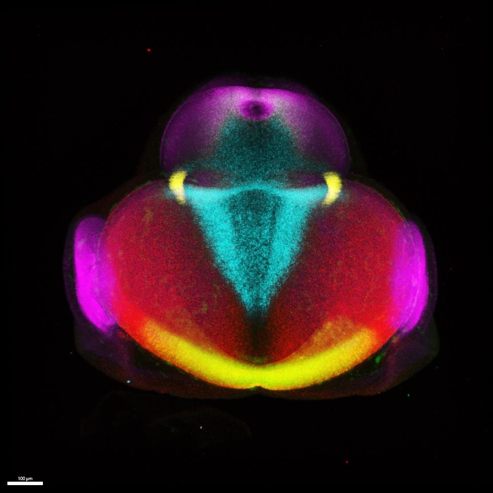
This week, it's a 3D image of a multiplex, in-situ of a chick forebrain by @danafd.bsky.social who studies early forebrain development in vertebrates & uses HCR to show the expression patterns of gene products instrumental in pallial/subpallial specification.

Donate link below⬇️


Donate link below⬇️
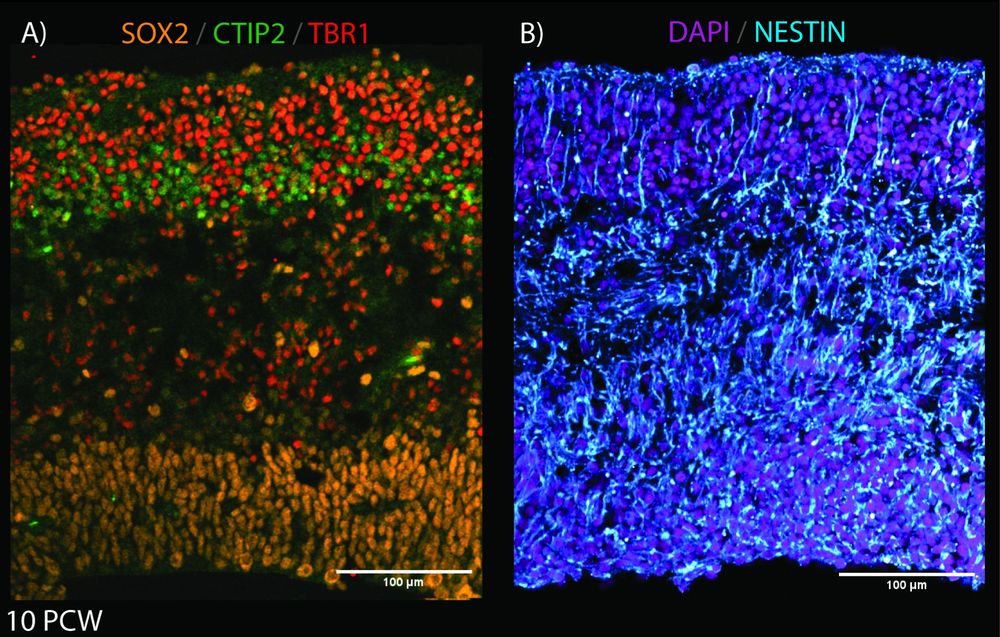
Here, José shows the building blocks of the developing cortex at 10pcw with SOX2 (apical progenitors), CTIP2 & TBR1(neuronal markers), NESTIN (radial glial fibers), and DAPI.
Here, José shows the building blocks of the developing cortex at 10pcw with SOX2 (apical progenitors), CTIP2 & TBR1(neuronal markers), NESTIN (radial glial fibers), and DAPI.
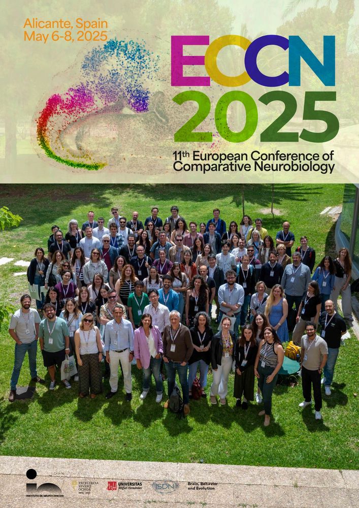
We had meaningful discussions and learned a lot from a vibrant community tackling tough questions on brain evolution and development! Super excited for what lies ahead!
We had meaningful discussions and learned a lot from a vibrant community tackling tough questions on brain evolution and development! Super excited for what lies ahead!
Malik Missaoui is using genetics to follow behaviour of endogenous proteins and crack their local functions.
@mrc-cndd.bsky.social @kingsioppn.bsky.social
In the lab, PhD student Malik Misssaoui looks for the expression patterns of the dkk genes in zebrafish. Dkk proteins are modulators of wnt singalling, and changes in their expression levels have been linked to neurodegenerative diseases like Alzheimers.
Malik Missaoui is using genetics to follow behaviour of endogenous proteins and crack their local functions.
@mrc-cndd.bsky.social @kingsioppn.bsky.social
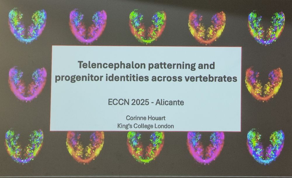
#ECCN2025 @borrell-lab.bsky.social @k4tj4.bsky.social
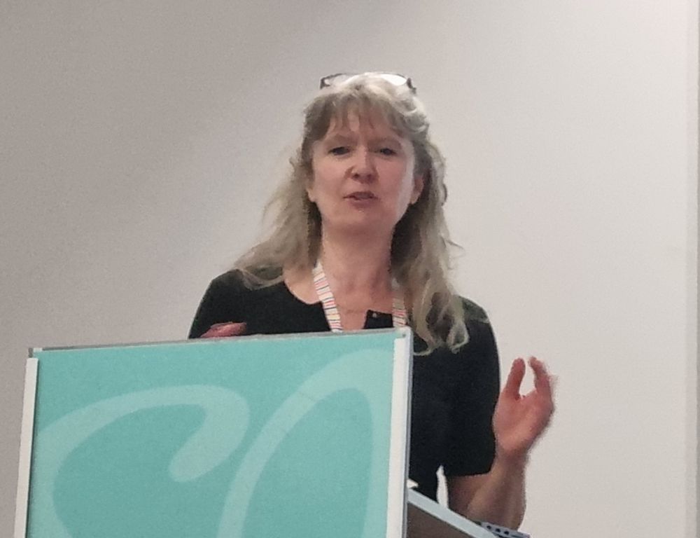
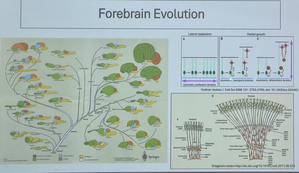
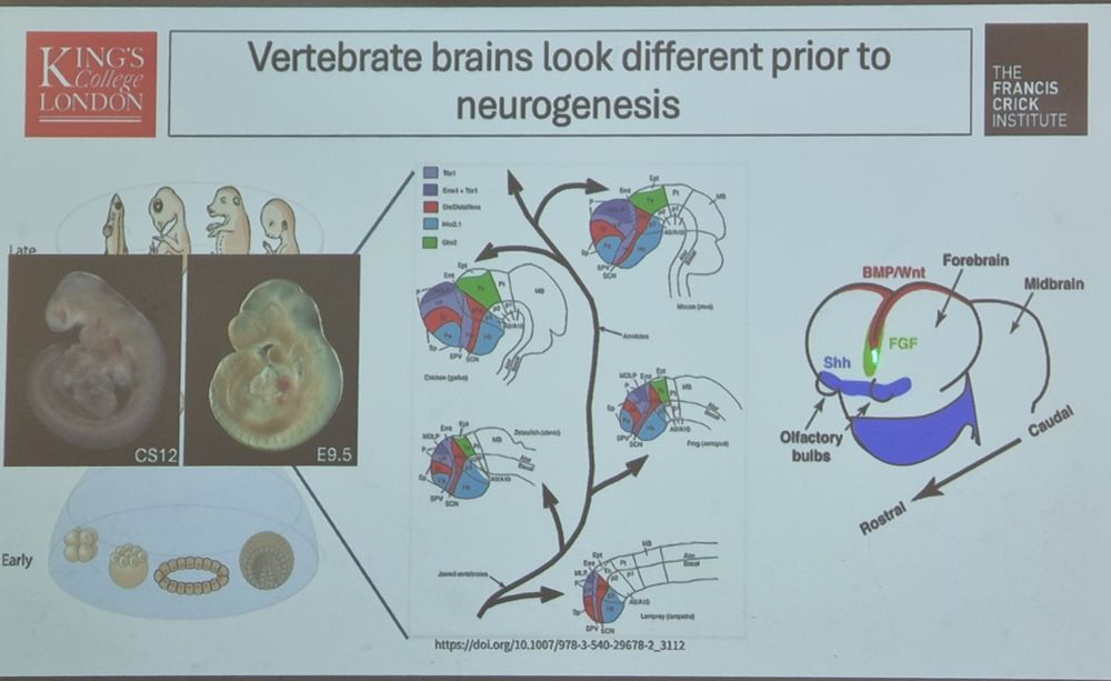
#ECCN2025 @borrell-lab.bsky.social @k4tj4.bsky.social


#ECCN2025 @borrell-lab.bsky.social @k4tj4.bsky.social
In the lab, PhD student Malik Misssaoui looks for the expression patterns of the dkk genes in zebrafish. Dkk proteins are modulators of wnt singalling, and changes in their expression levels have been linked to neurodegenerative diseases like Alzheimers.
In the lab, PhD student Malik Misssaoui looks for the expression patterns of the dkk genes in zebrafish. Dkk proteins are modulators of wnt singalling, and changes in their expression levels have been linked to neurodegenerative diseases like Alzheimers.
This week is a #zebrafish ‘kiss and cross’ of the left and right first axons of the optic nerve. Let’s see what next week is preparing for us!
@houartlab.bsky.social @kingsioppn.bsky.social
@crick.ac.uk
Image taken by @matthewbostock.bsky.social
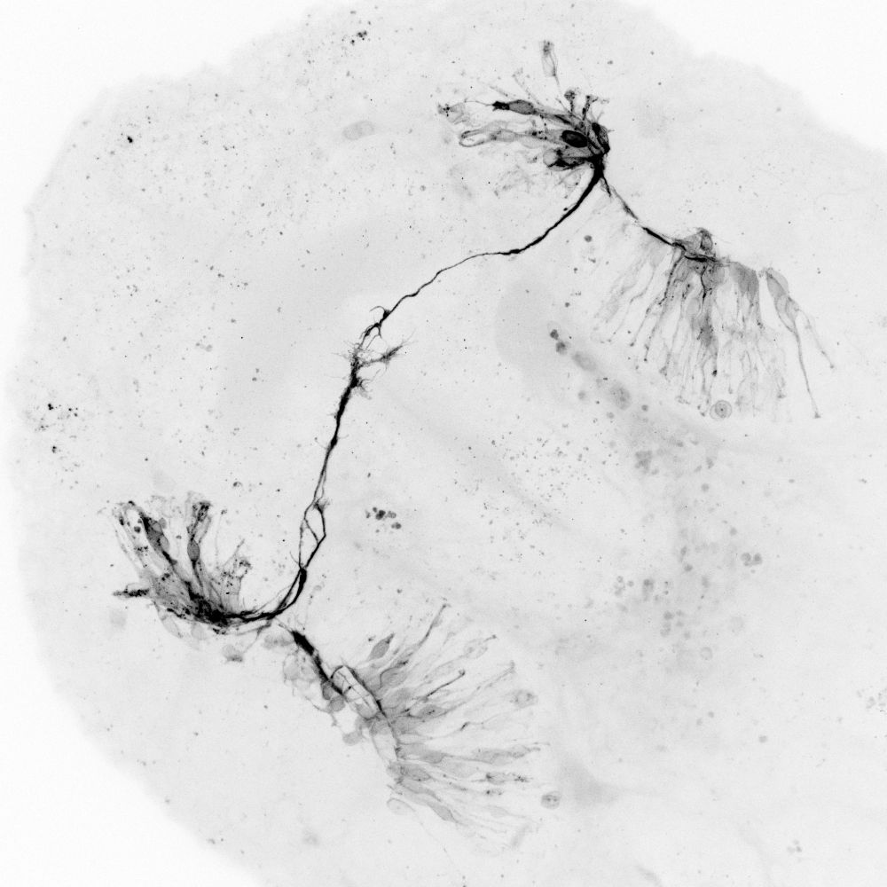
This week is a #zebrafish ‘kiss and cross’ of the left and right first axons of the optic nerve. Let’s see what next week is preparing for us!
@houartlab.bsky.social @kingsioppn.bsky.social
@crick.ac.uk
Image taken by @matthewbostock.bsky.social

Image taken by @matthewbostock.bsky.social

