


Houart Lab
@houartlab.bsky.social
Our lab studies fundamental mechanisms in early neurodevelopment and neurodegenerative diseases | Centre for Developmental Neurobiology @ King's College London | Satellite lab @ The Francis Crick Institute
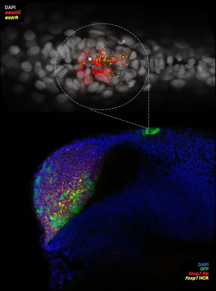
How does foxg1 impact pineal gland development? What are the implications for FOXG1 syndrome?
Lewis Hill, masters student, investigates this in #zebrafish. His image shows in-situ HCR + immuno in foxg1,Gal4;UAS,GFP zebrafish at 24hpf revealing foxg1 mRNA/protein with pineal markers aanat2 & exorh.
Lewis Hill, masters student, investigates this in #zebrafish. His image shows in-situ HCR + immuno in foxg1,Gal4;UAS,GFP zebrafish at 24hpf revealing foxg1 mRNA/protein with pineal markers aanat2 & exorh.
July 29, 2025 at 4:23 PM
How does foxg1 impact pineal gland development? What are the implications for FOXG1 syndrome?
Lewis Hill, masters student, investigates this in #zebrafish. His image shows in-situ HCR + immuno in foxg1,Gal4;UAS,GFP zebrafish at 24hpf revealing foxg1 mRNA/protein with pineal markers aanat2 & exorh.
Lewis Hill, masters student, investigates this in #zebrafish. His image shows in-situ HCR + immuno in foxg1,Gal4;UAS,GFP zebrafish at 24hpf revealing foxg1 mRNA/protein with pineal markers aanat2 & exorh.
Human cortical neurons growing in a dish, with their signal-receiving structures - dendrites - coloured in green by Richard Taylor.
Richard explores how translation of mRNAs into proteins is regulated in dendrites and whether disruptions in this process in dementia contribute to their degeneration.
Richard explores how translation of mRNAs into proteins is regulated in dendrites and whether disruptions in this process in dementia contribute to their degeneration.
July 18, 2025 at 2:59 PM
Human cortical neurons growing in a dish, with their signal-receiving structures - dendrites - coloured in green by Richard Taylor.
Richard explores how translation of mRNAs into proteins is regulated in dendrites and whether disruptions in this process in dementia contribute to their degeneration.
Richard explores how translation of mRNAs into proteins is regulated in dendrites and whether disruptions in this process in dementia contribute to their degeneration.
This week, post-doc Katie Adamson shares an incredible image of a #zebrafish embryo forebrain at 24hpf with membrane labelled tbr2+ excitatory neurons.
Just 24 hours post fertilisation, neuronal cell types are already being born and will come to form the forebrain of this magnificent beast.
Just 24 hours post fertilisation, neuronal cell types are already being born and will come to form the forebrain of this magnificent beast.
June 24, 2025 at 4:54 PM
This week, post-doc Katie Adamson shares an incredible image of a #zebrafish embryo forebrain at 24hpf with membrane labelled tbr2+ excitatory neurons.
Just 24 hours post fertilisation, neuronal cell types are already being born and will come to form the forebrain of this magnificent beast.
Just 24 hours post fertilisation, neuronal cell types are already being born and will come to form the forebrain of this magnificent beast.
A 3D view of apical radial glia finding their way in the developing human cortex.
A stunning video shared by @gessicagoncalves.bsky.social who cultures organotypic cortical slices to study cell dynamics during human cortex development.
A stunning video shared by @gessicagoncalves.bsky.social who cultures organotypic cortical slices to study cell dynamics during human cortex development.
June 20, 2025 at 1:59 PM
A 3D view of apical radial glia finding their way in the developing human cortex.
A stunning video shared by @gessicagoncalves.bsky.social who cultures organotypic cortical slices to study cell dynamics during human cortex development.
A stunning video shared by @gessicagoncalves.bsky.social who cultures organotypic cortical slices to study cell dynamics during human cortex development.
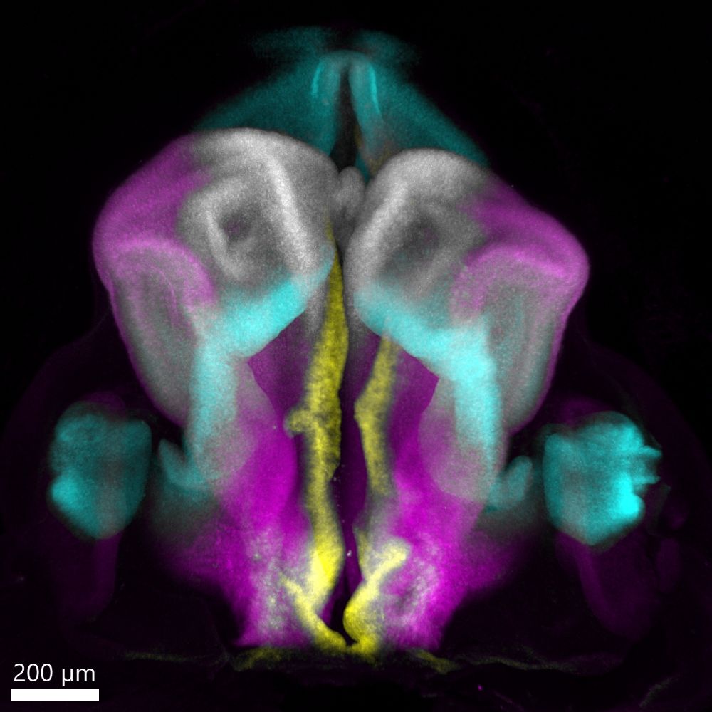
A look at the developing human embryonic forebrain in 3D!
Post-doc @stochastalive.bsky.social shares this week's image showing a dorsal view of the human embryonic forebrain at Carnegie Stage 16 (~6 weeks post conception). Major regions marked by FOXG1, WNT8B, PAX6, and SHH are displayed.
Post-doc @stochastalive.bsky.social shares this week's image showing a dorsal view of the human embryonic forebrain at Carnegie Stage 16 (~6 weeks post conception). Major regions marked by FOXG1, WNT8B, PAX6, and SHH are displayed.
June 11, 2025 at 4:54 PM
A look at the developing human embryonic forebrain in 3D!
Post-doc @stochastalive.bsky.social shares this week's image showing a dorsal view of the human embryonic forebrain at Carnegie Stage 16 (~6 weeks post conception). Major regions marked by FOXG1, WNT8B, PAX6, and SHH are displayed.
Post-doc @stochastalive.bsky.social shares this week's image showing a dorsal view of the human embryonic forebrain at Carnegie Stage 16 (~6 weeks post conception). Major regions marked by FOXG1, WNT8B, PAX6, and SHH are displayed.
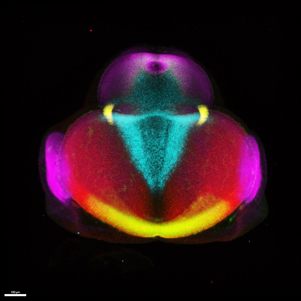
Another week, another Houart lab image🔬
This week, it's a 3D image of a multiplex, in-situ of a chick forebrain by @danafd.bsky.social who studies early forebrain development in vertebrates & uses HCR to show the expression patterns of gene products instrumental in pallial/subpallial specification.
This week, it's a 3D image of a multiplex, in-situ of a chick forebrain by @danafd.bsky.social who studies early forebrain development in vertebrates & uses HCR to show the expression patterns of gene products instrumental in pallial/subpallial specification.
June 3, 2025 at 1:28 PM
Another week, another Houart lab image🔬
This week, it's a 3D image of a multiplex, in-situ of a chick forebrain by @danafd.bsky.social who studies early forebrain development in vertebrates & uses HCR to show the expression patterns of gene products instrumental in pallial/subpallial specification.
This week, it's a 3D image of a multiplex, in-situ of a chick forebrain by @danafd.bsky.social who studies early forebrain development in vertebrates & uses HCR to show the expression patterns of gene products instrumental in pallial/subpallial specification.
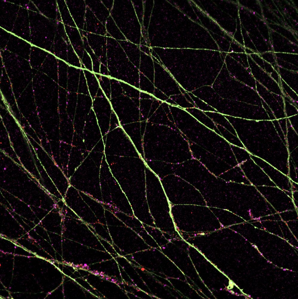
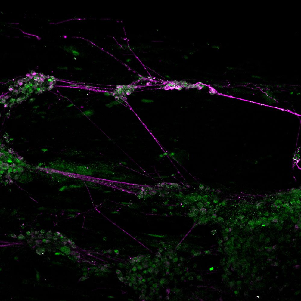
This week, Rachel Moore shares a gorgeous image from her #zebrafish neuron culture. In a dish, these neurons can grow long axons (magenta). If you look closely, you might be able to see some RNA splicing proteins (green) localised as small puncta along the axons.
May 28, 2025 at 8:41 AM
This week, Rachel Moore shares a gorgeous image from her #zebrafish neuron culture. In a dish, these neurons can grow long axons (magenta). If you look closely, you might be able to see some RNA splicing proteins (green) localised as small puncta along the axons.
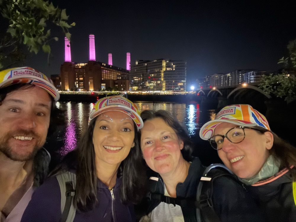
We are so proud of Triona, Stephania, Joao & Vicky for finishing the London Moonwalk for Marie Johansson, a dear member of our lab and the CDN, who lost her battle against breast cancer last June. Marie was a talented scientist and an exceptional colleague whom we miss terribly.
Donate link below⬇️
Donate link below⬇️


May 20, 2025 at 9:10 AM
We are so proud of Triona, Stephania, Joao & Vicky for finishing the London Moonwalk for Marie Johansson, a dear member of our lab and the CDN, who lost her battle against breast cancer last June. Marie was a talented scientist and an exceptional colleague whom we miss terribly.
Donate link below⬇️
Donate link below⬇️
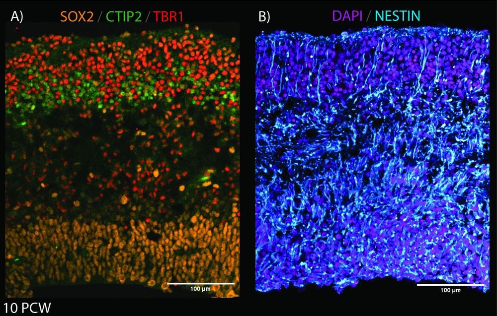
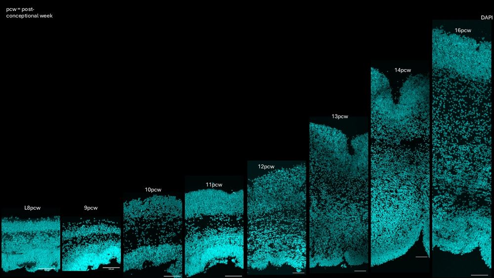
This week's image is by @joseriveraz.bsky.social who's tracing the lineage of progenitors in the human developing cortex.
Here, José shows the building blocks of the developing cortex at 10pcw with SOX2 (apical progenitors), CTIP2 & TBR1(neuronal markers), NESTIN (radial glial fibers), and DAPI.
Here, José shows the building blocks of the developing cortex at 10pcw with SOX2 (apical progenitors), CTIP2 & TBR1(neuronal markers), NESTIN (radial glial fibers), and DAPI.
May 17, 2025 at 1:15 PM
This week's image is by @joseriveraz.bsky.social who's tracing the lineage of progenitors in the human developing cortex.
Here, José shows the building blocks of the developing cortex at 10pcw with SOX2 (apical progenitors), CTIP2 & TBR1(neuronal markers), NESTIN (radial glial fibers), and DAPI.
Here, José shows the building blocks of the developing cortex at 10pcw with SOX2 (apical progenitors), CTIP2 & TBR1(neuronal markers), NESTIN (radial glial fibers), and DAPI.
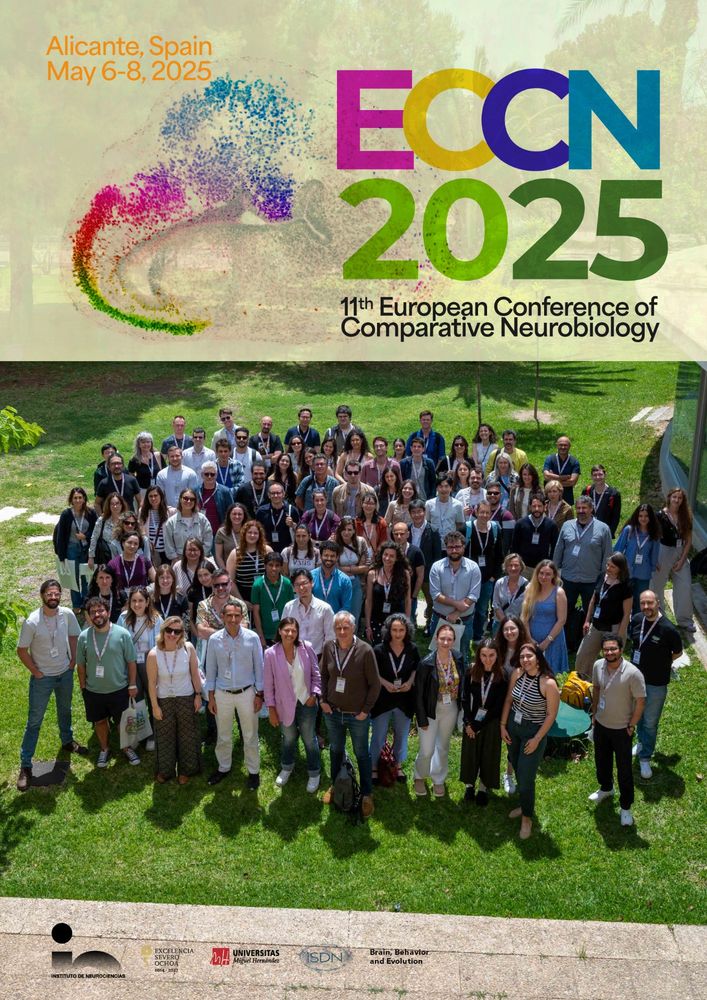
It has been an absolute pleasure for our lab to attend #ECCN2025 for the first time this year.
We had meaningful discussions and learned a lot from a vibrant community tackling tough questions on brain evolution and development! Super excited for what lies ahead!
We had meaningful discussions and learned a lot from a vibrant community tackling tough questions on brain evolution and development! Super excited for what lies ahead!
May 9, 2025 at 11:44 AM
It has been an absolute pleasure for our lab to attend #ECCN2025 for the first time this year.
We had meaningful discussions and learned a lot from a vibrant community tackling tough questions on brain evolution and development! Super excited for what lies ahead!
We had meaningful discussions and learned a lot from a vibrant community tackling tough questions on brain evolution and development! Super excited for what lies ahead!
🎣 Fishing for Dkk in #zebrafish 🎣
In the lab, PhD student Malik Misssaoui looks for the expression patterns of the dkk genes in zebrafish. Dkk proteins are modulators of wnt singalling, and changes in their expression levels have been linked to neurodegenerative diseases like Alzheimers.
In the lab, PhD student Malik Misssaoui looks for the expression patterns of the dkk genes in zebrafish. Dkk proteins are modulators of wnt singalling, and changes in their expression levels have been linked to neurodegenerative diseases like Alzheimers.
May 6, 2025 at 6:11 PM
🎣 Fishing for Dkk in #zebrafish 🎣
In the lab, PhD student Malik Misssaoui looks for the expression patterns of the dkk genes in zebrafish. Dkk proteins are modulators of wnt singalling, and changes in their expression levels have been linked to neurodegenerative diseases like Alzheimers.
In the lab, PhD student Malik Misssaoui looks for the expression patterns of the dkk genes in zebrafish. Dkk proteins are modulators of wnt singalling, and changes in their expression levels have been linked to neurodegenerative diseases like Alzheimers.
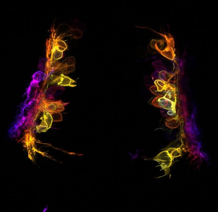
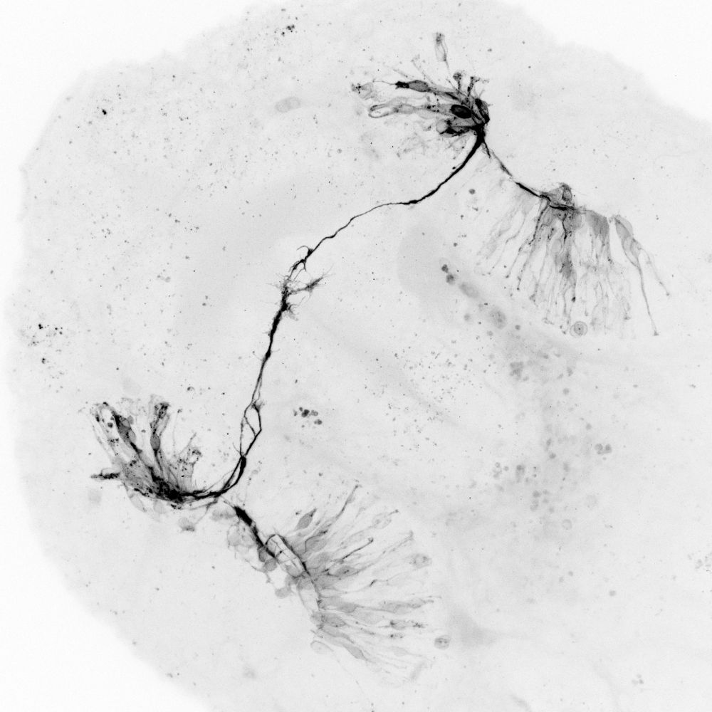
The first retinal ganglion cell axons reaching each other in the formation of the optic nerves in a #zebrafish embryo!
Image taken by @matthewbostock.bsky.social
Image taken by @matthewbostock.bsky.social
April 22, 2025 at 11:54 AM
The first retinal ganglion cell axons reaching each other in the formation of the optic nerves in a #zebrafish embryo!
Image taken by @matthewbostock.bsky.social
Image taken by @matthewbostock.bsky.social
Beautiful staining of the developing foetal cortex🤩. In her project, @gessicagoncalves.bsky.social in the lab is taking on the quest of exploring the cell dynamics of neuroprogenitors in the developing human cortex.
April 11, 2025 at 6:29 PM
Beautiful staining of the developing foetal cortex🤩. In her project, @gessicagoncalves.bsky.social in the lab is taking on the quest of exploring the cell dynamics of neuroprogenitors in the developing human cortex.
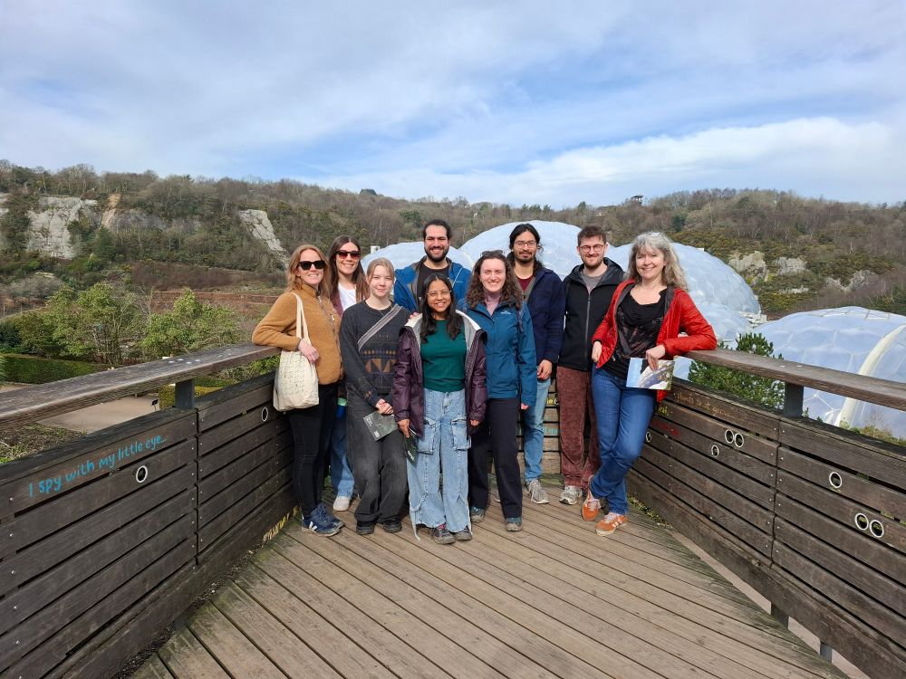
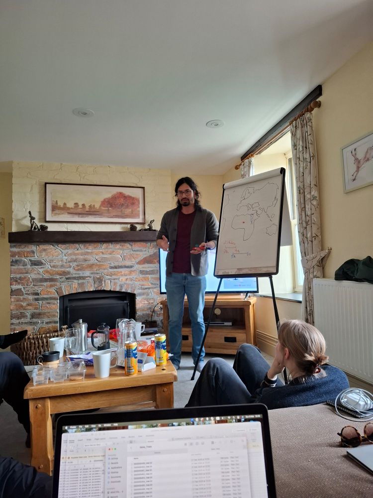
We've just wrapped up our annual lab retreat in Cornwall! A long weekend well spent engaging in scientific discussions, fun exploration of the Cornish countryside, and some delicious homemade cooking!


March 11, 2025 at 6:15 PM
We've just wrapped up our annual lab retreat in Cornwall! A long weekend well spent engaging in scientific discussions, fun exploration of the Cornish countryside, and some delicious homemade cooking!