


Eva-Maria Schentarra
@evaschentarra.bsky.social
Biotechnologist - PhD student @jungmannlab.bsky.social
Enjoying the little things in life using #DNAPAINT 🔬🧬
@mpibiochem.bsky.social
@lmumuenchen.bsky.social
@imprs-ml.bsky.social
Enjoying the little things in life using #DNAPAINT 🔬🧬
@mpibiochem.bsky.social
@lmumuenchen.bsky.social
@imprs-ml.bsky.social
Reposted by Eva-Maria Schentarra

🔬 Join Imaging Bites seminar 28 Jan at 12:30 at @kingscollegelondon.bsky.social.
Prof. Dr. Felipe Opazo @opazo.bsky.social presents #OneStep-IF, a simplified approach to highly #multiplexed IF using same-species primaries and Smart Secondaries® premixes.
Register here: www.kcl.ac.uk/events/imagi...
Prof. Dr. Felipe Opazo @opazo.bsky.social presents #OneStep-IF, a simplified approach to highly #multiplexed IF using same-species primaries and Smart Secondaries® premixes.
Register here: www.kcl.ac.uk/events/imagi...

January 21, 2026 at 11:25 AM
🔬 Join Imaging Bites seminar 28 Jan at 12:30 at @kingscollegelondon.bsky.social.
Prof. Dr. Felipe Opazo @opazo.bsky.social presents #OneStep-IF, a simplified approach to highly #multiplexed IF using same-species primaries and Smart Secondaries® premixes.
Register here: www.kcl.ac.uk/events/imagi...
Prof. Dr. Felipe Opazo @opazo.bsky.social presents #OneStep-IF, a simplified approach to highly #multiplexed IF using same-species primaries and Smart Secondaries® premixes.
Register here: www.kcl.ac.uk/events/imagi...
Reposted by Eva-Maria Schentarra
Our article "Ångström-resolution imaging of cell-surface glycans" made it to the cover of Nature Nanotechnology! 🤩 @natnano.nature.com
www.nature.com/nnano/volume...
www.nature.com/nnano/volume...

October 18, 2025 at 2:32 PM
Our article "Ångström-resolution imaging of cell-surface glycans" made it to the cover of Nature Nanotechnology! 🤩 @natnano.nature.com
www.nature.com/nnano/volume...
www.nature.com/nnano/volume...
Reposted by Eva-Maria Schentarra
Happy to share this article with my views on @elislenders.bsky.social and @vicidominilab.bsky.social work and discussing current developments in single-molecule localization combining structured excitation and detection. So many exciting perspectives in the field!
www.nature.com/articles/s41...
www.nature.com/articles/s41...

Localization of single molecules with structured illumination and structured detection - Light: Science & Applications
Light: Science & Applications - Localization of single molecules with structured illumination and structured detection
www.nature.com
September 30, 2025 at 6:17 PM
Happy to share this article with my views on @elislenders.bsky.social and @vicidominilab.bsky.social work and discussing current developments in single-molecule localization combining structured excitation and detection. So many exciting perspectives in the field!
www.nature.com/articles/s41...
www.nature.com/articles/s41...
Mirror, mirror on the wall... 🪞 I'm super proud to have contributed to our latest work on left-handed DNA-PAINT! Here we introduce 6 new sequences using the left-handed enantiomers of our speed-optimized DNA-PAINT palette, enabling out-of-the-box 12-plex imaging without extra hybridization steps. 🌈🔬
Highly efficient 12-color multiplexing with speed-optimized DNA-PAINT. We are excited to share our latest paper in @natcomms.nature.com, using left-handed DNA to extend speed-optimized DNA-PAINT to 12 targets in a simple and straightforward way! 🧬👈🚀https://www.nature.com/articles/s41467-025-64228-x
October 2, 2025 at 11:58 AM
Mirror, mirror on the wall... 🪞 I'm super proud to have contributed to our latest work on left-handed DNA-PAINT! Here we introduce 6 new sequences using the left-handed enantiomers of our speed-optimized DNA-PAINT palette, enabling out-of-the-box 12-plex imaging without extra hybridization steps. 🌈🔬
Reposted by Eva-Maria Schentarra
Our new preprint is up! This is the main postdoc work of @wiesner-t.bsky.social focusing on exocytosis along the axon shaft and its regulation by the sub membrane actin-spectrin scaffold: www.biorxiv.org/content/10.1...
Read the thread below for a summary of our findings 🧵1/11
Read the thread below for a summary of our findings 🧵1/11

September 17, 2025 at 2:55 PM
Our new preprint is up! This is the main postdoc work of @wiesner-t.bsky.social focusing on exocytosis along the axon shaft and its regulation by the sub membrane actin-spectrin scaffold: www.biorxiv.org/content/10.1...
Read the thread below for a summary of our findings 🧵1/11
Read the thread below for a summary of our findings 🧵1/11
Reposted by Eva-Maria Schentarra

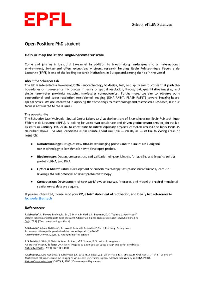
🚨 2 × PhD positions @EPFL! 🚨
Help us push the boundaries of fluorescence microscopy - DNA nanotech, custom optics & spatial omics in Lausanne 🇨🇭. Start Jan 2026. Send CV + motivation + 2 refs → fschueder@ethz.ch
#PhD #Hiring #microscopy #SuperResolution #SpatialOmics #DNAPAINT #FLASHPAINT
Help us push the boundaries of fluorescence microscopy - DNA nanotech, custom optics & spatial omics in Lausanne 🇨🇭. Start Jan 2026. Send CV + motivation + 2 refs → fschueder@ethz.ch
#PhD #Hiring #microscopy #SuperResolution #SpatialOmics #DNAPAINT #FLASHPAINT
July 31, 2025 at 8:56 AM
🚨 2 × PhD positions @EPFL! 🚨
Help us push the boundaries of fluorescence microscopy - DNA nanotech, custom optics & spatial omics in Lausanne 🇨🇭. Start Jan 2026. Send CV + motivation + 2 refs → fschueder@ethz.ch
#PhD #Hiring #microscopy #SuperResolution #SpatialOmics #DNAPAINT #FLASHPAINT
Help us push the boundaries of fluorescence microscopy - DNA nanotech, custom optics & spatial omics in Lausanne 🇨🇭. Start Jan 2026. Send CV + motivation + 2 refs → fschueder@ethz.ch
#PhD #Hiring #microscopy #SuperResolution #SpatialOmics #DNAPAINT #FLASHPAINT
Reposted by Eva-Maria Schentarra

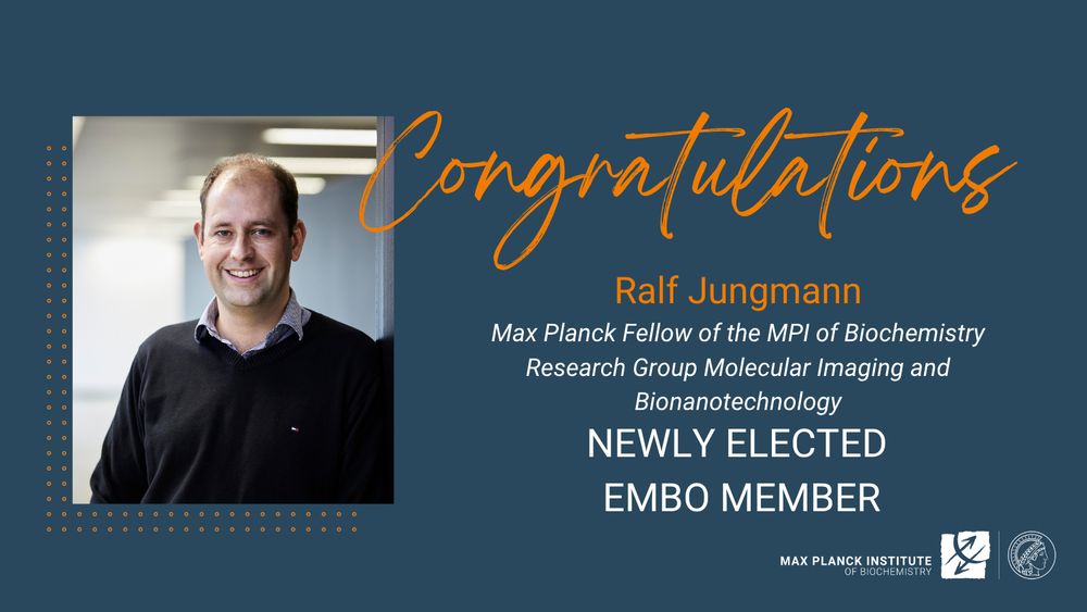
Congratulations to Ralf on your election as a new EMBO member:
❕Original press release from @embo.org : www.embo.org/press-releas...
❕Original press release from @embo.org : www.embo.org/press-releas...
July 3, 2025 at 8:57 AM
Congratulations to Ralf on your election as a new EMBO member:
❕Original press release from @embo.org : www.embo.org/press-releas...
❕Original press release from @embo.org : www.embo.org/press-releas...
Reposted by Eva-Maria Schentarra

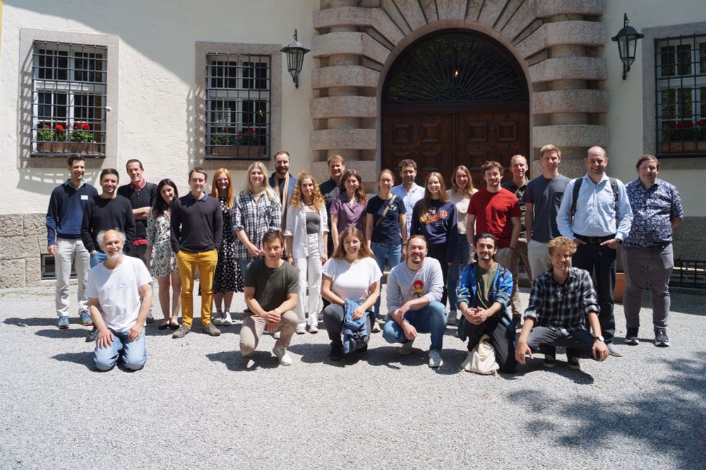
Great science, great company and stunning views at our Lab retreat on Schloss Ringberg 🏰🧬🔬. Big thanks to our guests Sabrina Simoncelli, Sebastian Kobold, Thomas Schlichthärle, @massivephotonics.bsky.social & students from the @lfmilles.bsky.social and @mlsb-borgwardt.bsky.social Labs for joining!
June 14, 2025 at 6:31 PM
Great science, great company and stunning views at our Lab retreat on Schloss Ringberg 🏰🧬🔬. Big thanks to our guests Sabrina Simoncelli, Sebastian Kobold, Thomas Schlichthärle, @massivephotonics.bsky.social & students from the @lfmilles.bsky.social and @mlsb-borgwardt.bsky.social Labs for joining!
Reposted by Eva-Maria Schentarra
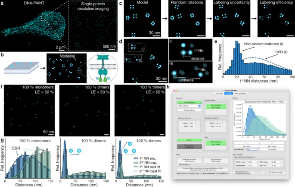
Very excited to present our latest work: SPINNA, an analysis framework and software package for single-protein resolution data! 🖥️🤩
We can directly quantify stoichiometry and oligomerization from super-res (DNA-PAINT, RESI) images!! 🧬🎨
We can directly quantify stoichiometry and oligomerization from super-res (DNA-PAINT, RESI) images!! 🧬🎨
Spatial and stoichiometric in situ analysis of biomolecular oligomerization at single-protein resolution
We are excited to present our latest work published in @natcomms.nature.com
www.nature.com/articles/s41...
We are excited to present our latest work published in @natcomms.nature.com
www.nature.com/articles/s41...
Spatial and stoichiometric in situ analysis of biomolecular oligomerization at single-protein resolution - Nature Communications
Extracting quantitative information on biomolecular oligomerisation with high resolution remains a significant challenge. Here, the authors propose SPINNA, a framework that compares nearest-neighbour ...
www.nature.com
May 7, 2025 at 2:56 PM
Very excited to present our latest work: SPINNA, an analysis framework and software package for single-protein resolution data! 🖥️🤩
We can directly quantify stoichiometry and oligomerization from super-res (DNA-PAINT, RESI) images!! 🧬🎨
We can directly quantify stoichiometry and oligomerization from super-res (DNA-PAINT, RESI) images!! 🧬🎨
Reposted by Eva-Maria Schentarra
Spatial and stoichiometric in situ analysis of biomolecular oligomerization at single-protein resolution
We are excited to present our latest work published in @natcomms.nature.com
www.nature.com/articles/s41...
We are excited to present our latest work published in @natcomms.nature.com
www.nature.com/articles/s41...
Spatial and stoichiometric in situ analysis of biomolecular oligomerization at single-protein resolution - Nature Communications
Extracting quantitative information on biomolecular oligomerisation with high resolution remains a significant challenge. Here, the authors propose SPINNA, a framework that compares nearest-neighbour ...
www.nature.com
May 7, 2025 at 2:43 PM
Spatial and stoichiometric in situ analysis of biomolecular oligomerization at single-protein resolution
We are excited to present our latest work published in @natcomms.nature.com
www.nature.com/articles/s41...
We are excited to present our latest work published in @natcomms.nature.com
www.nature.com/articles/s41...
Reposted by Eva-Maria Schentarra

Excited for our latest work! We developed nanobinders for the luminal domain of Synaptotagmin 1 (calcium sensor for synaptic vesicles). Thanks among others to @verstrekenlab.bsky.social @opazo.bsky.social @meunierlab.bsky.social @volkerhaucke-lab.bsky.social www.biorxiv.org/content/10.1...

Nanobinders for Synaptotagmin 1 enable the analysis of synaptic vesicle dynamics in rodent and human models.
Synaptic neurotransmission is a critical hallmark of brain activity and one of the first processes to be affected in neural diseases. Monitoring this process, and in particular synaptic vesicle recycl...
www.biorxiv.org
April 22, 2025 at 9:27 AM
Excited for our latest work! We developed nanobinders for the luminal domain of Synaptotagmin 1 (calcium sensor for synaptic vesicles). Thanks among others to @verstrekenlab.bsky.social @opazo.bsky.social @meunierlab.bsky.social @volkerhaucke-lab.bsky.social www.biorxiv.org/content/10.1...
Reposted by Eva-Maria Schentarra
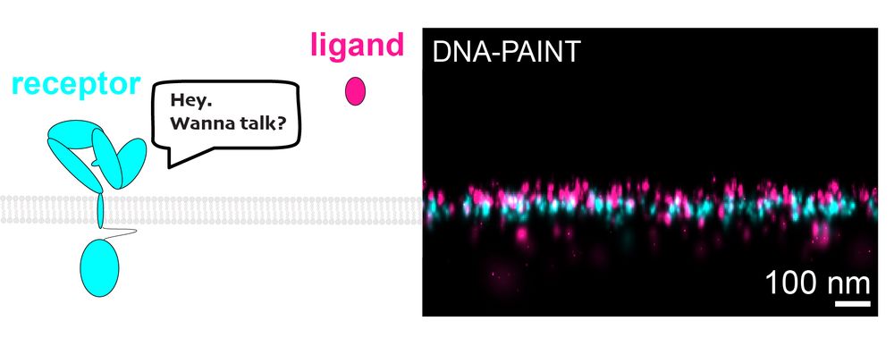
IMAGING LIGAND-RECEPTOR INTERACTIONS AT SINGLE-PROTEIN RESOLUTION WITH DNA-PAINT🔬
Ever wonder how cells "talk"? It starts when ligands bind to receptors on cell surfaces. We have cracked the challenge of imaging small ligands on cell surfaces. #DNAPAINT #celltalk 💬
doi.org/10.1002/smtd...
Ever wonder how cells "talk"? It starts when ligands bind to receptors on cell surfaces. We have cracked the challenge of imaging small ligands on cell surfaces. #DNAPAINT #celltalk 💬
doi.org/10.1002/smtd...
April 10, 2025 at 11:40 AM
IMAGING LIGAND-RECEPTOR INTERACTIONS AT SINGLE-PROTEIN RESOLUTION WITH DNA-PAINT🔬
Ever wonder how cells "talk"? It starts when ligands bind to receptors on cell surfaces. We have cracked the challenge of imaging small ligands on cell surfaces. #DNAPAINT #celltalk 💬
doi.org/10.1002/smtd...
Ever wonder how cells "talk"? It starts when ligands bind to receptors on cell surfaces. We have cracked the challenge of imaging small ligands on cell surfaces. #DNAPAINT #celltalk 💬
doi.org/10.1002/smtd...
One month ago today, I published my first paper with the @jungmannlab.bsky.social 🥳
What better way to celebrate #MicroscopyMonday than with this STAR Protocol on SUM-PAINT spatial proteomic imaging: a guide for highly multiplexed DNA-PAINT imaging in neurons. star-protocols.cell.com/protocols/4066
What better way to celebrate #MicroscopyMonday than with this STAR Protocol on SUM-PAINT spatial proteomic imaging: a guide for highly multiplexed DNA-PAINT imaging in neurons. star-protocols.cell.com/protocols/4066

Cell Press: STAR Protocols
STAR Protocols is an open access, peer-reviewed journal from Cell Press. We offer structured, transparent, accessible, and repeatable step-by-step experimental and computational protocols from all are...
star-protocols.cell.com
April 7, 2025 at 7:58 PM
One month ago today, I published my first paper with the @jungmannlab.bsky.social 🥳
What better way to celebrate #MicroscopyMonday than with this STAR Protocol on SUM-PAINT spatial proteomic imaging: a guide for highly multiplexed DNA-PAINT imaging in neurons. star-protocols.cell.com/protocols/4066
What better way to celebrate #MicroscopyMonday than with this STAR Protocol on SUM-PAINT spatial proteomic imaging: a guide for highly multiplexed DNA-PAINT imaging in neurons. star-protocols.cell.com/protocols/4066
Reposted by Eva-Maria Schentarra
My main PhD paper is on bioRxiv! 🥳 We used motor-PAINT, expansion microscopy, and live-cell imaging of StableMARK to map out how the microtubule cytoskeleton reorganizes during neuronal development.
www.biorxiv.org/content/10.1...
www.biorxiv.org/content/10.1...

Polarity reversal of stable microtubules during neuronal development
Neurons critically depend on long-distance transport orchestrated by motor proteins walking over their highly asymmetric microtubule cytoskeleton. These microtubules are organized uniformly in axons w...
www.biorxiv.org
February 9, 2025 at 1:24 PM
My main PhD paper is on bioRxiv! 🥳 We used motor-PAINT, expansion microscopy, and live-cell imaging of StableMARK to map out how the microtubule cytoskeleton reorganizes during neuronal development.
www.biorxiv.org/content/10.1...
www.biorxiv.org/content/10.1...
Reposted by Eva-Maria Schentarra
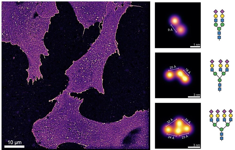
ÅNGSTRÖM-RESOLUTION IMAGING OF CELL-SURFACE GLYCANS 🧬🎨🍬
The glycocalyx, our cells' sugar coat, holds secrets in immunology, cancer, viral infections, and more. Visualizing its molecular architecture was impossible… until now. #glycotime #microscopy
www.biorxiv.org/content/10.1...
The glycocalyx, our cells' sugar coat, holds secrets in immunology, cancer, viral infections, and more. Visualizing its molecular architecture was impossible… until now. #glycotime #microscopy
www.biorxiv.org/content/10.1...
February 10, 2025 at 8:21 AM
ÅNGSTRÖM-RESOLUTION IMAGING OF CELL-SURFACE GLYCANS 🧬🎨🍬
The glycocalyx, our cells' sugar coat, holds secrets in immunology, cancer, viral infections, and more. Visualizing its molecular architecture was impossible… until now. #glycotime #microscopy
www.biorxiv.org/content/10.1...
The glycocalyx, our cells' sugar coat, holds secrets in immunology, cancer, viral infections, and more. Visualizing its molecular architecture was impossible… until now. #glycotime #microscopy
www.biorxiv.org/content/10.1...
Do you know what I love most about my PhD @JungmannLab? Actually seeing the things I learned from biology textbooks come to life🪄. Check out this DNA-PAINT image of the Golgi, showing both cis (GM130) and trans (TGN38) faces together with peroxisomes. Happy
#FluorescenceFriday😊
#FluorescenceFriday😊
January 24, 2025 at 1:44 PM
Do you know what I love most about my PhD @JungmannLab? Actually seeing the things I learned from biology textbooks come to life🪄. Check out this DNA-PAINT image of the Golgi, showing both cis (GM130) and trans (TGN38) faces together with peroxisomes. Happy
#FluorescenceFriday😊
#FluorescenceFriday😊
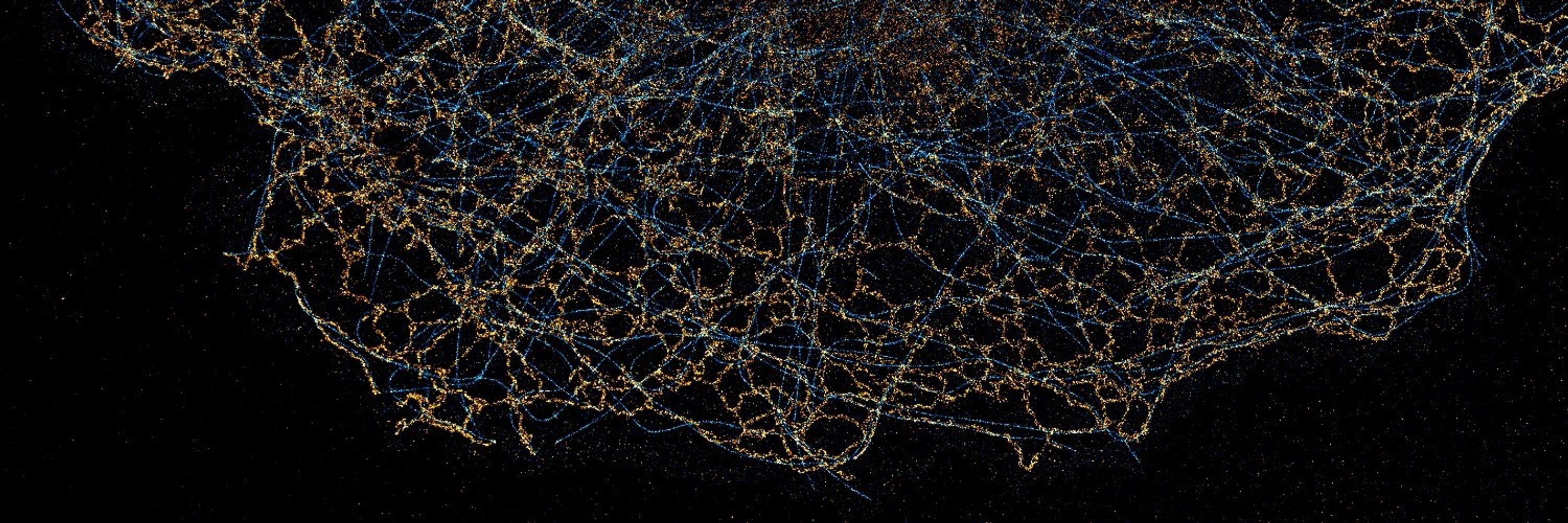
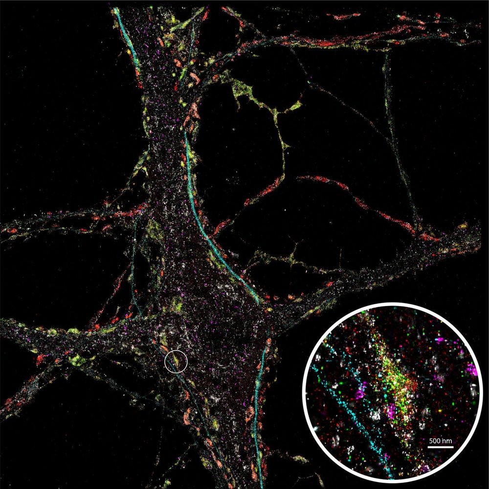
Looking at this beautifully detailed neuron, stained for 9 different proteins using SUM-PAINT, feels like stepping into one of David S. Goodsell's paintings illustrating the crowded cellular environment. #FluorescenceFriday #phdlife @JungmannLab
January 24, 2025 at 1:33 PM
Looking at this beautifully detailed neuron, stained for 9 different proteins using SUM-PAINT, feels like stepping into one of David S. Goodsell's paintings illustrating the crowded cellular environment. #FluorescenceFriday #phdlife @JungmannLab
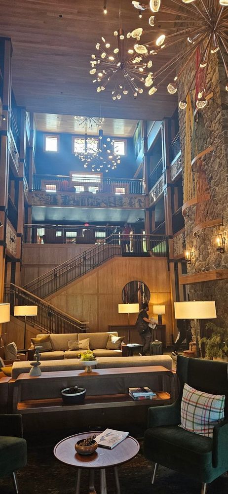
Excited to attend my first ever conference on single molecule approaches to biology by @GordonConf in beautiful Maine!
Caught a fun 'Where's Waldo' moment in the hotel lobby - can you spot Ralf Jungmann and Luciano Masullo waving at me in the background? 🔍 @JungmannLab @l_masu
Caught a fun 'Where's Waldo' moment in the hotel lobby - can you spot Ralf Jungmann and Luciano Masullo waving at me in the background? 🔍 @JungmannLab @l_masu
January 24, 2025 at 1:33 PM
Excited to attend my first ever conference on single molecule approaches to biology by @GordonConf in beautiful Maine!
Caught a fun 'Where's Waldo' moment in the hotel lobby - can you spot Ralf Jungmann and Luciano Masullo waving at me in the background? 🔍 @JungmannLab @l_masu
Caught a fun 'Where's Waldo' moment in the hotel lobby - can you spot Ralf Jungmann and Luciano Masullo waving at me in the background? 🔍 @JungmannLab @l_masu
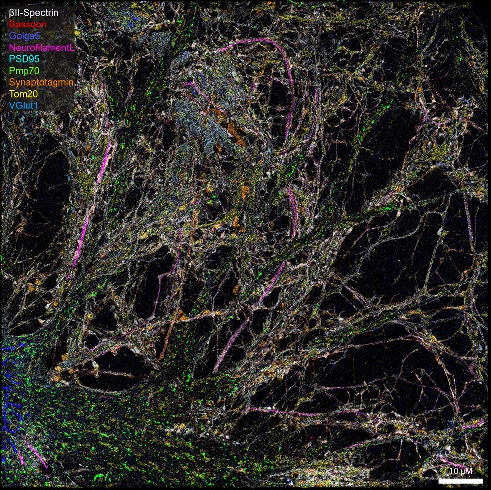
Happy Friday! To wrap up another exciting week of learning highly multiplexed DNA-PAINT in the @JungmannLab, here's a nice #cellfie of a rat hippocampal neuron stained for synaptic vesicle markers (Vamp2, VGlut1 and synaptotagmin), clathrin, neurofilament and peroxisomes.
January 24, 2025 at 1:33 PM
Happy Friday! To wrap up another exciting week of learning highly multiplexed DNA-PAINT in the @JungmannLab, here's a nice #cellfie of a rat hippocampal neuron stained for synaptic vesicle markers (Vamp2, VGlut1 and synaptotagmin), clathrin, neurofilament and peroxisomes.
Here it is - fresh off the press. Tracking substeps of kinesin in mildly fixed neurons using MINFLUX 🔬.
I am proud to see my master thesis in close work together with Otto Wirth finally published!
nature.com/articles/s4200…
I am proud to see my master thesis in close work together with Otto Wirth finally published!
nature.com/articles/s4200…
Uncovering kinesin dynamics in neurites with MINFLUX
Communications Biology - A MINFLUX tracking study of a truncated kinesin-1 mutant in gently fixed primary rat hippocampal neurons revealed 8 nm substeps and a rotation of its head during the...
www.nature.com
January 24, 2025 at 1:33 PM
Here it is - fresh off the press. Tracking substeps of kinesin in mildly fixed neurons using MINFLUX 🔬.
I am proud to see my master thesis in close work together with Otto Wirth finally published!
nature.com/articles/s4200…
I am proud to see my master thesis in close work together with Otto Wirth finally published!
nature.com/articles/s4200…
My first preprint as a joint first author together with Otto Wirth and I couldn't be more excited! 📚 🔬
Exploring kinesin with MINFLUX during my Master's thesis was a blast while working with such talented minds in the Hell Lab.
x.com/Stefan_W_Hell/… biorxiv.org/content/10.110…
Exploring kinesin with MINFLUX during my Master's thesis was a blast while working with such talented minds in the Hell Lab.
x.com/Stefan_W_Hell/… biorxiv.org/content/10.110…
Uncovering kinesin dynamics in neurites with MINFLUX
Neurons grow neurites of several tens of micrometers in length, necessitating active transport from the cell body by motor proteins. By tracking fluorophores as minimally invasive labels, MINFLUX is able to quantify the motion of those proteins with nanometer/millisecond resolution. Here we study the substeps of a truncated kinesin-1 mutant in primary rat hippocampal neurons, which have so far been mainly observed on microtubules polymerized on glass coverslips. A gentle fixation protocol largely maintains the structure and surface modifications of the microtubules in the cell. By analyzing the time between the substeps, we identify the ATP-binding state of kinesin-1 and observe the associated rotation of the kinesin-1 head in neurites. We also observed kinesin-1 switching microtubules mid-walk, highlighting the potential of MINFLUX to study the details of active cellular transport. ### Competing Interest Statement S.W.H. is inventor on patent applications WO 2013/072273 and WO 2015/097000 filed by the Max Planck Society that cover basic principles and arrangements of MINFLUX, including single-molecule tracking. S.W.H. is inventor on patent application WO 2020/064108 submitted by the Max Planck Society that covers principles and arrangements of the phase/amplitude modulator for shifting the intensity minimum. S.W.H. is a cofounder of the company Abberior Instruments, which commercializes MINFLUX microscopes. The remaining authors declare no competing interests.
www.biorxiv.org
January 24, 2025 at 1:33 PM
My first preprint as a joint first author together with Otto Wirth and I couldn't be more excited! 📚 🔬
Exploring kinesin with MINFLUX during my Master's thesis was a blast while working with such talented minds in the Hell Lab.
x.com/Stefan_W_Hell/… biorxiv.org/content/10.110…
Exploring kinesin with MINFLUX during my Master's thesis was a blast while working with such talented minds in the Hell Lab.
x.com/Stefan_W_Hell/… biorxiv.org/content/10.110…