


Biological Electron Microscopy
@electronmicroscopy.bsky.social
All about basic and solid electron microscopy.
Pinned

Quantifying Golgi structure using EM: combining volume-SEM and stereology for higher throughput - Histochemistry and Cell Biology
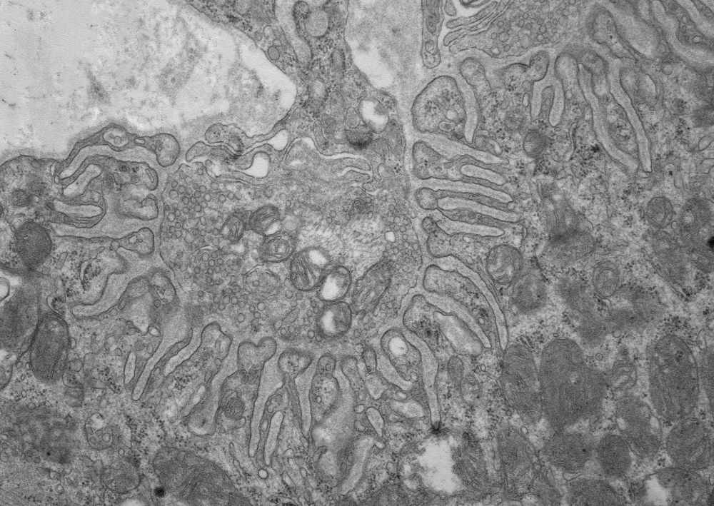
Investigating organelles such as the Golgi complex depends increasingly on high-throughput quantitative morphological analyses from multiple experimental or genetic conditions. Light microscopy (LM) h...
doi.org
Tired of segmenting your volume data sets? Overbooked time slots for automatic #segmentation on the workstation? Concerned about conclusions based on single or few reconstructed organelles? #Stereology effortlessly generates precise quantitative data of the entire population. doi.org/10.1007/s004...
Reposted by Biological Electron Microscopy

(1/n) DNA-PAINT imaging inside the nucleus at single antibody resolution using TIRF? Ultrathin sectioning makes it happen!
Grateful to share my postdoctoral work introducing “tomographic & kinetically-enhanced DNA-PAINT” or in brief: tkPAINT. Out in @pnas.org!
doi.org/10.1073/pnas...
👇🧵
Grateful to share my postdoctoral work introducing “tomographic & kinetically-enhanced DNA-PAINT” or in brief: tkPAINT. Out in @pnas.org!
doi.org/10.1073/pnas...
👇🧵

August 13, 2025 at 2:19 PM
(1/n) DNA-PAINT imaging inside the nucleus at single antibody resolution using TIRF? Ultrathin sectioning makes it happen!
Grateful to share my postdoctoral work introducing “tomographic & kinetically-enhanced DNA-PAINT” or in brief: tkPAINT. Out in @pnas.org!
doi.org/10.1073/pnas...
👇🧵
Grateful to share my postdoctoral work introducing “tomographic & kinetically-enhanced DNA-PAINT” or in brief: tkPAINT. Out in @pnas.org!
doi.org/10.1073/pnas...
👇🧵
Reposted by Biological Electron Microscopy
I am excited to share our first Pt replica EM images. It took us a little while, but now we have establish the unroofing, drying and Pt coating workflow 🎉 Great work by our postdoc Luis Wong Dilworth! The image below shows the cytosolic membrane leaflet of a fibroblast 👇
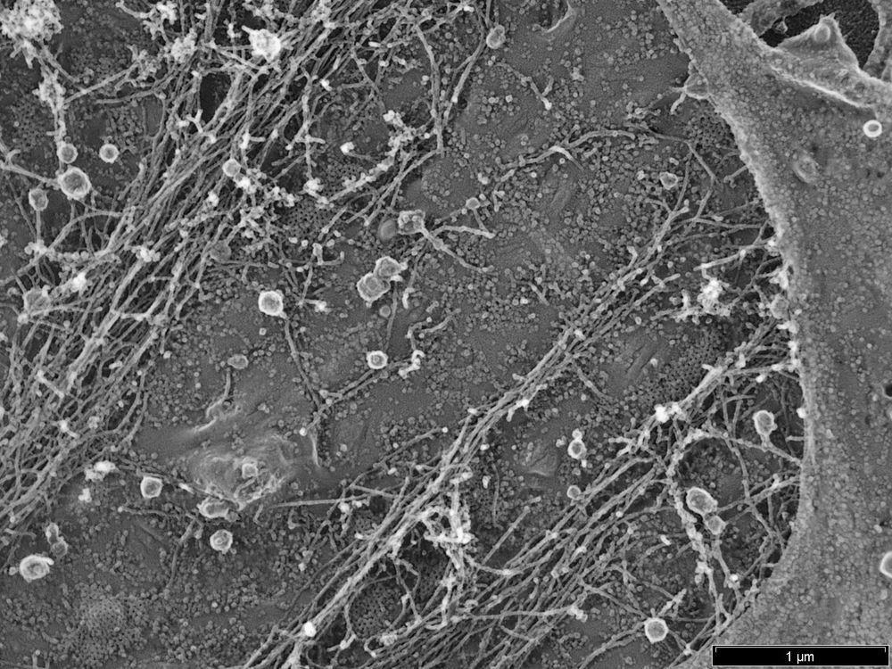
February 13, 2025 at 2:34 PM
I am excited to share our first Pt replica EM images. It took us a little while, but now we have establish the unroofing, drying and Pt coating workflow 🎉 Great work by our postdoc Luis Wong Dilworth! The image below shows the cytosolic membrane leaflet of a fibroblast 👇
Reposted by Biological Electron Microscopy

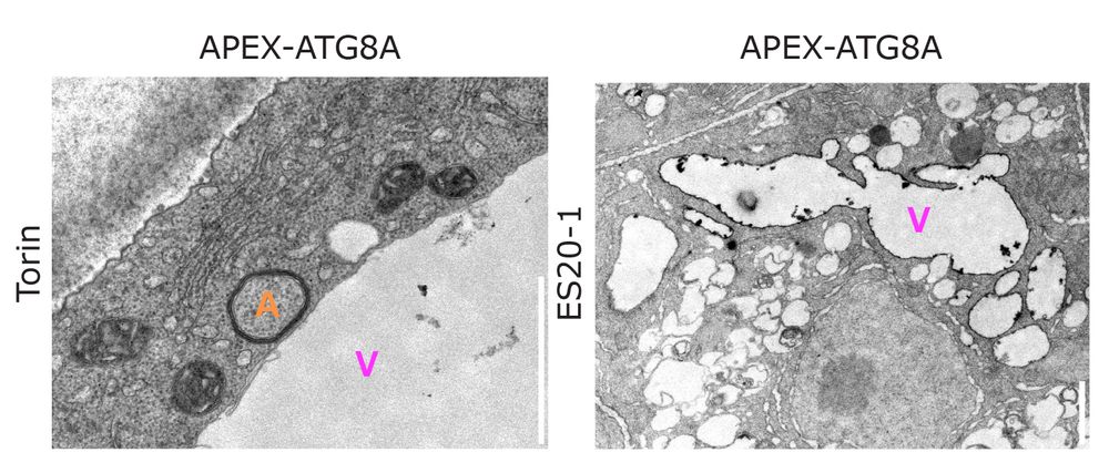
For the first time in plants, we used APEX-based electron microscopy to map the precise localization of ATG8 at the vacuolar membrane after stress! 🚀
Pushing the boundaries of plant cell biology—one EM image at a time. ⚡👀
#PlantScience #ElectronMicroscopy #Autophagy #Vacuole #NaturePlants
Pushing the boundaries of plant cell biology—one EM image at a time. ⚡👀
#PlantScience #ElectronMicroscopy #Autophagy #Vacuole #NaturePlants
February 7, 2025 at 11:11 AM
For the first time in plants, we used APEX-based electron microscopy to map the precise localization of ATG8 at the vacuolar membrane after stress! 🚀
Pushing the boundaries of plant cell biology—one EM image at a time. ⚡👀
#PlantScience #ElectronMicroscopy #Autophagy #Vacuole #NaturePlants
Pushing the boundaries of plant cell biology—one EM image at a time. ⚡👀
#PlantScience #ElectronMicroscopy #Autophagy #Vacuole #NaturePlants
Reposted by Biological Electron Microscopy
Zooming in on the ultimate connection! 🔬 💥Feast your eyes on this stunning electron microscopy image of a neuromuscular junction #EMFriday #ElectronMicroscopy
January 10, 2025 at 4:04 PM
Zooming in on the ultimate connection! 🔬 💥Feast your eyes on this stunning electron microscopy image of a neuromuscular junction #EMFriday #ElectronMicroscopy
Reposted by Biological Electron Microscopy
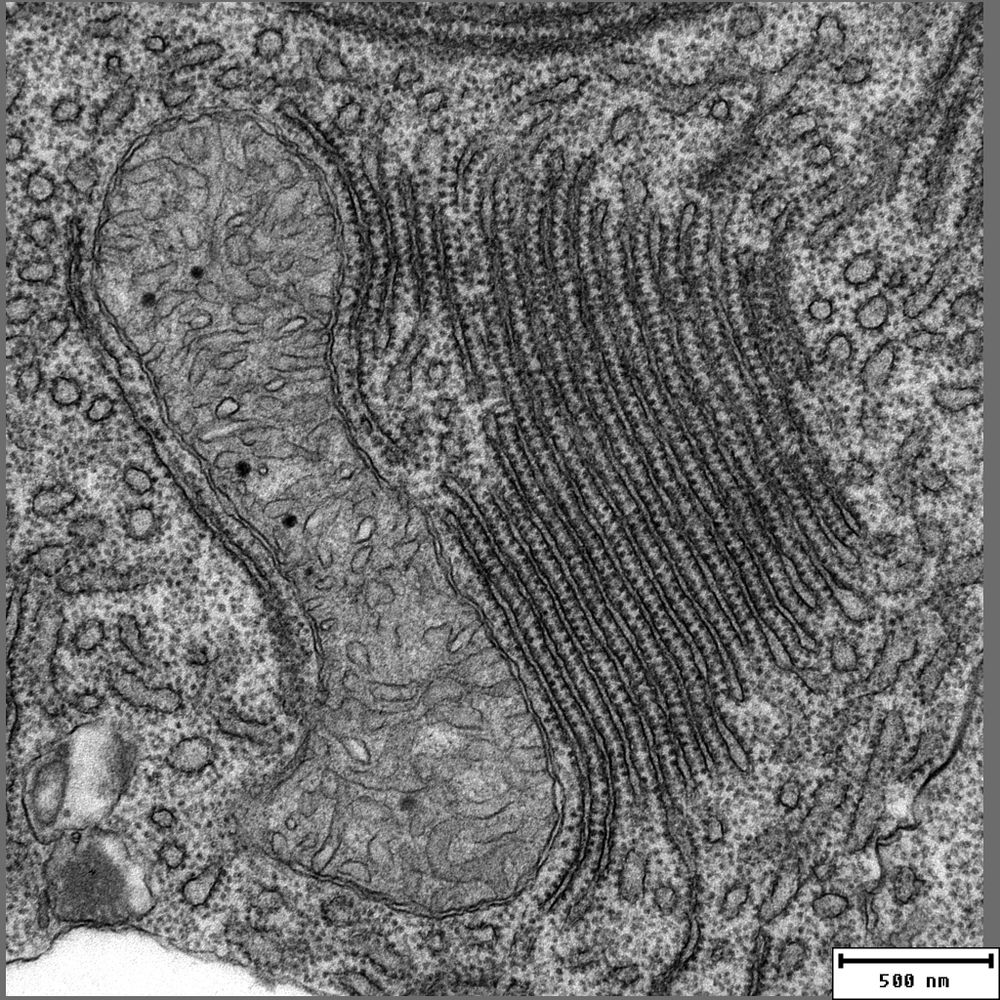
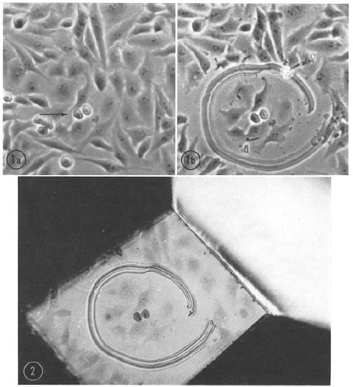
#Correlative Light and Electron Microscopy, #CLEM, is a modern #method combining light and #electronmicroscopy data. Although it is considered “new”, it was already used in the early 1960s, for instance to study the #ultrastructure of mitotic cells in this beautiful example: doi.org/10.1083/jcb....
January 13, 2025 at 7:47 AM
#Correlative Light and Electron Microscopy, #CLEM, is a modern #method combining light and #electronmicroscopy data. Although it is considered “new”, it was already used in the early 1960s, for instance to study the #ultrastructure of mitotic cells in this beautiful example: doi.org/10.1083/jcb....
Reposted by Biological Electron Microscopy
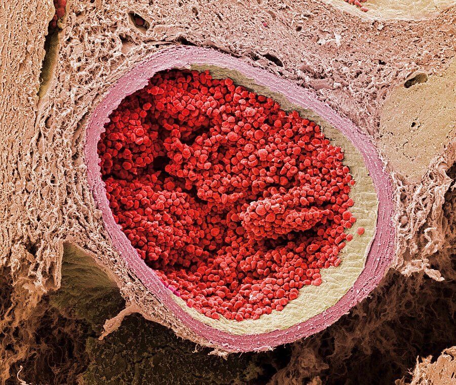
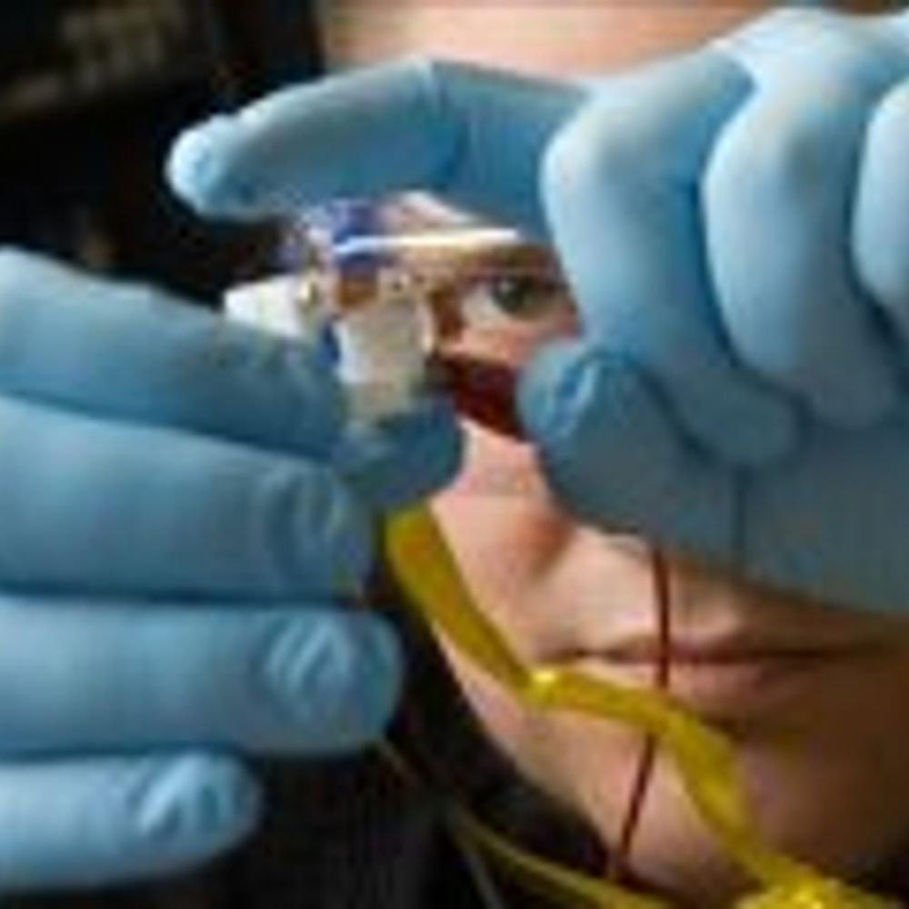
electron microscope image of a section through a foetal aorta filled with red blood cells
January 12, 2025 at 10:09 PM
electron microscope image of a section through a foetal aorta filled with red blood cells
Reposted by Biological Electron Microscopy

I fear there is deep truth here.

January 11, 2025 at 8:47 PM
I fear there is deep truth here.
Reposted by Biological Electron Microscopy
“One of the challenges in science journalism is the oversimplification of research findings to attract attention. Headlines such as “scientists find cure for cancer” or “ozone layer is healing” are designed to attract readers’ attention but often misrepresent the complexity of scientific research.”

Beyond misalignment of science in the news and in schools
Almost 40 years ago, the American astronomer, planetary scientist, and science communicator, Carl Sagan, reflected on the role of mass media in science communication. “How much science and technology ...
www.science.org
January 9, 2025 at 3:56 PM
“One of the challenges in science journalism is the oversimplification of research findings to attract attention. Headlines such as “scientists find cure for cancer” or “ozone layer is healing” are designed to attract readers’ attention but often misrepresent the complexity of scientific research.”
Reposted by Biological Electron Microscopy
I always wanted to name a protein after a Greek goddess or nymph and here we go: journals.plos.org/plospathogen...
Happy holidays everyone
Happy holidays everyone
APEX-based proximity labeling in Plasmodium identifies a membrane protein with dual functions during mosquito infection
Author summary Malaria parasites are transmitted by and to mosquitoes. Blocking either of these transmission steps would stop the parasite life cycle. Hence, identification of new proteins that are es...
journals.plos.org
December 20, 2024 at 2:38 PM
I always wanted to name a protein after a Greek goddess or nymph and here we go: journals.plos.org/plospathogen...
Happy holidays everyone
Happy holidays everyone
Reposted by Biological Electron Microscopy
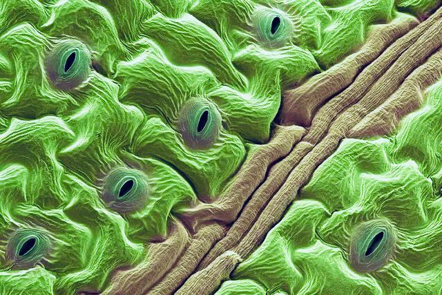
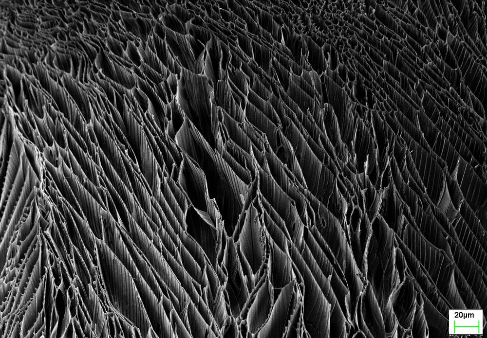
electron microscope image of a coriander leaf
January 8, 2025 at 8:42 PM
electron microscope image of a coriander leaf
Reposted by Biological Electron Microscopy

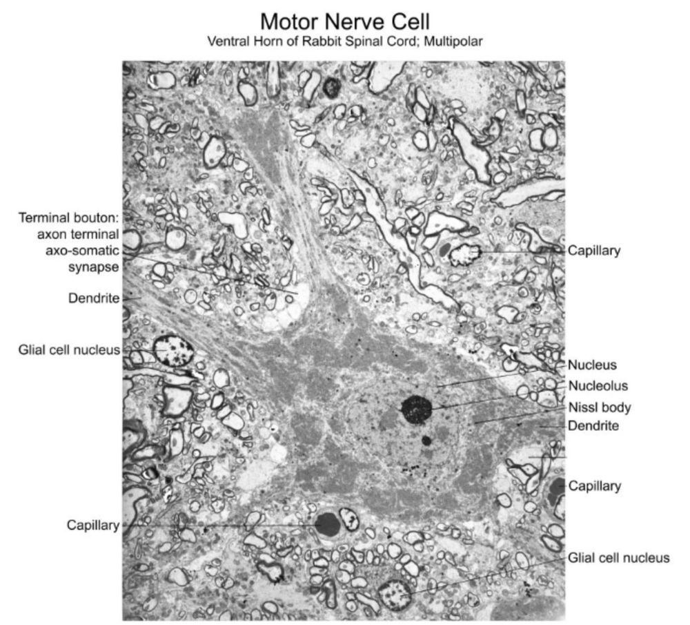
Images and instruction span from the area level down to the subcellular. Check out this rabbit motor neuron! Again, the text walks students through what they should be learning and appreciating from the image.
3/n
3/n
January 7, 2025 at 11:10 AM
Images and instruction span from the area level down to the subcellular. Check out this rabbit motor neuron! Again, the text walks students through what they should be learning and appreciating from the image.
3/n
3/n
Reposted by Biological Electron Microscopy
Finding my bearings here on BlueSky. Figuring on dropping more cool "sciencey" stuff on here. I have done quite a bit of electron microscopy... nothing scientifically earth shattering, but I found so many of the images so darned pretty so I'm going to start posting ones I like from time to time.
January 7, 2025 at 7:34 PM
Finding my bearings here on BlueSky. Figuring on dropping more cool "sciencey" stuff on here. I have done quite a bit of electron microscopy... nothing scientifically earth shattering, but I found so many of the images so darned pretty so I'm going to start posting ones I like from time to time.
Reposted by Biological Electron Microscopy

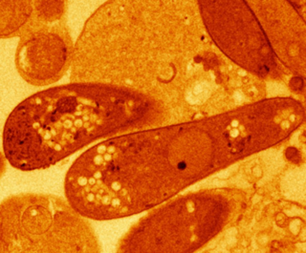
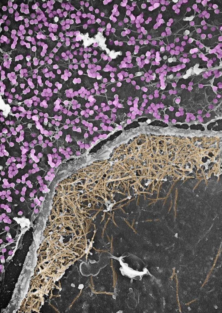
Just published. Check the astonishing diversity of bacteria that can not only form intracellular nanomagnets but also amorphous calcium carbonates that litterally fill the cells. + beautiful EM and cryo-X-ray microscopy images + genomes; follow the link academic.oup.com/ismej/advanc...
January 8, 2025 at 8:27 PM
Just published. Check the astonishing diversity of bacteria that can not only form intracellular nanomagnets but also amorphous calcium carbonates that litterally fill the cells. + beautiful EM and cryo-X-ray microscopy images + genomes; follow the link academic.oup.com/ismej/advanc...
Reposted by Biological Electron Microscopy

Gene discovery sheds light on ‘glue ear’ in people with Down syndrome, paving the way for future targeted therapies.
https://buff.ly/4h6P8eF
https://buff.ly/4h6P8eF
January 8, 2025 at 9:01 PM
Gene discovery sheds light on ‘glue ear’ in people with Down syndrome, paving the way for future targeted therapies.
https://buff.ly/4h6P8eF
https://buff.ly/4h6P8eF
Reposted by Biological Electron Microscopy
Still our favorite #review! Unfortunately, as relevant today as when it was published, it is an excellent illustration of the #problems arising from the neglect of #ultrastructure and #electronmicroscopy in #cell-biology and the over-reliance on #fluorescence #microscopy.
doi.org/10.1016/0962...
doi.org/10.1016/0962...
January 8, 2025 at 8:18 AM
Still our favorite #review! Unfortunately, as relevant today as when it was published, it is an excellent illustration of the #problems arising from the neglect of #ultrastructure and #electronmicroscopy in #cell-biology and the over-reliance on #fluorescence #microscopy.
doi.org/10.1016/0962...
doi.org/10.1016/0962...
Reposted by Biological Electron Microscopy

Tanmay now also combining electronmicroscopy with metabolic imaging, see their preprint with @kiranrpatil.bsky.social @lisamaierlab.bsky.social and colleagues
www.biorxiv.org/content/10.1...
#MultiBac2025
www.biorxiv.org/content/10.1...
#MultiBac2025

Extensive PFAS accumulation by human gut bacteria
Per- and polyfluoroalkyl Substances (PFAS) – the so-called ‘forever chemicals’ – are a major cause of environmental and health concern due to their toxicity and long-term persistence[1][1],[2][2]. Yet...
www.biorxiv.org
January 7, 2025 at 10:45 AM
Tanmay now also combining electronmicroscopy with metabolic imaging, see their preprint with @kiranrpatil.bsky.social @lisamaierlab.bsky.social and colleagues
www.biorxiv.org/content/10.1...
#MultiBac2025
www.biorxiv.org/content/10.1...
#MultiBac2025
Reposted by Biological Electron Microscopy

Nonvesicular lipid transfer is key for myelin growth! Oligodendrocytes' ER enriches myelin development. Huge lipid synthesis for myelin biogenesis—volume electron microscopy shows it all! PMID:39528474, Nat Commun 2024 @NatureComms doi.org/10.1038/s414...
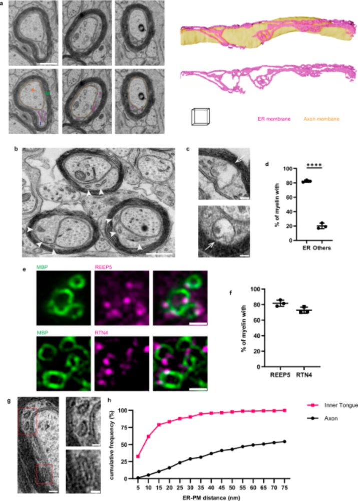
Nonvesicular lipid transfer drives myelin growth in the central nervous system - Nature Communications
To generate myelin, oligodendrocytes must greatly expand their membrane. Here, the authors show that oligodendroglial endoplasmic reticulum-myelin contact sites and nonvesicular glycolipid transport a...
doi.org
December 18, 2024 at 7:14 AM
Nonvesicular lipid transfer is key for myelin growth! Oligodendrocytes' ER enriches myelin development. Huge lipid synthesis for myelin biogenesis—volume electron microscopy shows it all! PMID:39528474, Nat Commun 2024 @NatureComms doi.org/10.1038/s414...
Reposted by Biological Electron Microscopy

Potential new invasive species of #bryozoa for Norway alert!
On their way to our #NorDigBryo workshop in Sletvik in October, the people from Bergen did some sampling in some harbors/marinas to look for invasive species and lo and behold:
On their way to our #NorDigBryo workshop in Sletvik in October, the people from Bergen did some sampling in some harbors/marinas to look for invasive species and lo and behold:
December 20, 2024 at 2:13 PM
Potential new invasive species of #bryozoa for Norway alert!
On their way to our #NorDigBryo workshop in Sletvik in October, the people from Bergen did some sampling in some harbors/marinas to look for invasive species and lo and behold:
On their way to our #NorDigBryo workshop in Sletvik in October, the people from Bergen did some sampling in some harbors/marinas to look for invasive species and lo and behold:
Reposted by Biological Electron Microscopy

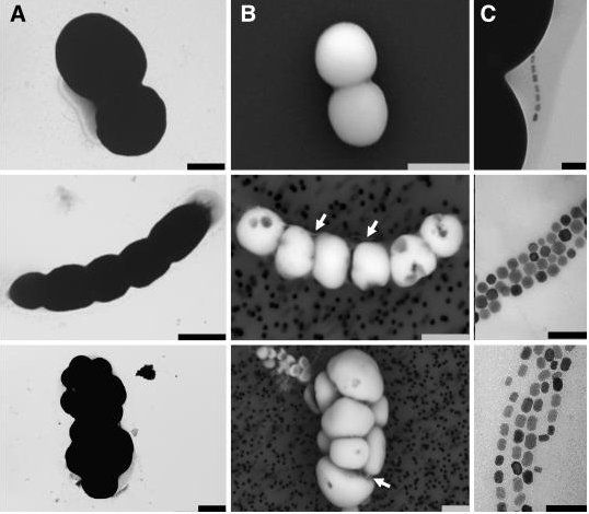
I wanted to end 2024 with these cool Transmission electron microscopy images from my PhD research on raw skin tissue!
Can anyone guess what this structure might be?
#collagen #skinscience
Can anyone guess what this structure might be?
#collagen #skinscience
December 20, 2024 at 8:03 PM
I wanted to end 2024 with these cool Transmission electron microscopy images from my PhD research on raw skin tissue!
Can anyone guess what this structure might be?
#collagen #skinscience
Can anyone guess what this structure might be?
#collagen #skinscience
Reposted by Biological Electron Microscopy

New #ISEPpapers! The nature of ‘jaws’: a new predatory representative of #Provora and the ultrastructure of nibbling protists royalsocietypublishing.org/doi/10.1098/...
#Protists #Microbes #Biology #TreeOfLife #Microscopy
#Protists #Microbes #Biology #TreeOfLife #Microscopy
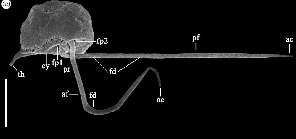
December 21, 2024 at 6:51 PM
New #ISEPpapers! The nature of ‘jaws’: a new predatory representative of #Provora and the ultrastructure of nibbling protists royalsocietypublishing.org/doi/10.1098/...
#Protists #Microbes #Biology #TreeOfLife #Microscopy
#Protists #Microbes #Biology #TreeOfLife #Microscopy
Reposted by Biological Electron Microscopy

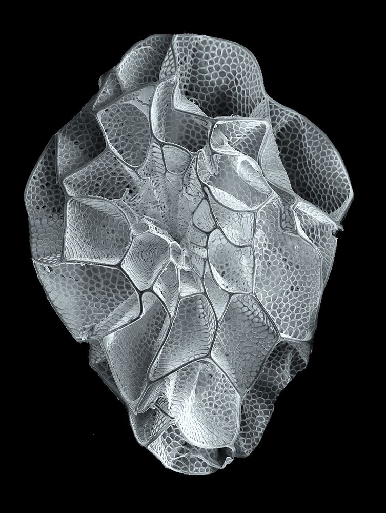
Ending the year with micro-morphology for nerdy taxonomy #1 - Scanning Electron Microscopy never gets old @ Prince of Songkla University, Hat Yai!
ps: Any Diatom people around? We're finding a lot of funky stuff..
ps: Any Diatom people around? We're finding a lot of funky stuff..
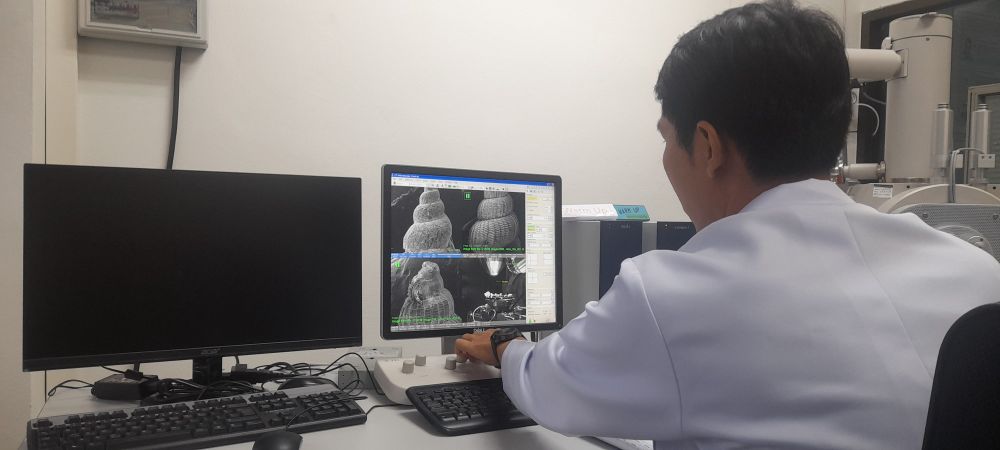
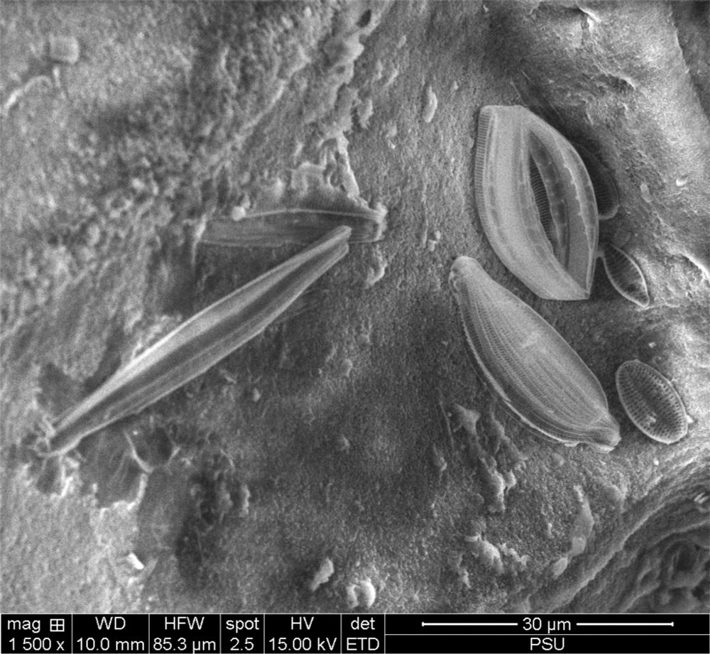
December 24, 2024 at 6:37 AM
Ending the year with micro-morphology for nerdy taxonomy #1 - Scanning Electron Microscopy never gets old @ Prince of Songkla University, Hat Yai!
ps: Any Diatom people around? We're finding a lot of funky stuff..
ps: Any Diatom people around? We're finding a lot of funky stuff..
Reposted by Biological Electron Microscopy
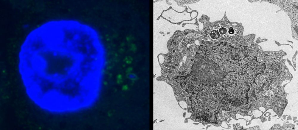
Using electron microscopy, the research team identified an accumulation of “dark microglia”, a subset of microglia associated with cellular stress and neuro-degeneration, in postmortem brain tissues from Alzheimer’s patients. The cells were present at twice the levels seen in healthy-aged people.
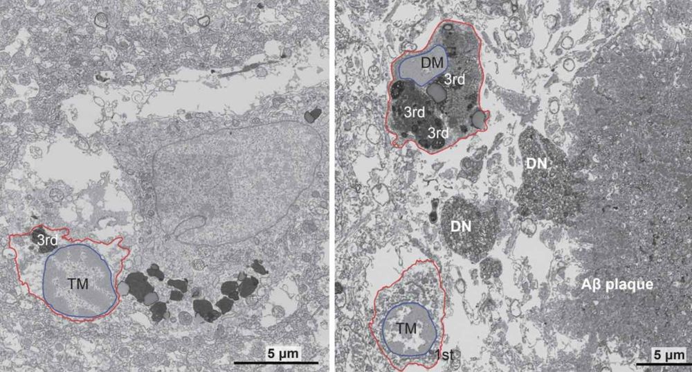
Key Alzheimer’s Breakthrough Identifies Stress-Related Cellular Mechanism Driving the Disease
CUNY scientists made a key Alzheimer's disease breakthrough, identifying a vital mechanism in the brain’s immune cells that drive dementia.
www.goodnewsnetwork.org
December 31, 2024 at 6:11 PM
Using electron microscopy, the research team identified an accumulation of “dark microglia”, a subset of microglia associated with cellular stress and neuro-degeneration, in postmortem brain tissues from Alzheimer’s patients. The cells were present at twice the levels seen in healthy-aged people.
Reposted by Biological Electron Microscopy

Today my colleague shared with me the most EXQUISITE image of a seed the size of a dust particle, collected from a rare plant called Xylanche in Nepal, and photographed using scanning electron microscopy. Wow.
January 3, 2025 at 3:22 PM
Today my colleague shared with me the most EXQUISITE image of a seed the size of a dust particle, collected from a rare plant called Xylanche in Nepal, and photographed using scanning electron microscopy. Wow.
Reposted by Biological Electron Microscopy
#Science in the age of #selfies
A short #opinion article that we think hits a very important point!
Albert #Einstein: “an academic career, in which a person is forced to produce #scientific writings in great amounts, creates a #danger of intellectual superficiality”
www.pnas.org/doi/10.1073/...
A short #opinion article that we think hits a very important point!
Albert #Einstein: “an academic career, in which a person is forced to produce #scientific writings in great amounts, creates a #danger of intellectual superficiality”
www.pnas.org/doi/10.1073/...
Science in the age of selfies | PNAS
Science in the age of selfies
www.pnas.org
January 6, 2025 at 7:35 AM
#Science in the age of #selfies
A short #opinion article that we think hits a very important point!
Albert #Einstein: “an academic career, in which a person is forced to produce #scientific writings in great amounts, creates a #danger of intellectual superficiality”
www.pnas.org/doi/10.1073/...
A short #opinion article that we think hits a very important point!
Albert #Einstein: “an academic career, in which a person is forced to produce #scientific writings in great amounts, creates a #danger of intellectual superficiality”
www.pnas.org/doi/10.1073/...
Reposted by Biological Electron Microscopy
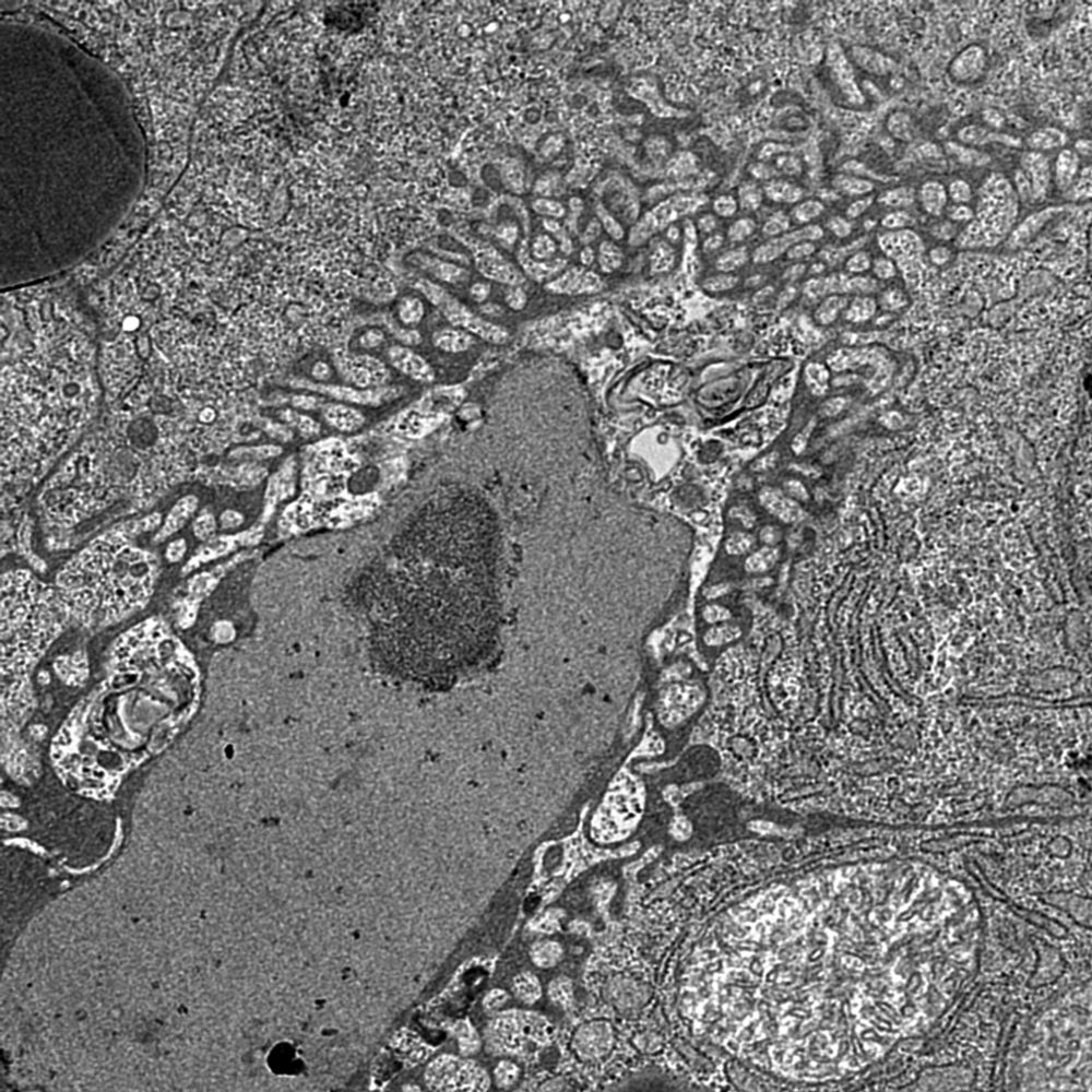
Dynamic duo at the cell's edge: Caveolae and the actin cytoskeleton work together to sense and respond to mechanical stress. Caveolae buffer tension, while actin provides structure and force. Ultimate biomechanics team. #CellBiology #Mechanotransduction
December 21, 2024 at 5:38 PM
Dynamic duo at the cell's edge: Caveolae and the actin cytoskeleton work together to sense and respond to mechanical stress. Caveolae buffer tension, while actin provides structure and force. Ultimate biomechanics team. #CellBiology #Mechanotransduction