


Joe McKellar
@viroscope.bsky.social
Post-doc at @IGMM Montpellier
Studying Deltaviruses through the lens of a microscope 🔬
Studying Deltaviruses through the lens of a microscope 🔬
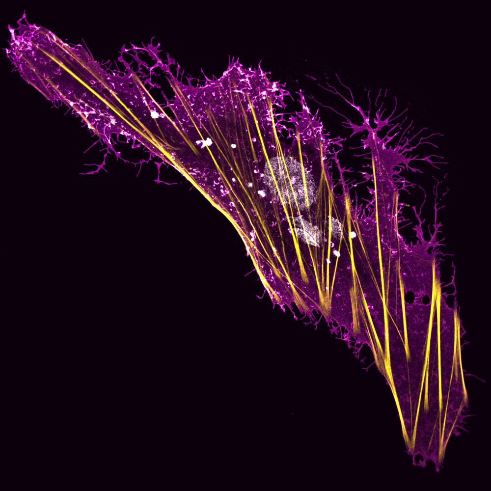
A Boa Constrictor brain-derived cell expressing a viral glycoprotein (magenta) and stained for actin (yellow) and DNA (white).
#FluorescenceFriday
#FluorescenceFriday
June 27, 2025 at 11:20 AM
A Boa Constrictor brain-derived cell expressing a viral glycoprotein (magenta) and stained for actin (yellow) and DNA (white).
#FluorescenceFriday
#FluorescenceFriday
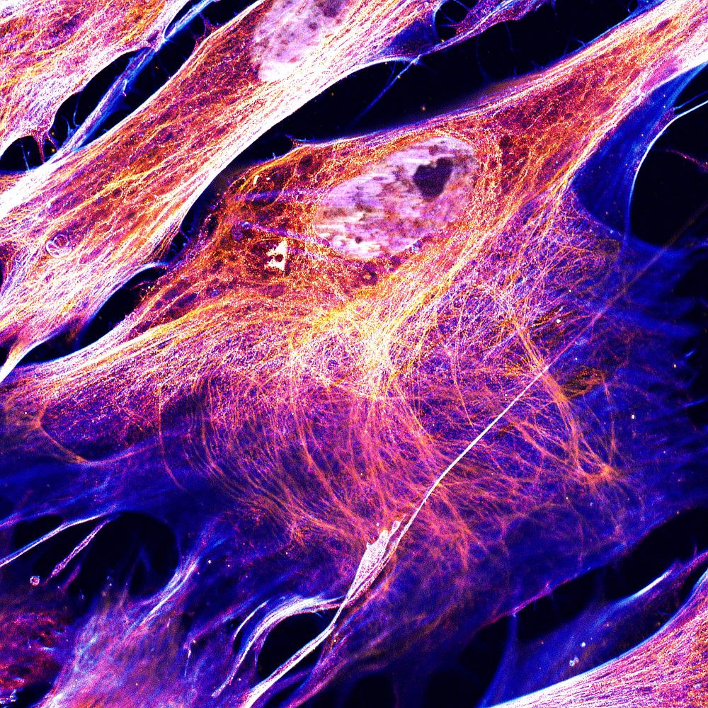
HFF cells stained for DNA ⚪, intermediate filaments 🟠, microtubules 🔴 and actin 🔵.
#FluorescenceFriday
#SciArt
#FluorescenceFriday
#SciArt
May 30, 2025 at 2:18 PM
HFF cells stained for DNA ⚪, intermediate filaments 🟠, microtubules 🔴 and actin 🔵.
#FluorescenceFriday
#SciArt
#FluorescenceFriday
#SciArt
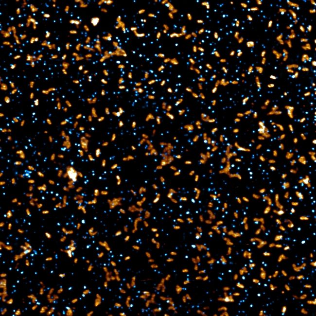
For #FluorescenceFriday, a field of individual VSV (rod-shaped, in 🟧) and Deltavirus (dot-shaped, in 🔵) particles imaged by STED (20nm per pixel).
We now know that Deltaviruses can package inside particles of other viruses.
Can you find any of these dual particles in this field?
We now know that Deltaviruses can package inside particles of other viruses.
Can you find any of these dual particles in this field?
May 23, 2025 at 12:05 PM
For #FluorescenceFriday, a field of individual VSV (rod-shaped, in 🟧) and Deltavirus (dot-shaped, in 🔵) particles imaged by STED (20nm per pixel).
We now know that Deltaviruses can package inside particles of other viruses.
Can you find any of these dual particles in this field?
We now know that Deltaviruses can package inside particles of other viruses.
Can you find any of these dual particles in this field?
Viruses in the forest of the lungs.
Human airway epithelia stained for actin 🟢 and SARS-CoV-2 RNA 🟣.
#FluorescenceFriday
#SciArt
Human airway epithelia stained for actin 🟢 and SARS-CoV-2 RNA 🟣.
#FluorescenceFriday
#SciArt

May 16, 2025 at 11:01 AM
Viruses in the forest of the lungs.
Human airway epithelia stained for actin 🟢 and SARS-CoV-2 RNA 🟣.
#FluorescenceFriday
#SciArt
Human airway epithelia stained for actin 🟢 and SARS-CoV-2 RNA 🟣.
#FluorescenceFriday
#SciArt
This opens up many questions, as to whether deltaviruses may be causative agents of disease in humans, associating with helper viruses of different origins.

May 10, 2025 at 8:12 AM
This opens up many questions, as to whether deltaviruses may be causative agents of disease in humans, associating with helper viruses of different origins.
To conclude, we evidence a novel mode of viral transmission – spread through a viral Trojan Horse.

May 10, 2025 at 8:12 AM
To conclude, we evidence a novel mode of viral transmission – spread through a viral Trojan Horse.
We show that snake deltavirus can use a reptarenavirus (UGV-1) as a viral Trojan Horse, being found inside of their particles, and that these Trojan Horse particles are over 100-fold more infectious than free deltavirus particles!

May 10, 2025 at 8:12 AM
We show that snake deltavirus can use a reptarenavirus (UGV-1) as a viral Trojan Horse, being found inside of their particles, and that these Trojan Horse particles are over 100-fold more infectious than free deltavirus particles!
Finally, the only animal deltavirus - helper virus association accepted to date is that of snake deltavirus and reptarenavirus, found in boa constrictors suffering from Boid Inclusion Body Disease.

May 10, 2025 at 8:12 AM
Finally, the only animal deltavirus - helper virus association accepted to date is that of snake deltavirus and reptarenavirus, found in boa constrictors suffering from Boid Inclusion Body Disease.
Furthermore, we show the tissue- and species-shifting capabilities that Trojan Horse particles allow, by showing infection of human neuronal cells with HSV-1 Trojan Horse particles!
Neuronal marker in 🟣 HSV-1 in 🟡, deltavirus in 🔵 and DNA in ⚪.
Neuronal marker in 🟣 HSV-1 in 🟡, deltavirus in 🔵 and DNA in ⚪.
May 10, 2025 at 8:12 AM
Furthermore, we show the tissue- and species-shifting capabilities that Trojan Horse particles allow, by showing infection of human neuronal cells with HSV-1 Trojan Horse particles!
Neuronal marker in 🟣 HSV-1 in 🟡, deltavirus in 🔵 and DNA in ⚪.
Neuronal marker in 🟣 HSV-1 in 🟡, deltavirus in 🔵 and DNA in ⚪.
Separating the Trojan Horse particles from the free deltavirus particles showed us that deltaviruses are actually unable to steal the glycoproteins of HSV-1 to decorate their own free particles and that the Trojan Horse particles are the only possible infectious unit when HSV-1 is a helper.

May 10, 2025 at 8:12 AM
Separating the Trojan Horse particles from the free deltavirus particles showed us that deltaviruses are actually unable to steal the glycoproteins of HSV-1 to decorate their own free particles and that the Trojan Horse particles are the only possible infectious unit when HSV-1 is a helper.
We next showed that this mode of viral transmission is not limited to Rhabdoviruses, as we could also see deltaviruses inside of the viral particles of Herpes Simplex Virus 1 (HSV-1).

May 10, 2025 at 8:12 AM
We next showed that this mode of viral transmission is not limited to Rhabdoviruses, as we could also see deltaviruses inside of the viral particles of Herpes Simplex Virus 1 (HSV-1).
We therefore called these hybrid particles viral Trojan Horse particles, as this mode of viral transmission strongly reminds of the classical Greek invasion of Troy, hiding inside of the Trojan Horse!

May 10, 2025 at 8:12 AM
We therefore called these hybrid particles viral Trojan Horse particles, as this mode of viral transmission strongly reminds of the classical Greek invasion of Troy, hiding inside of the Trojan Horse!
Cryo-EM revealed that the deltavirus ribonucleoprotein (vRNP) was in fact found inside of the VSV virions!
Found between the Matrix and viral envelope, it locally elevates the membrane, forming the characteristic“bumps” we were observing in other techniques.
Found between the Matrix and viral envelope, it locally elevates the membrane, forming the characteristic“bumps” we were observing in other techniques.

May 10, 2025 at 8:12 AM
Cryo-EM revealed that the deltavirus ribonucleoprotein (vRNP) was in fact found inside of the VSV virions!
Found between the Matrix and viral envelope, it locally elevates the membrane, forming the characteristic“bumps” we were observing in other techniques.
Found between the Matrix and viral envelope, it locally elevates the membrane, forming the characteristic“bumps” we were observing in other techniques.
Atomic Force Microscopy, within the BSL-3 environment @cemipai.bsky.social with AFM wizard Sébastien Lyonnais, allowed us to probe native infectious particle topology at the single virion level, revealing that these hybrid viral particles exist at the native state in viral supernatants.

May 10, 2025 at 8:12 AM
Atomic Force Microscopy, within the BSL-3 environment @cemipai.bsky.social with AFM wizard Sébastien Lyonnais, allowed us to probe native infectious particle topology at the single virion level, revealing that these hybrid viral particles exist at the native state in viral supernatants.
Are these small particles deltaviruses?
We confirmed that they are indeed deltavirus particles using orthogonal super-resolution imaging approaches, including STED, Airyscan and immunogold-EM.
We confirmed that they are indeed deltavirus particles using orthogonal super-resolution imaging approaches, including STED, Airyscan and immunogold-EM.

May 10, 2025 at 8:12 AM
Are these small particles deltaviruses?
We confirmed that they are indeed deltavirus particles using orthogonal super-resolution imaging approaches, including STED, Airyscan and immunogold-EM.
We confirmed that they are indeed deltavirus particles using orthogonal super-resolution imaging approaches, including STED, Airyscan and immunogold-EM.
First, using Vesicular Stomatitis Virus (VSV), we show that upon superinfection, deltaviruses are released from the host cell in their own viral particles (inset 2) but that, unexpectedly, they also seem to be associated with VSV particles themselves (inset 3)!

May 10, 2025 at 8:12 AM
First, using Vesicular Stomatitis Virus (VSV), we show that upon superinfection, deltaviruses are released from the host cell in their own viral particles (inset 2) but that, unexpectedly, they also seem to be associated with VSV particles themselves (inset 3)!
Our team and others have shown that deltaviruses can steal glycoproteins from different viral origins if over-expressed in a cell replicating a deltavirus, but what exactly happens during a real helper virus superinfection of deltavirus-replicating cells?

May 10, 2025 at 8:12 AM
Our team and others have shown that deltaviruses can steal glycoproteins from different viral origins if over-expressed in a cell replicating a deltavirus, but what exactly happens during a real helper virus superinfection of deltavirus-replicating cells?
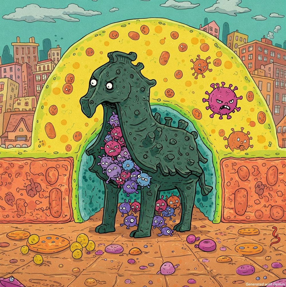
⚠️ Discovery of a new mode of viral transmission:
Deltaviruses spread through a viral Trojan Horse !
These viral satellites that do not encode their own glycoproteins, are in fact physically encapsulated within their helper virus particles!
www.biorxiv.org/content/10.1...
A thread 1/14
Deltaviruses spread through a viral Trojan Horse !
These viral satellites that do not encode their own glycoproteins, are in fact physically encapsulated within their helper virus particles!
www.biorxiv.org/content/10.1...
A thread 1/14
May 10, 2025 at 8:12 AM
⚠️ Discovery of a new mode of viral transmission:
Deltaviruses spread through a viral Trojan Horse !
These viral satellites that do not encode their own glycoproteins, are in fact physically encapsulated within their helper virus particles!
www.biorxiv.org/content/10.1...
A thread 1/14
Deltaviruses spread through a viral Trojan Horse !
These viral satellites that do not encode their own glycoproteins, are in fact physically encapsulated within their helper virus particles!
www.biorxiv.org/content/10.1...
A thread 1/14
The beauty and chaos of structure.
HFF cells stained for cytoskeletal proteins Actin (⚪), Septin (🟡), Tubulin (🟣) and DNA (🔵).
#FluorescenceFriday
#SciArt
HFF cells stained for cytoskeletal proteins Actin (⚪), Septin (🟡), Tubulin (🟣) and DNA (🔵).
#FluorescenceFriday
#SciArt

May 9, 2025 at 3:25 PM
The beauty and chaos of structure.
HFF cells stained for cytoskeletal proteins Actin (⚪), Septin (🟡), Tubulin (🟣) and DNA (🔵).
#FluorescenceFriday
#SciArt
HFF cells stained for cytoskeletal proteins Actin (⚪), Septin (🟡), Tubulin (🟣) and DNA (🔵).
#FluorescenceFriday
#SciArt
A culture of boa constrictor cells expressing a fusogenic viral protein, leading to cell fusion and giant multinucleated cells!
Actin is in gold, membranes in white and DNA in blue.
#FluorescenceFriday #SciArt
Actin is in gold, membranes in white and DNA in blue.
#FluorescenceFriday #SciArt

April 11, 2025 at 11:23 AM
A culture of boa constrictor cells expressing a fusogenic viral protein, leading to cell fusion and giant multinucleated cells!
Actin is in gold, membranes in white and DNA in blue.
#FluorescenceFriday #SciArt
Actin is in gold, membranes in white and DNA in blue.
#FluorescenceFriday #SciArt
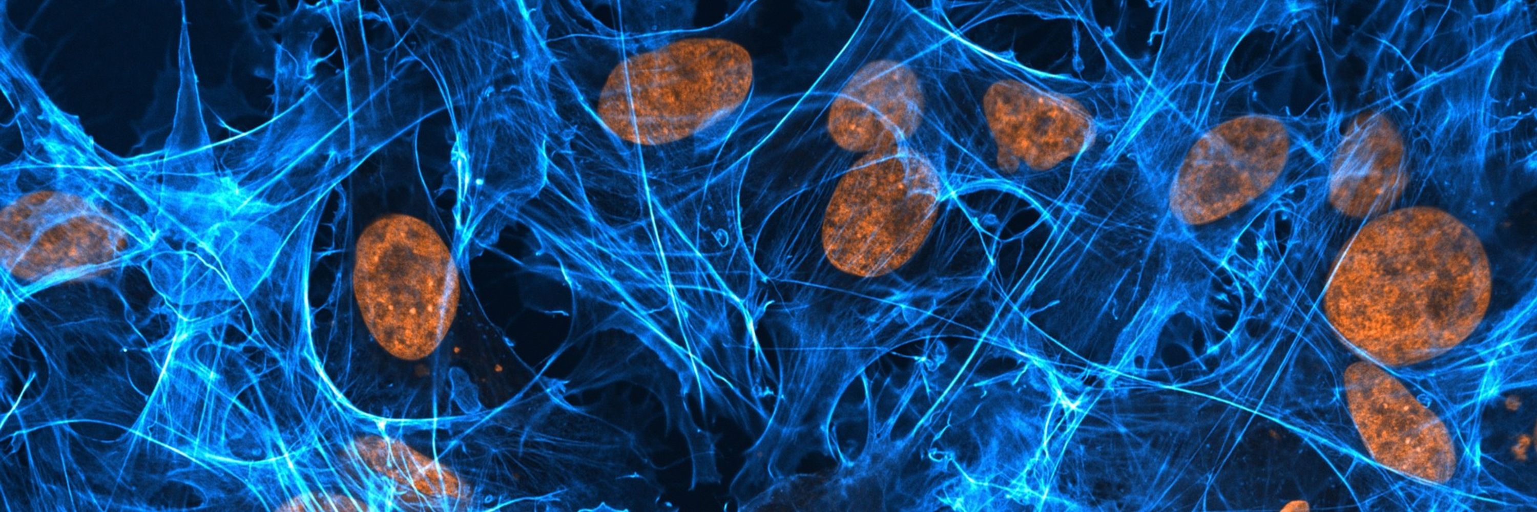
A human lung cell expressing the antiviral protein MX1.
Actin is in cyan, MX1 in orange and DNA in yellow.
#FluorescenceFriday #SciArt
Actin is in cyan, MX1 in orange and DNA in yellow.
#FluorescenceFriday #SciArt

April 4, 2025 at 5:27 PM
A human lung cell expressing the antiviral protein MX1.
Actin is in cyan, MX1 in orange and DNA in yellow.
#FluorescenceFriday #SciArt
Actin is in cyan, MX1 in orange and DNA in yellow.
#FluorescenceFriday #SciArt
Micro-rearrangement of DNA and proteins is a highly dynamic process!
As shown here with cell nucleus (DNA in gray) expressing a fluorescent protein (orange).
1 image acquired every 2 seconds for 2 minutes.
#FluorescenceFriday
#SciArt
As shown here with cell nucleus (DNA in gray) expressing a fluorescent protein (orange).
1 image acquired every 2 seconds for 2 minutes.
#FluorescenceFriday
#SciArt
March 28, 2025 at 2:59 PM
Micro-rearrangement of DNA and proteins is a highly dynamic process!
As shown here with cell nucleus (DNA in gray) expressing a fluorescent protein (orange).
1 image acquired every 2 seconds for 2 minutes.
#FluorescenceFriday
#SciArt
As shown here with cell nucleus (DNA in gray) expressing a fluorescent protein (orange).
1 image acquired every 2 seconds for 2 minutes.
#FluorescenceFriday
#SciArt
New cell line = New cell morphologies to explore!
This hepatic cell line has fully invested into membrane protrusions!
Actin is shown in 🟦 and DNA in 🟧
#FluorescenceFriday
#SciArt
This hepatic cell line has fully invested into membrane protrusions!
Actin is shown in 🟦 and DNA in 🟧
#FluorescenceFriday
#SciArt

March 21, 2025 at 1:05 PM
New cell line = New cell morphologies to explore!
This hepatic cell line has fully invested into membrane protrusions!
Actin is shown in 🟦 and DNA in 🟧
#FluorescenceFriday
#SciArt
This hepatic cell line has fully invested into membrane protrusions!
Actin is shown in 🟦 and DNA in 🟧
#FluorescenceFriday
#SciArt
A Spiky Cell
A boa constrictor brain-derived cell stained for membranes (cyan), actin (yellow) and DNA (pink)
#FluorescenceFriday
#SciArt
A boa constrictor brain-derived cell stained for membranes (cyan), actin (yellow) and DNA (pink)
#FluorescenceFriday
#SciArt

March 7, 2025 at 2:09 PM
A Spiky Cell
A boa constrictor brain-derived cell stained for membranes (cyan), actin (yellow) and DNA (pink)
#FluorescenceFriday
#SciArt
A boa constrictor brain-derived cell stained for membranes (cyan), actin (yellow) and DNA (pink)
#FluorescenceFriday
#SciArt
Proteins can do weird things sometimes...!
Anyone else heard of cytoophidia? A cool and understudied cell biology process!
In red is a mutant of an antiviral protein, in cyan is an influenza A virus protein and in yellow are cell nuclei.
#FluorescenceFriday
#SciArt
Anyone else heard of cytoophidia? A cool and understudied cell biology process!
In red is a mutant of an antiviral protein, in cyan is an influenza A virus protein and in yellow are cell nuclei.
#FluorescenceFriday
#SciArt

February 28, 2025 at 5:36 PM
Proteins can do weird things sometimes...!
Anyone else heard of cytoophidia? A cool and understudied cell biology process!
In red is a mutant of an antiviral protein, in cyan is an influenza A virus protein and in yellow are cell nuclei.
#FluorescenceFriday
#SciArt
Anyone else heard of cytoophidia? A cool and understudied cell biology process!
In red is a mutant of an antiviral protein, in cyan is an influenza A virus protein and in yellow are cell nuclei.
#FluorescenceFriday
#SciArt