Sumanta Das
@sumantadas7.bsky.social
MD PATHOLOGY | PGY 3+4! SPECIAL INTEREST IN BONE AND SOFT TISSUE PATHOLOGY, NEUROPATHOLOGY.
Reposted by Sumanta Das

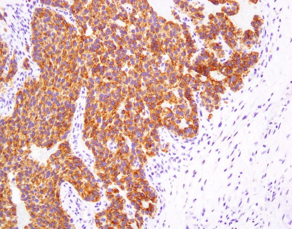
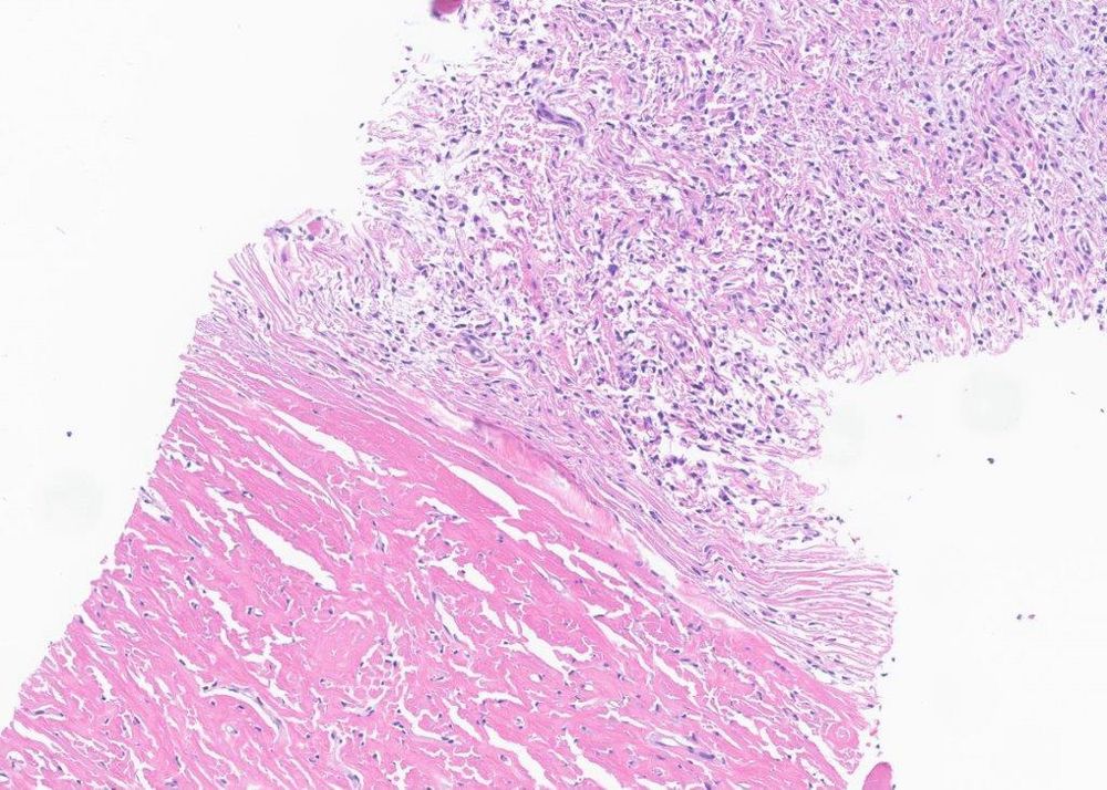
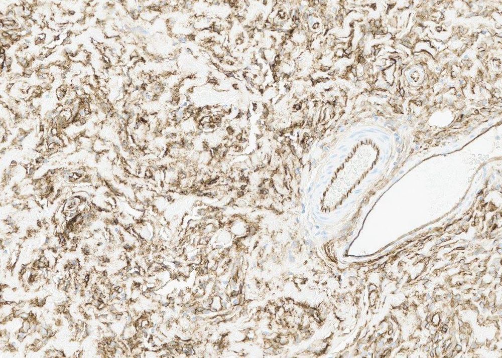
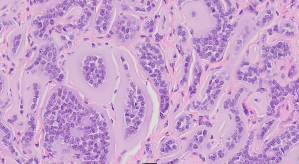
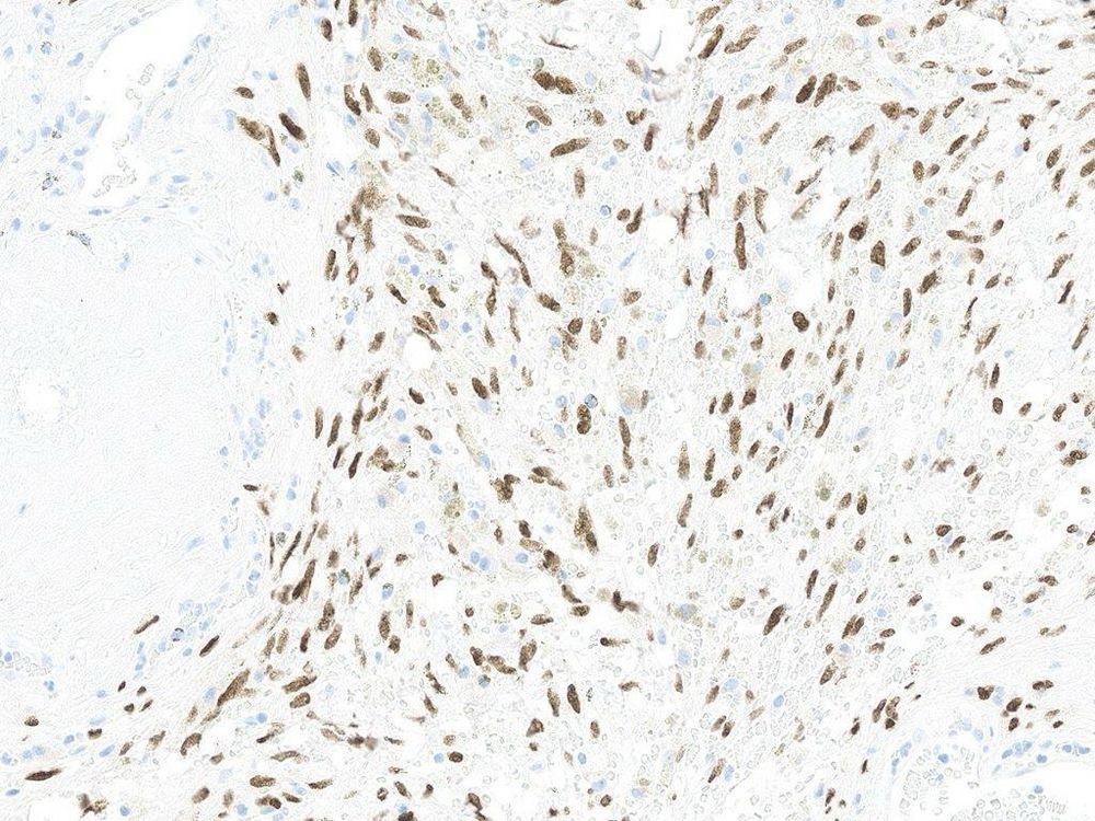
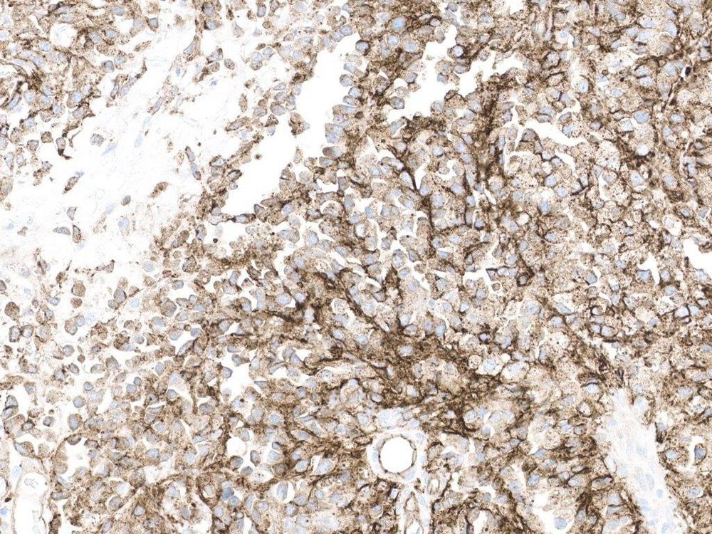
#PathSky 64M, upper chest wall skin lesion. There was some discussion of whether this strikingly “packeted”, strongly S100 protein-positive lesion might be a traumatic neuroma.


March 20, 2025 at 3:26 PM
#PathSky 64M, upper chest wall skin lesion. There was some discussion of whether this strikingly “packeted”, strongly S100 protein-positive lesion might be a traumatic neuroma.
Reposted by Sumanta Das
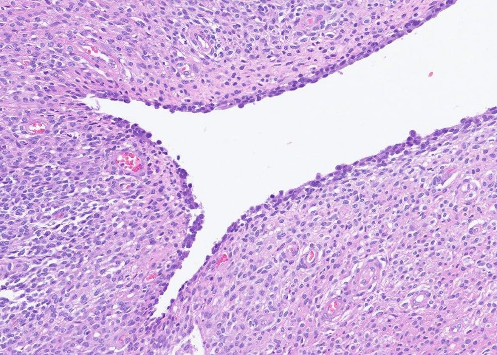
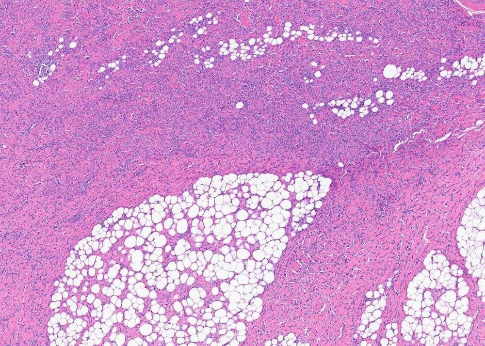
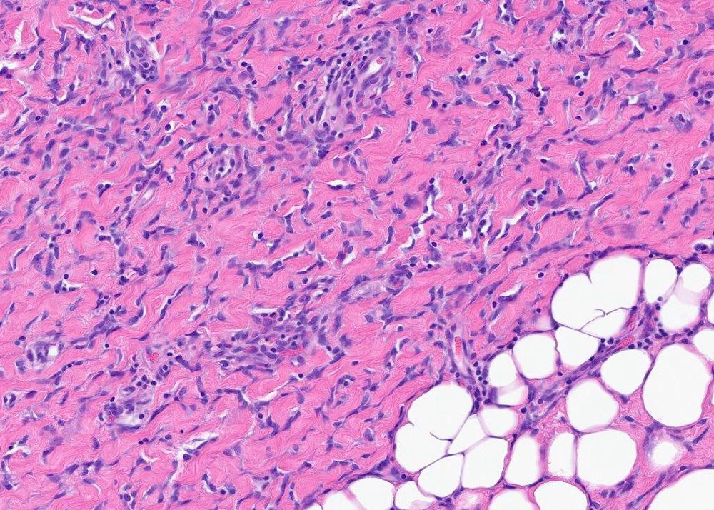
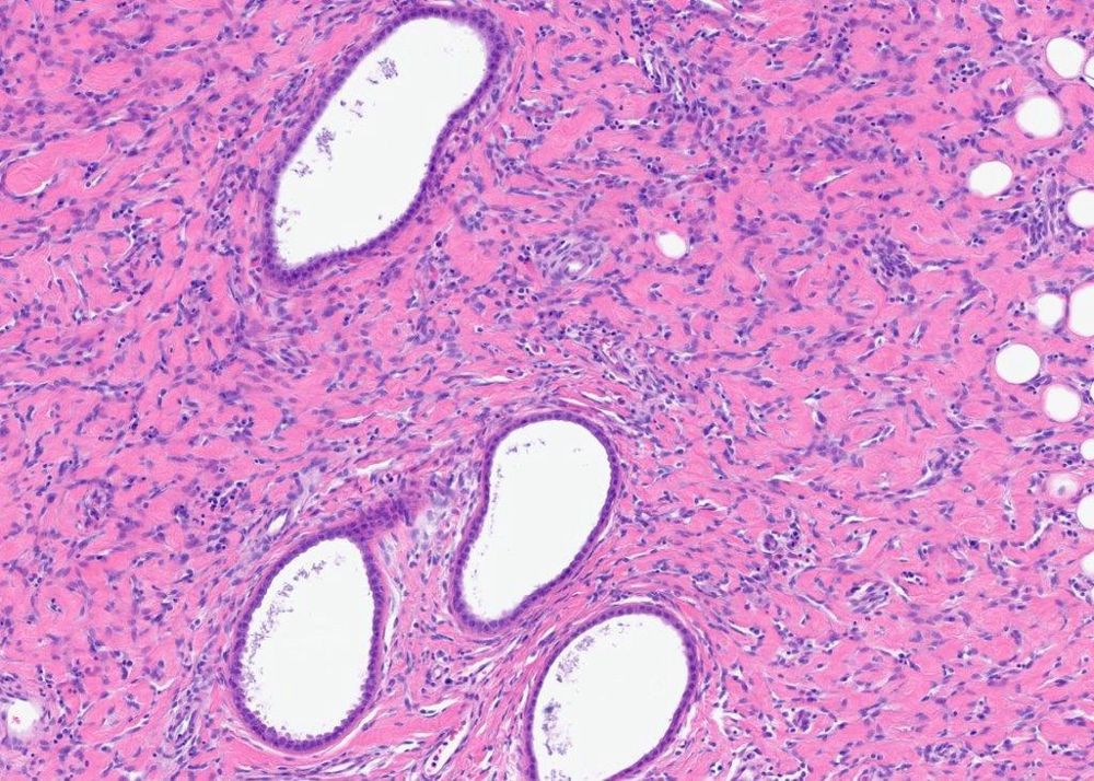
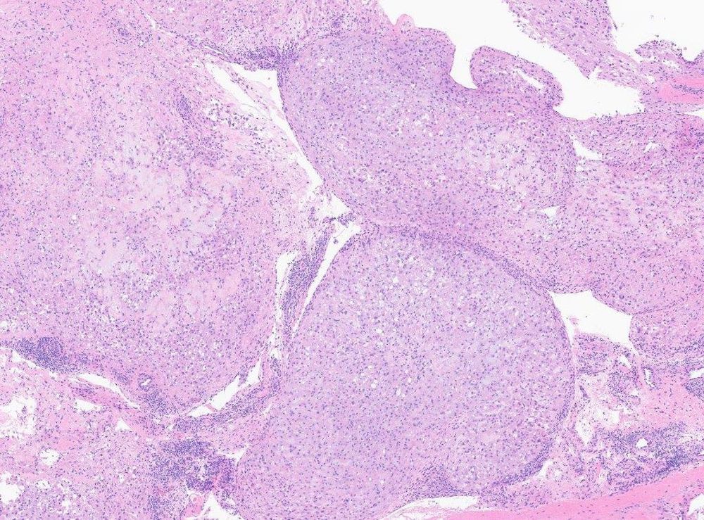
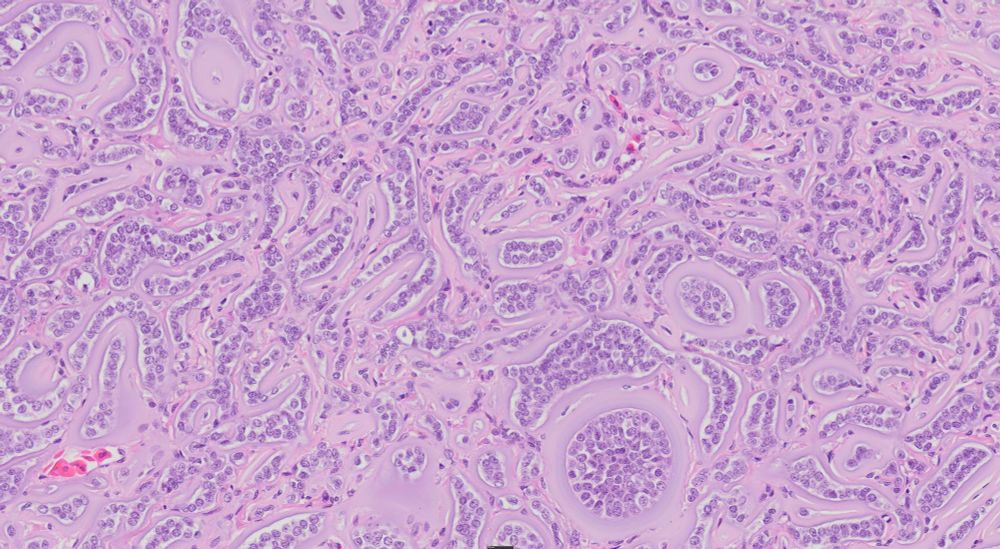
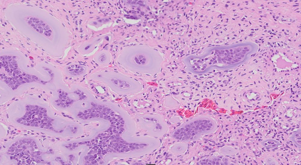
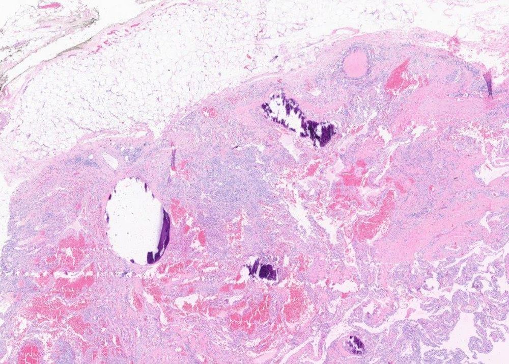
#PathSky Abdominal wall mall mass, 65M. Multinodular with thick fibrous septa. Bland spindled to ovoid cells surrounding prominent vessels. Microcystic, pseudoglandular spaces.
April 2, 2025 at 7:42 PM
#PathSky Abdominal wall mall mass, 65M. Multinodular with thick fibrous septa. Bland spindled to ovoid cells surrounding prominent vessels. Microcystic, pseudoglandular spaces.
Reposted by Sumanta Das
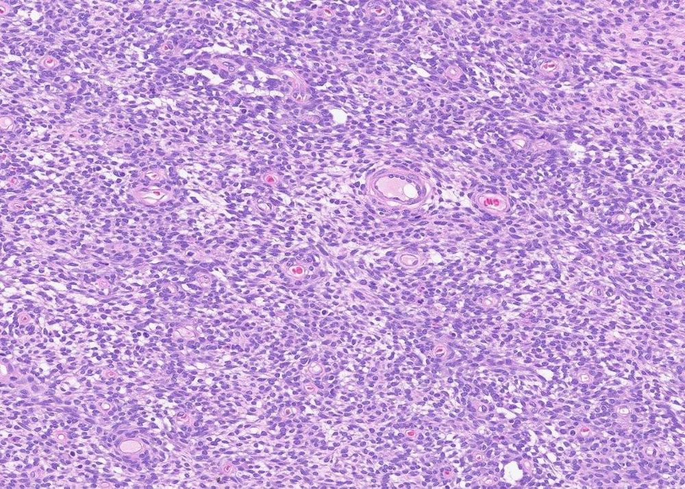
#PathSky Middle aged woman, leg mass. It all looks like this. More nuclear palisading/ Verocay bodies than you’ve seen in your entire career. Nerve sheath? S100/Sox10 negative. LMS? Negative for all the muscle markers. Any other ideas?


May 20, 2025 at 9:30 PM
#PathSky Middle aged woman, leg mass. It all looks like this. More nuclear palisading/ Verocay bodies than you’ve seen in your entire career. Nerve sheath? S100/Sox10 negative. LMS? Negative for all the muscle markers. Any other ideas?
Reposted by Sumanta Das

Reposted by Sumanta Das
#PathSky 30F with shoulder mass. Nice example of low-grade fibromyxoid sarcoma, with heavily collagenized areas and a very abrupt transition to more cellular nodules with a whorling pattern and a well-developed vasculature. MUC4 IHC is confirmatory. This case has another helpful H+E feature (below)


February 20, 2025 at 9:44 PM
#PathSky 30F with shoulder mass. Nice example of low-grade fibromyxoid sarcoma, with heavily collagenized areas and a very abrupt transition to more cellular nodules with a whorling pattern and a well-developed vasculature. MUC4 IHC is confirmatory. This case has another helpful H+E feature (below)
Reposted by Sumanta Das
#PathSky I usually like to show cases where the morphology makes sense of the molecular, but here’s one where you kind of need the molecular. Axillary mass, 9 month male. Very hyalinized, infiltration of fat, cracking artifact. Strongly CD34-pos. Giant cell fibroblastoma?
February 24, 2025 at 8:32 PM
#PathSky I usually like to show cases where the morphology makes sense of the molecular, but here’s one where you kind of need the molecular. Axillary mass, 9 month male. Very hyalinized, infiltration of fat, cracking artifact. Strongly CD34-pos. Giant cell fibroblastoma?
Reposted by Sumanta Das

Looking for opinions on this (alleged) ovarian mass from a garter snake. I presume the amphophilic material is redundant basement membrane. Could this be a granulosa cell tumor? We have seen those in snakes (incl garter snakes) but this one is odd.
#pathsky #vetpath #veterinary #tumor #repro
#pathsky #vetpath #veterinary #tumor #repro
February 27, 2025 at 11:59 PM
Looking for opinions on this (alleged) ovarian mass from a garter snake. I presume the amphophilic material is redundant basement membrane. Could this be a granulosa cell tumor? We have seen those in snakes (incl garter snakes) but this one is odd.
#pathsky #vetpath #veterinary #tumor #repro
#pathsky #vetpath #veterinary #tumor #repro
Reposted by Sumanta Das
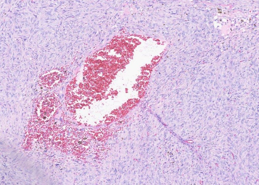
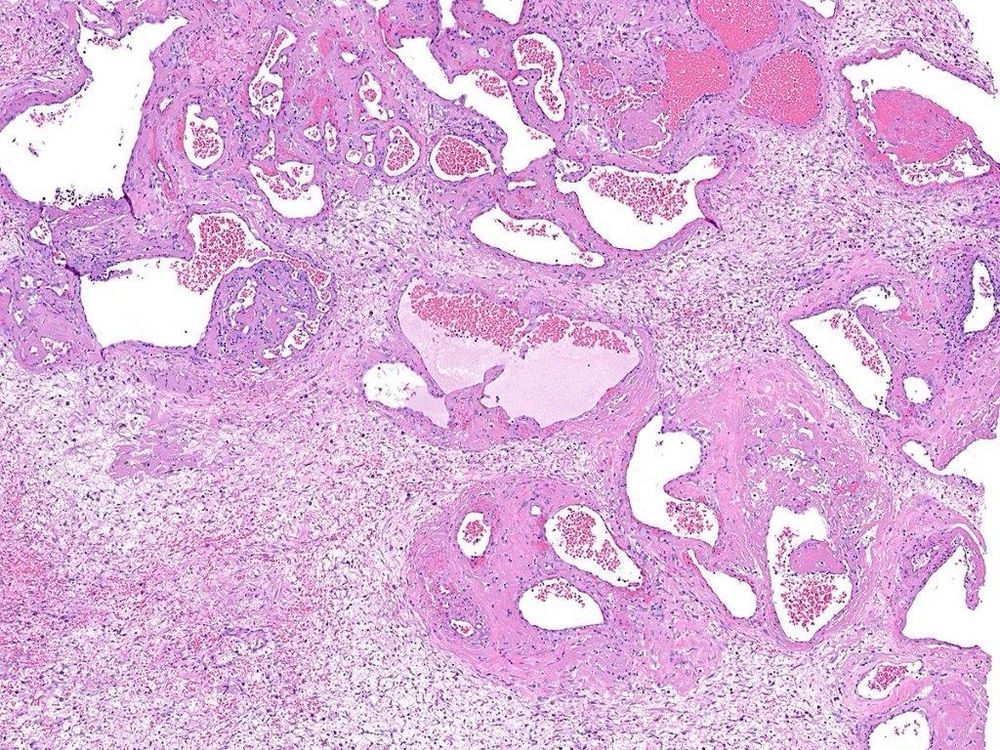
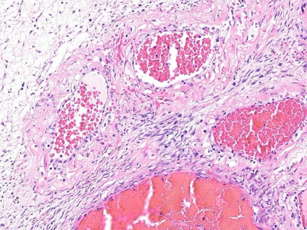
#PathSky Slowly growing superficial leg mass, 38M. Well-circumscribed, phleboliths, thrombosed vessels, cellular spindle cell areas, cavernous vascular channels. Dx?
February 5, 2025 at 6:57 PM
#PathSky Slowly growing superficial leg mass, 38M. Well-circumscribed, phleboliths, thrombosed vessels, cellular spindle cell areas, cavernous vascular channels. Dx?
Reposted by Sumanta Das
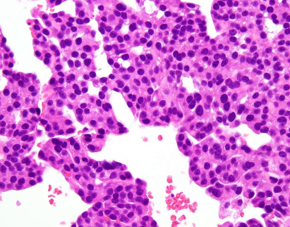
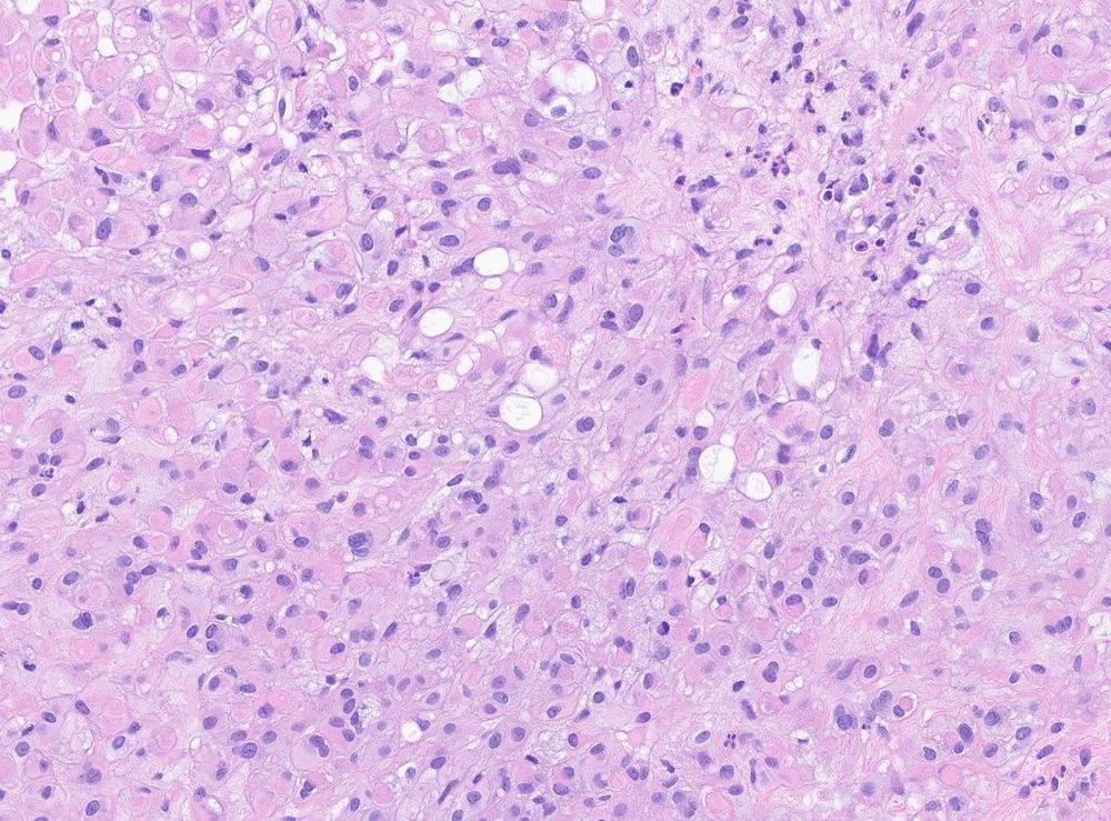
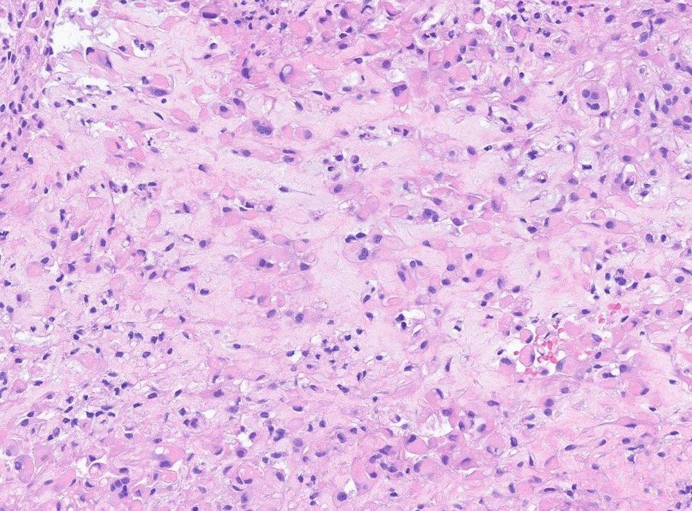
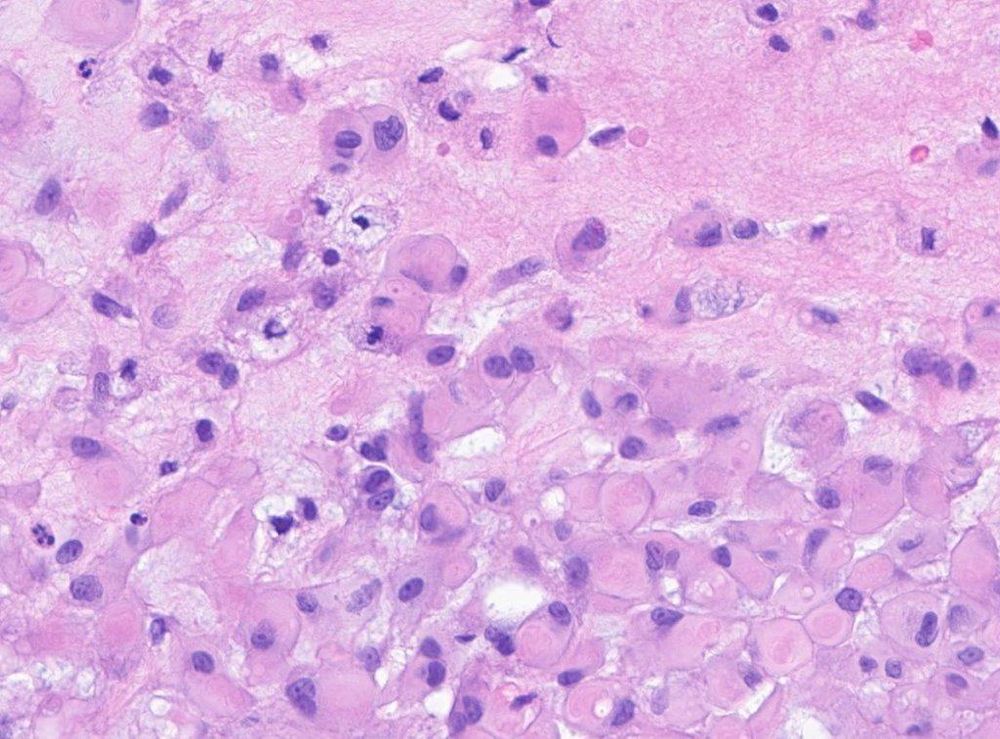
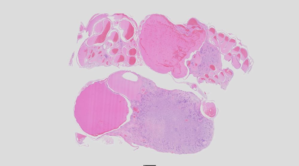
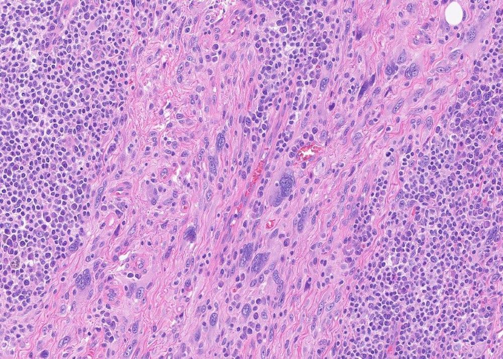
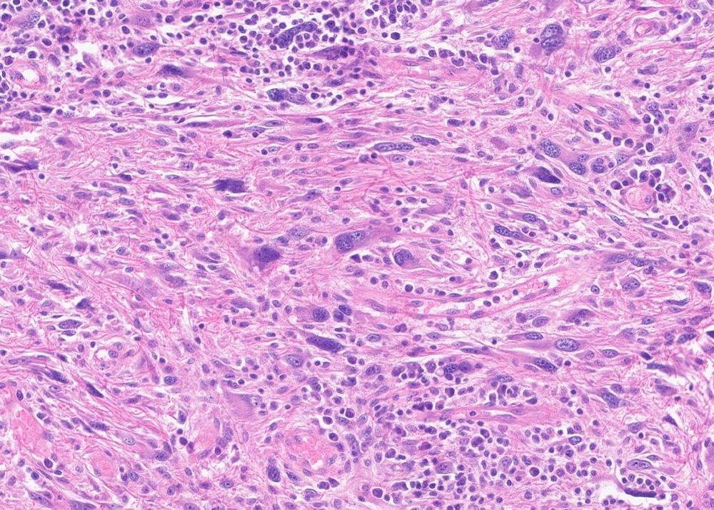
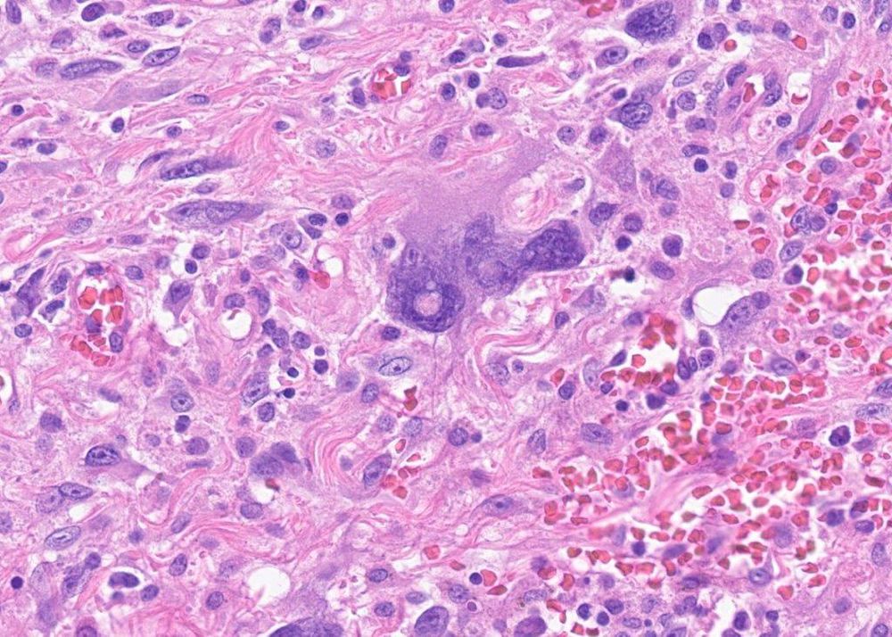
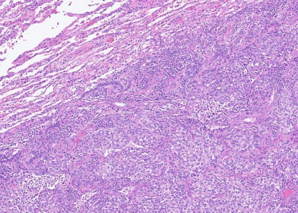
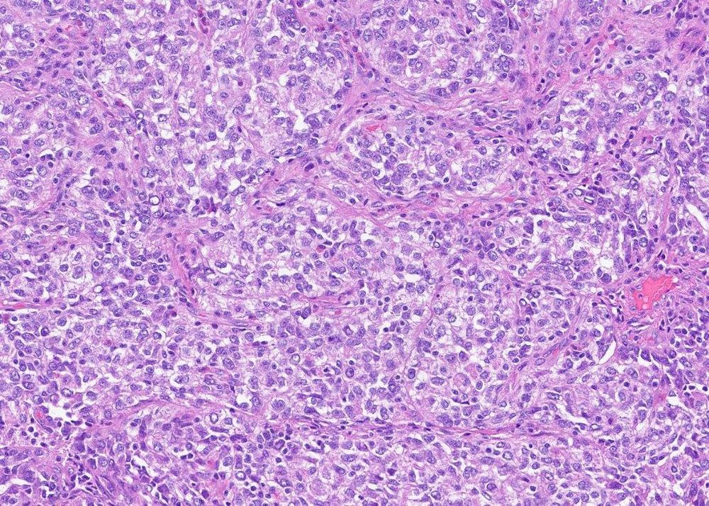
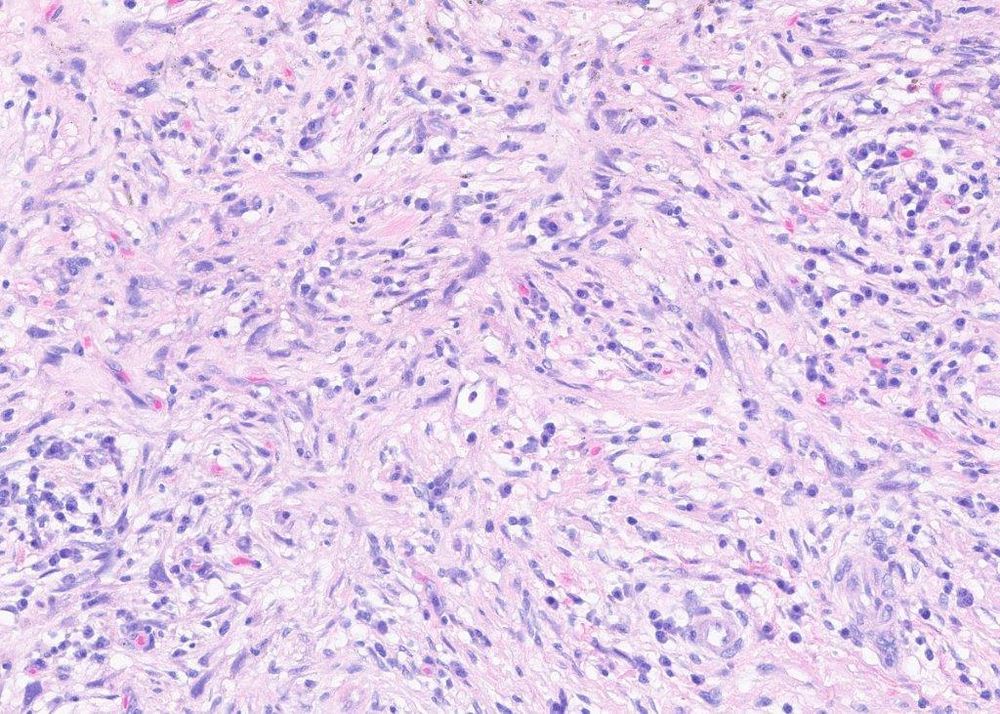
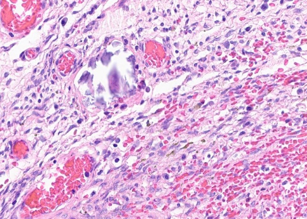
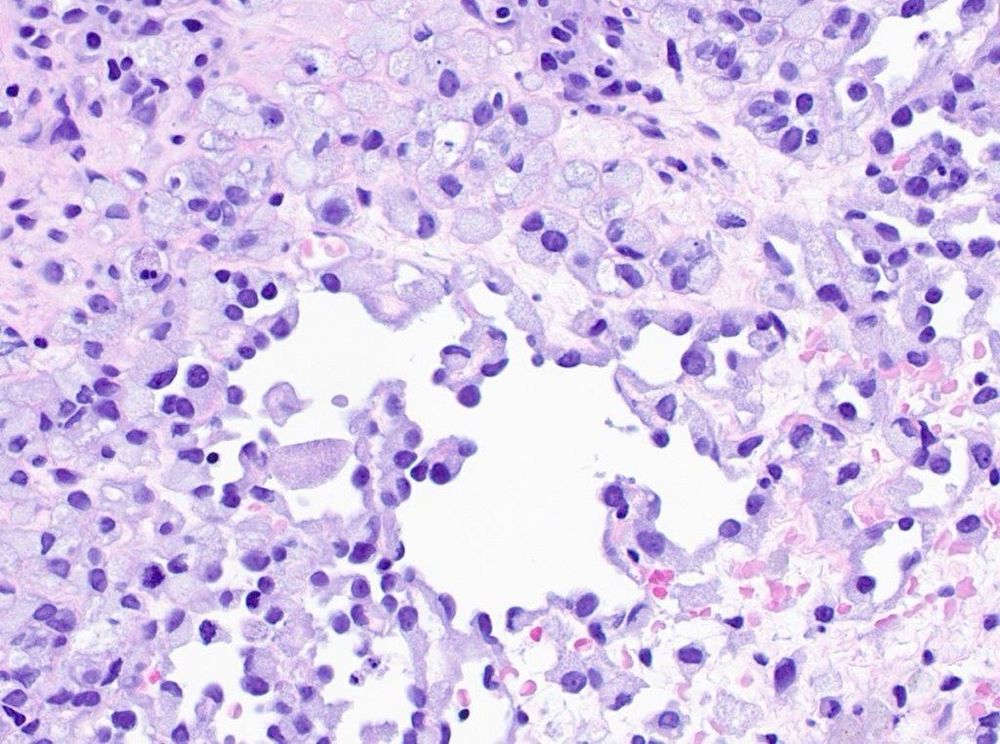
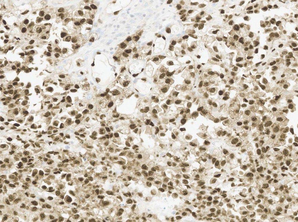
#PathSky 79M with a superficial neck mass. Wildly pleomorphic, quite cellular- undifferentiated pleomorphic sarcoma, right? There are basically no mitoses though, the cells have abundant granular-glassy cytoplasm, there is a lot of inflammation, and many bizarre cells have nuclear inclusions.
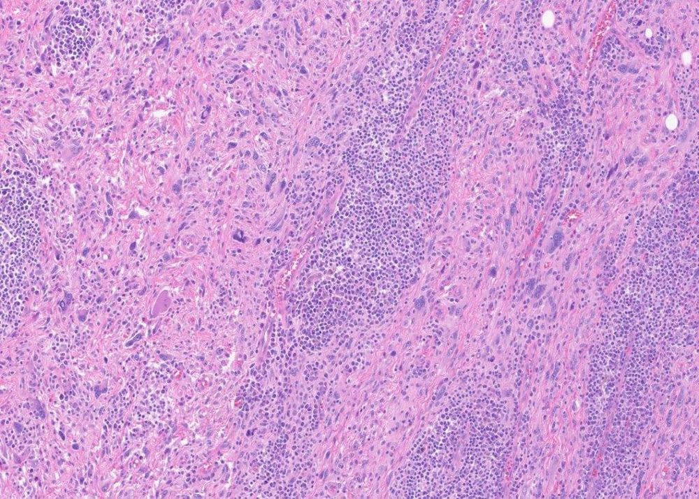
February 5, 2025 at 7:08 PM
#PathSky 79M with a superficial neck mass. Wildly pleomorphic, quite cellular- undifferentiated pleomorphic sarcoma, right? There are basically no mitoses though, the cells have abundant granular-glassy cytoplasm, there is a lot of inflammation, and many bizarre cells have nuclear inclusions.
Reposted by Sumanta Das
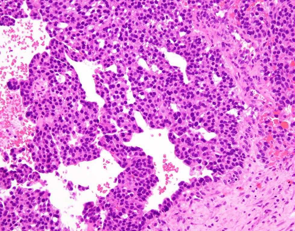
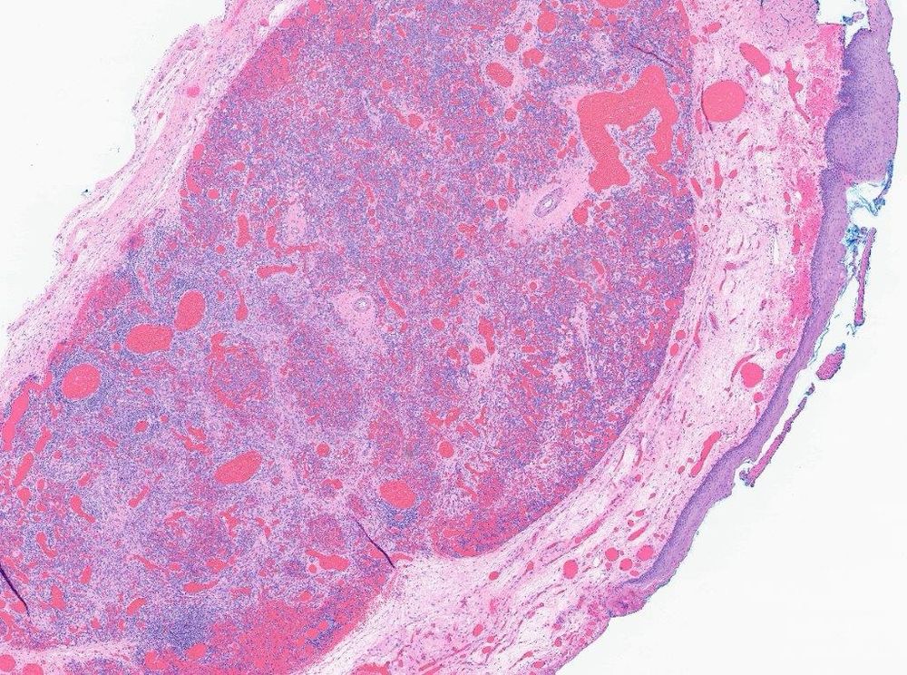
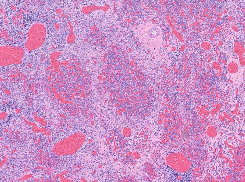
#PathSky 43F with a solitary lung mass. Relatively circumscribed, nests and interesting micropapillae of lightly eosinophilic epithelioid cells. Negative for everything except TFE3. Dx?
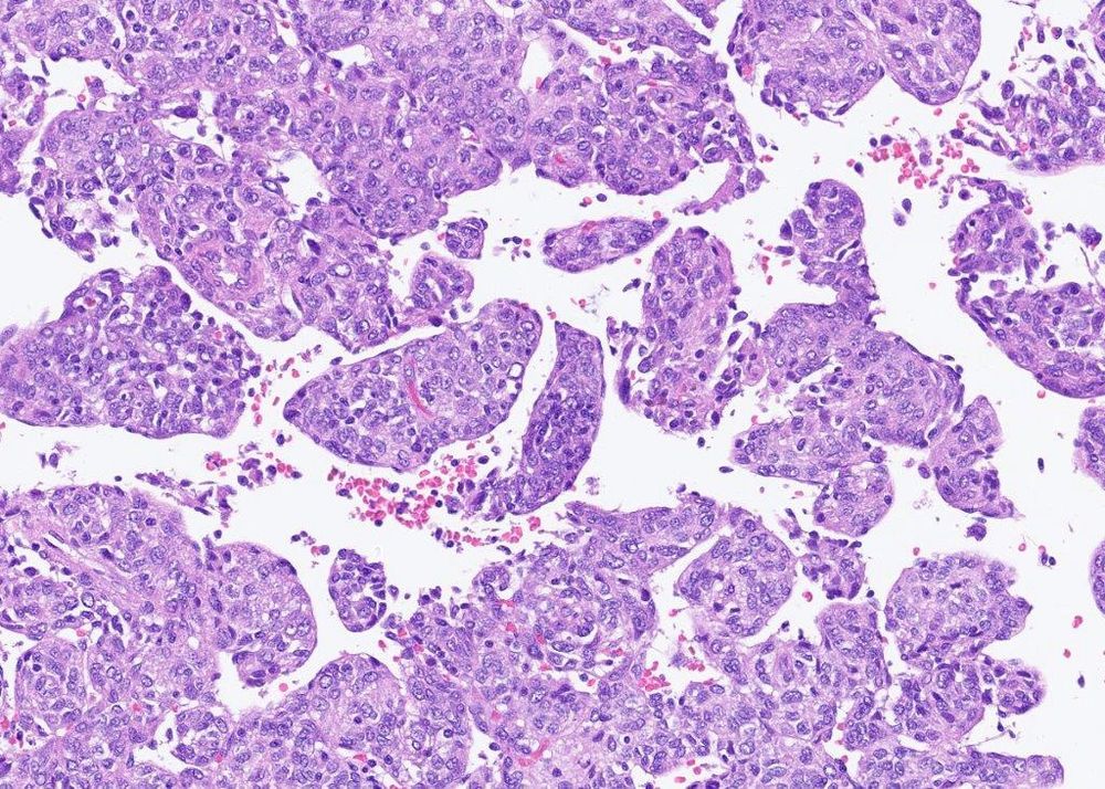
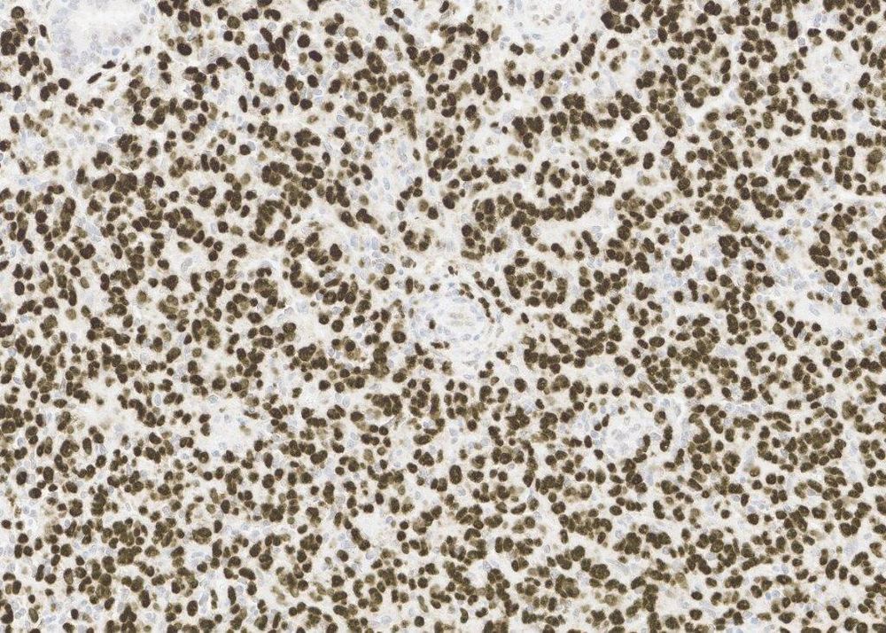
February 6, 2025 at 9:41 PM
#PathSky 43F with a solitary lung mass. Relatively circumscribed, nests and interesting micropapillae of lightly eosinophilic epithelioid cells. Negative for everything except TFE3. Dx?
Reposted by Sumanta Das
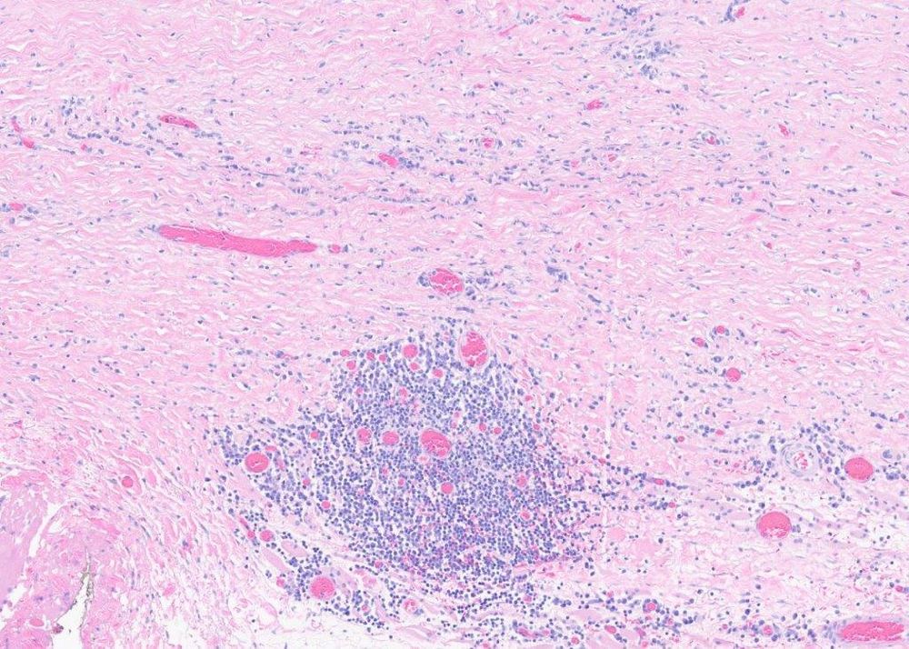
#PathSky 37M with a large retroperitoneal mass. Big, fatty thing. The outside quite rightly worried about well-differentiated LPS and a lipomatous angiomyolipoma, but MDM2, SMA and melanocytic markers were negative. Some WDL are MDM2-neg/MDM4-pos. Should we do more molecular ? Or something else?


January 13, 2025 at 3:02 PM
#PathSky 37M with a large retroperitoneal mass. Big, fatty thing. The outside quite rightly worried about well-differentiated LPS and a lipomatous angiomyolipoma, but MDM2, SMA and melanocytic markers were negative. Some WDL are MDM2-neg/MDM4-pos. Should we do more molecular ? Or something else?

Reposted by Sumanta Das
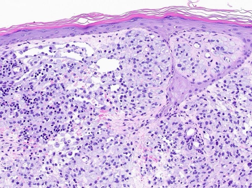
#PathSky Infant with leg mass. Looks like a circumscribed deep mass, but there is subtle involvement of the superficial subcutaneous fat. Small, infiltrating fascicles of rather bland spindled cells. The nodule itself is more cellular, fascicular to storiform, but still bland.


January 23, 2025 at 5:53 PM
#PathSky Infant with leg mass. Looks like a circumscribed deep mass, but there is subtle involvement of the superficial subcutaneous fat. Small, infiltrating fascicles of rather bland spindled cells. The nodule itself is more cellular, fascicular to storiform, but still bland.
Reposted by Sumanta Das
#PathSky Older adult with a large, prolapsing esophageal mass. An excellent example of a “giant fibrovascular polyp” of the esophagus. Except that essentially 100% of these turn out to be well-differentiated ( or less often dedifferentiated) liposarcomas.
pubmed.ncbi.nlm.nih.gov/28984298/
pubmed.ncbi.nlm.nih.gov/28984298/


January 24, 2025 at 8:37 PM
#PathSky Older adult with a large, prolapsing esophageal mass. An excellent example of a “giant fibrovascular polyp” of the esophagus. Except that essentially 100% of these turn out to be well-differentiated ( or less often dedifferentiated) liposarcomas.
pubmed.ncbi.nlm.nih.gov/28984298/
pubmed.ncbi.nlm.nih.gov/28984298/
Reposted by Sumanta Das
#PathSky. Since I’m old enough to remember K-type questions, here is one (answer in comment): Which of these events is this retroperitoneal mass likely to have? A) MDM2 rearrangement, B)MDM2 amplification, C)DDIT3 rearrangement, D) DDIT3 amp?
A only, B only, C only, A and C, B and D, all, none.
A only, B only, C only, A and C, B and D, all, none.


November 26, 2024 at 5:37 PM
#PathSky. Since I’m old enough to remember K-type questions, here is one (answer in comment): Which of these events is this retroperitoneal mass likely to have? A) MDM2 rearrangement, B)MDM2 amplification, C)DDIT3 rearrangement, D) DDIT3 amp?
A only, B only, C only, A and C, B and D, all, none.
A only, B only, C only, A and C, B and D, all, none.
Reposted by Sumanta Das
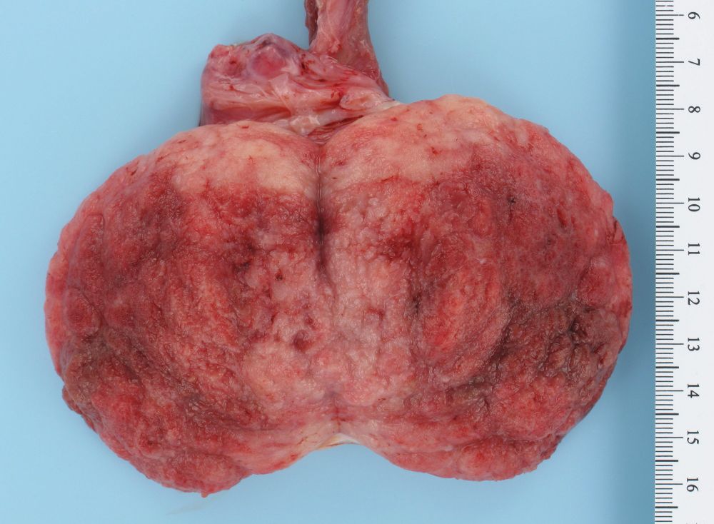
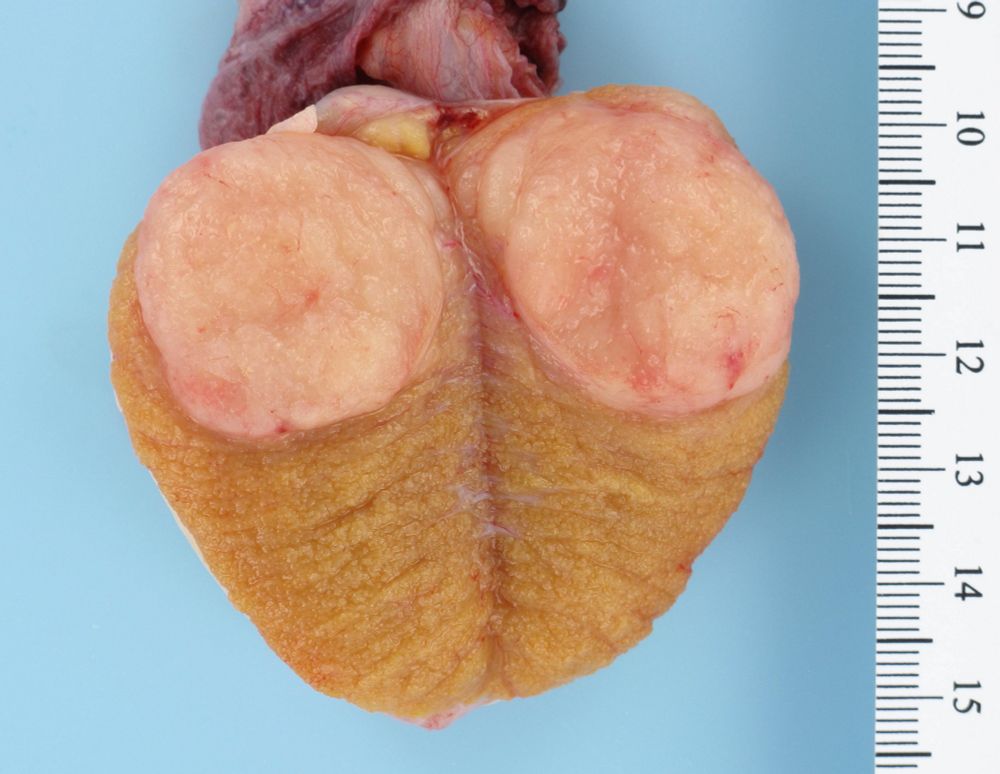
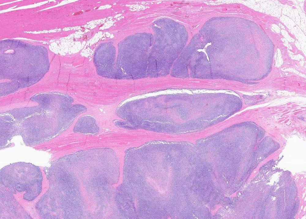
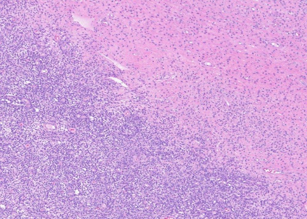
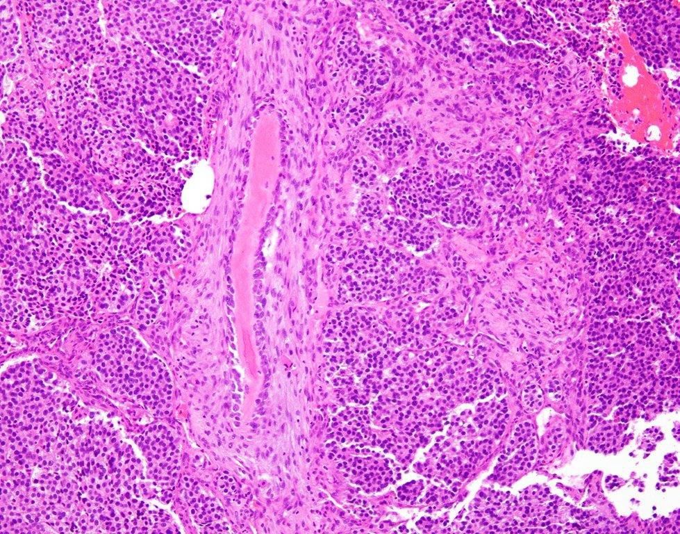
#PathSky. Mass next to knee, 27F. A hypocellular tumor with striking vascular changes, like we see in pleomorphic hyalinizing angiectatic tumor or schwannoma. But also some small fascicles of monomorphic spindled cells and a calcification. SS18::SSX fusion antibody +. Sneaky synovial sarcoma.
November 25, 2024 at 9:16 PM
#PathSky. Mass next to knee, 27F. A hypocellular tumor with striking vascular changes, like we see in pleomorphic hyalinizing angiectatic tumor or schwannoma. But also some small fascicles of monomorphic spindled cells and a calcification. SS18::SSX fusion antibody +. Sneaky synovial sarcoma.
Reposted by Sumanta Das

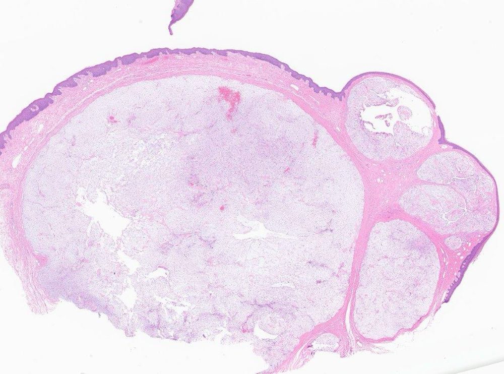
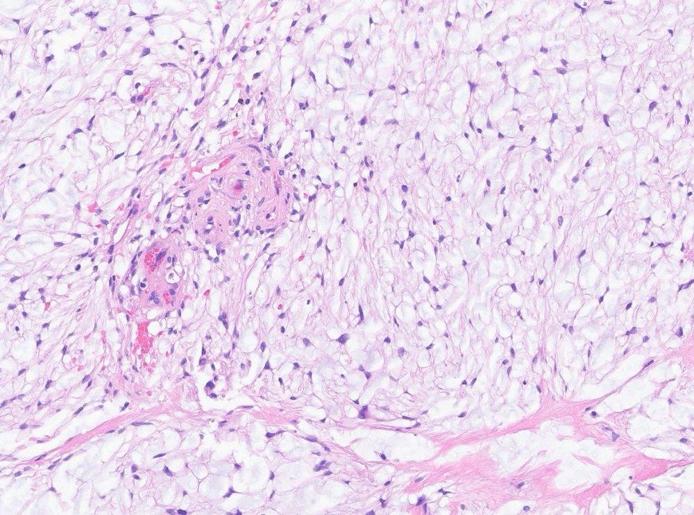
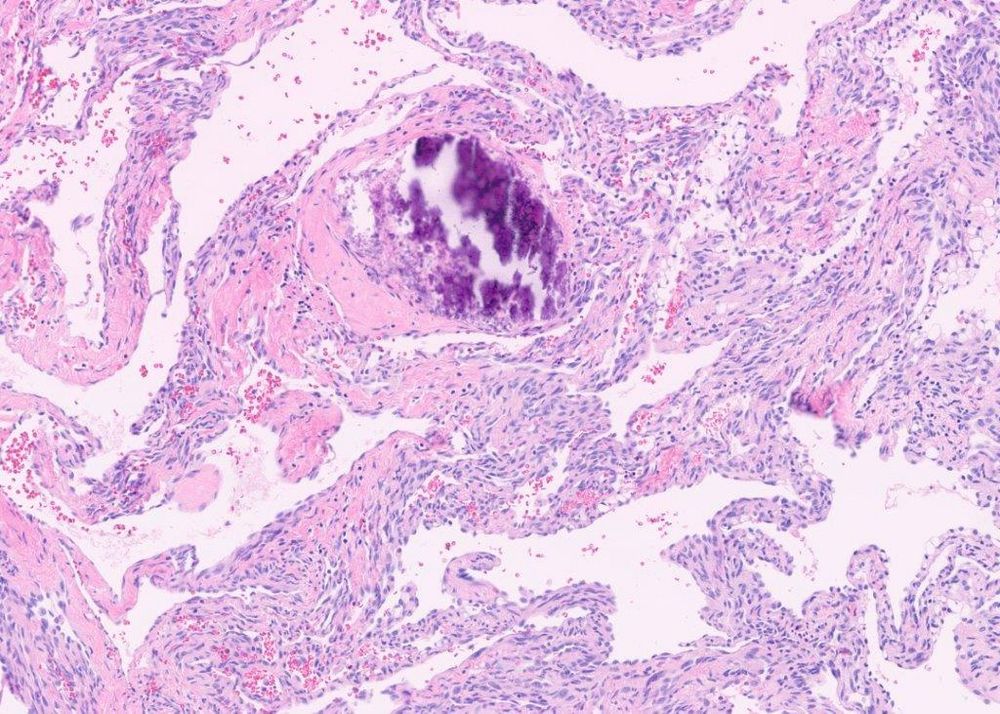
New series. Infectious disease pathology
1. Pneumocystis pneumonia
If you can spare a moment, please add a teaching point about this infection in the comments
#pathology #pulmpath #pathsky #pathbugs #crittersonbluesky
1. Pneumocystis pneumonia
If you can spare a moment, please add a teaching point about this infection in the comments
#pathology #pulmpath #pathsky #pathbugs #crittersonbluesky

November 24, 2024 at 10:39 PM
New series. Infectious disease pathology
1. Pneumocystis pneumonia
If you can spare a moment, please add a teaching point about this infection in the comments
#pathology #pulmpath #pathsky #pathbugs #crittersonbluesky
1. Pneumocystis pneumonia
If you can spare a moment, please add a teaching point about this infection in the comments
#pathology #pulmpath #pathsky #pathbugs #crittersonbluesky
Reposted by Sumanta Das
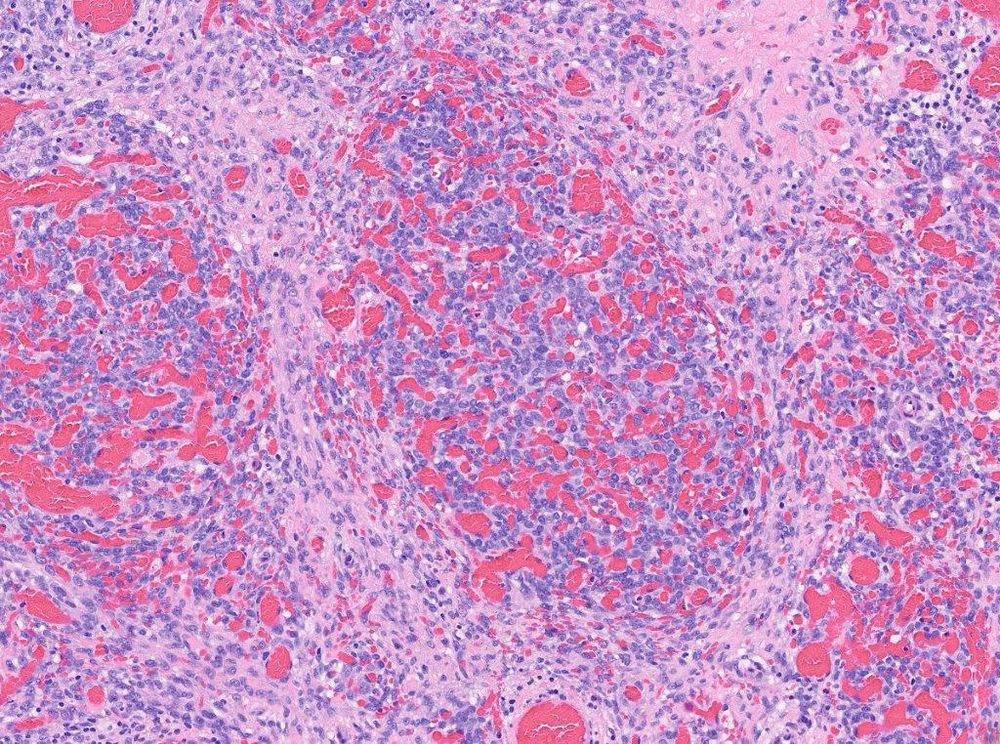
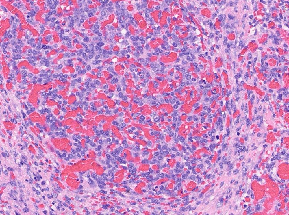
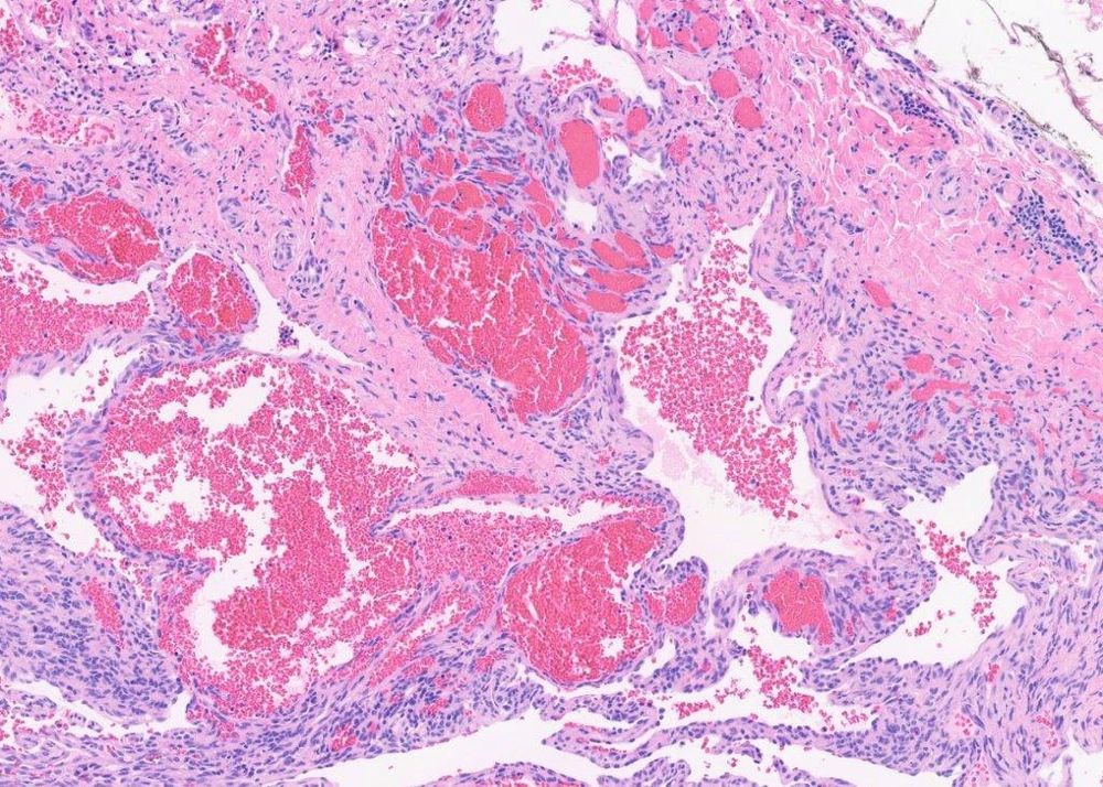
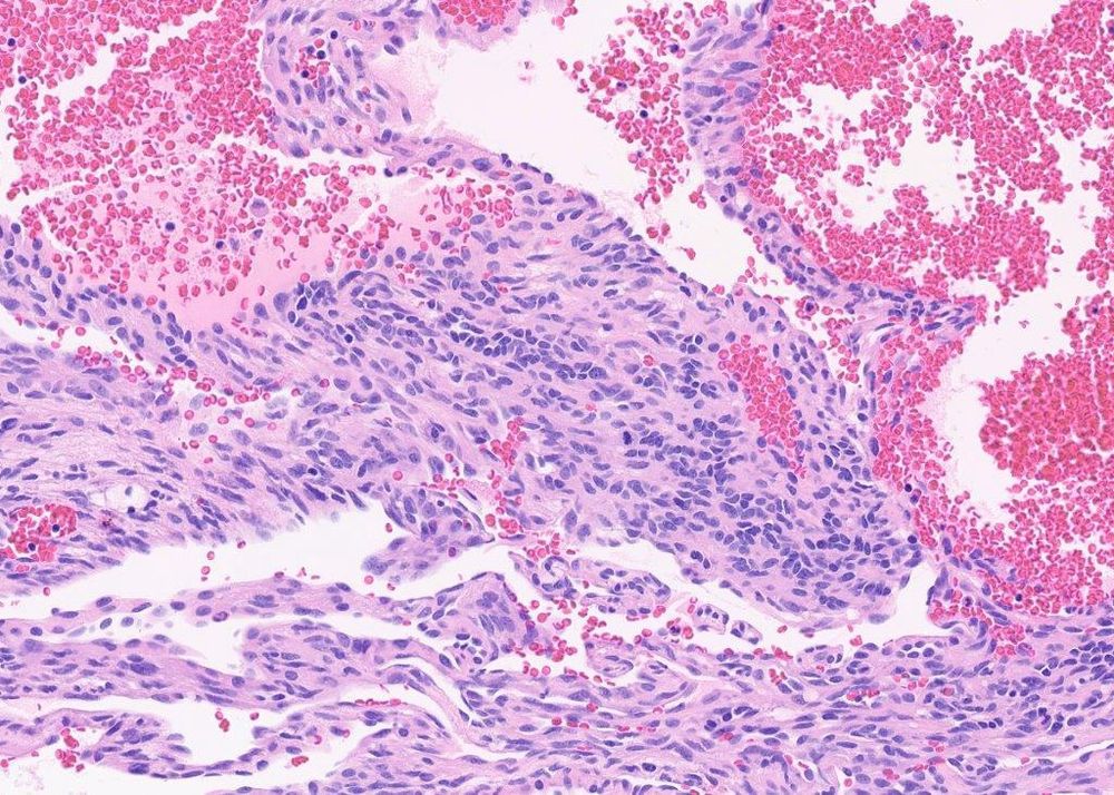
#PathSky. As long as I’m sitting here in Sunday doing a frozen….
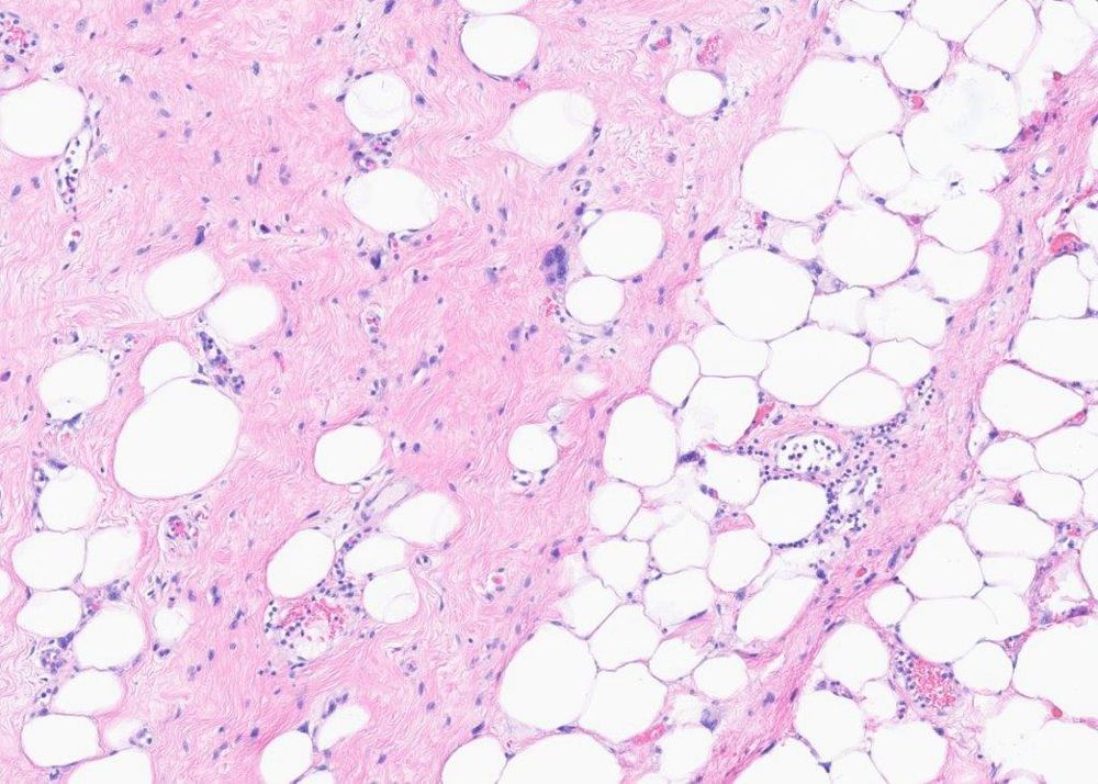
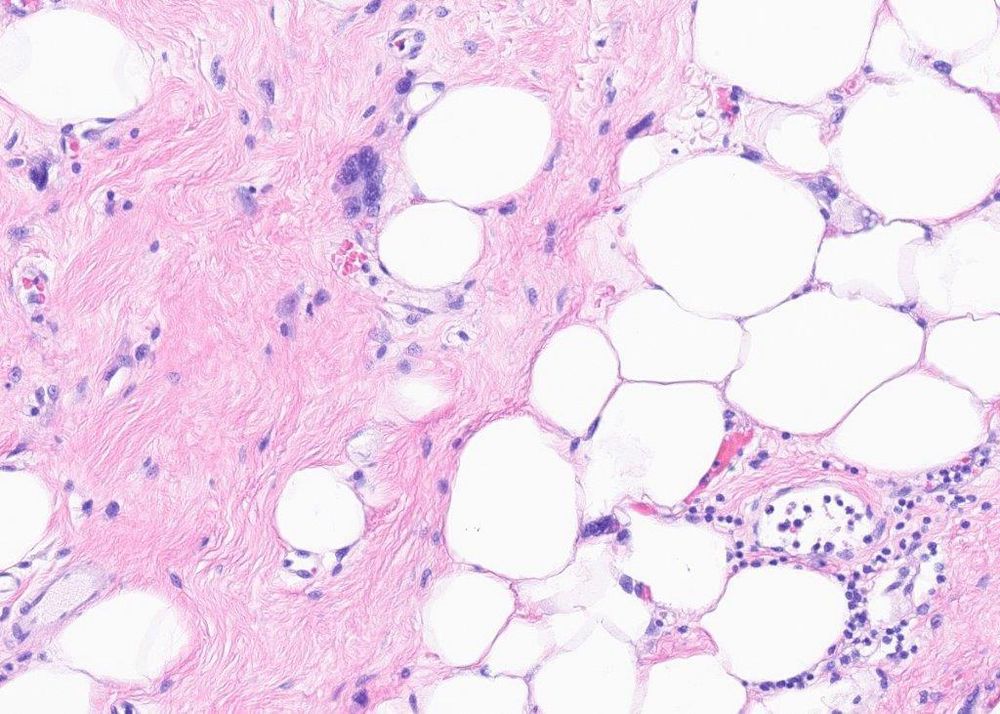
73F, arm. Foamy cell angiosarcoma- a very difficult, very rare subtype.
pubmed.ncbi.nlm.nih.gov/20175826/
73F, arm. Foamy cell angiosarcoma- a very difficult, very rare subtype.
pubmed.ncbi.nlm.nih.gov/20175826/
November 24, 2024 at 8:29 PM
#PathSky. As long as I’m sitting here in Sunday doing a frozen….
73F, arm. Foamy cell angiosarcoma- a very difficult, very rare subtype.
pubmed.ncbi.nlm.nih.gov/20175826/
73F, arm. Foamy cell angiosarcoma- a very difficult, very rare subtype.
pubmed.ncbi.nlm.nih.gov/20175826/