

Luciano A. Masullo
@lumasullo.bsky.social
Physicist | single molecules, bioscience, biotech, nanotech, optics, fluorescence | Project Leader at @jungmannlab.bsky.social Max Planck Institute of Biochemistry @mpibiochem.bsky.social
Our article "Ångström-resolution imaging of cell-surface glycans" made it to the cover of Nature Nanotechnology! 🤩 @natnano.nature.com
www.nature.com/nnano/volume...
www.nature.com/nnano/volume...

October 18, 2025 at 2:32 PM
Our article "Ångström-resolution imaging of cell-surface glycans" made it to the cover of Nature Nanotechnology! 🤩 @natnano.nature.com
www.nature.com/nnano/volume...
www.nature.com/nnano/volume...
I had a blast visiting the lab of Prof. Peng Xi at Peking University!!
Prof. Xi and his team are doing amazing work on various type of SIM and other imaging modalities!
Thanks so much for the warm welcome, the stimulating discussions, and the campus tour!!
Prof. Xi and his team are doing amazing work on various type of SIM and other imaging modalities!
Thanks so much for the warm welcome, the stimulating discussions, and the campus tour!!


April 30, 2025 at 7:43 AM
I had a blast visiting the lab of Prof. Peng Xi at Peking University!!
Prof. Xi and his team are doing amazing work on various type of SIM and other imaging modalities!
Thanks so much for the warm welcome, the stimulating discussions, and the campus tour!!
Prof. Xi and his team are doing amazing work on various type of SIM and other imaging modalities!
Thanks so much for the warm welcome, the stimulating discussions, and the campus tour!!
Amazing visit to the lab of Prof. Wei Ji at the Institute of Biophysics (CAS) in Beijing today!
They’re doing top-notch research, including awesome interferometric single-molecule localization methods (ROSE)
thanks so much for the invitation, warm welcome and hospitality!! 😍🤩
They’re doing top-notch research, including awesome interferometric single-molecule localization methods (ROSE)
thanks so much for the invitation, warm welcome and hospitality!! 😍🤩


April 28, 2025 at 11:51 AM
Amazing visit to the lab of Prof. Wei Ji at the Institute of Biophysics (CAS) in Beijing today!
They’re doing top-notch research, including awesome interferometric single-molecule localization methods (ROSE)
thanks so much for the invitation, warm welcome and hospitality!! 😍🤩
They’re doing top-notch research, including awesome interferometric single-molecule localization methods (ROSE)
thanks so much for the invitation, warm welcome and hospitality!! 😍🤩

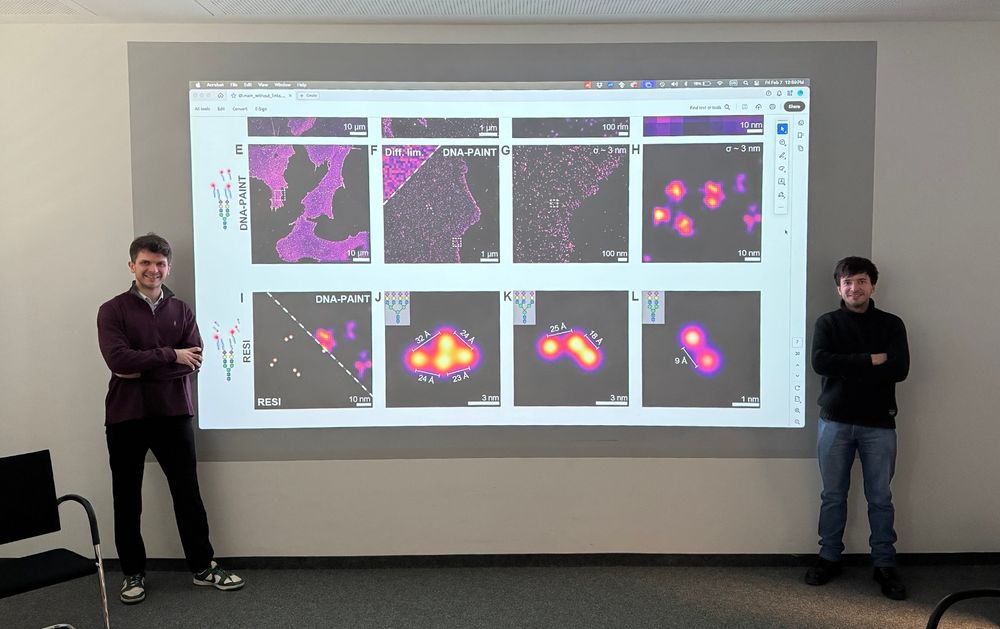
This has been an amazing collaboration between @lmoeckl.bsky.social Lab and Ralf Jungmann Lab led by first authors @karimalmahayni.bsky.social (left) and me (right), big congrats!!!
February 10, 2025 at 8:21 AM
This has been an amazing collaboration between @lmoeckl.bsky.social Lab and Ralf Jungmann Lab led by first authors @karimalmahayni.bsky.social (left) and me (right), big congrats!!!
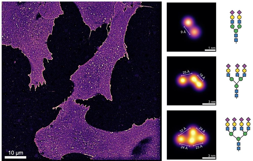
We then further applied downstream spatial analysis workflows to quantify the molecular architecture of the glycocalyx and its constituents for different kinds of sugars obtaining spatial fingerprints compatible with glycosylation patterns! 🍬 6/6

February 10, 2025 at 8:21 AM
We then further applied downstream spatial analysis workflows to quantify the molecular architecture of the glycocalyx and its constituents for different kinds of sugars obtaining spatial fingerprints compatible with glycosylation patterns! 🍬 6/6
We imaged Ångström-scale sugar structures compatible with branched glycans and this is not done in a small field of view but rather over several cells in a large area. A whole new view of the cellular glycocalyx at unprecedented resolution! 5/6
February 10, 2025 at 8:21 AM
We imaged Ångström-scale sugar structures compatible with branched glycans and this is not done in a small field of view but rather over several cells in a large area. A whole new view of the cellular glycocalyx at unprecedented resolution! 5/6
We pushed light microscopy from the diffraction limit (~250 nm) to the Ångström scale, resolving single sugars in glycans. Thanks to our bioorthogonal click-chemistry approach, this marks the first-ever optical Ångström-resolution imaging of biomolecules in cells! 4/6

February 10, 2025 at 8:21 AM
We pushed light microscopy from the diffraction limit (~250 nm) to the Ångström scale, resolving single sugars in glycans. Thanks to our bioorthogonal click-chemistry approach, this marks the first-ever optical Ångström-resolution imaging of biomolecules in cells! 4/6
We tackled this by combining metabolic labeling with our recently developed Ångström-resolution fluorescence microscopy (RESI 🧬). This lets us resolve individual sugars in glycans on the cell surface for the first time! 🔬✨🍬 3/6

February 10, 2025 at 8:21 AM
We tackled this by combining metabolic labeling with our recently developed Ångström-resolution fluorescence microscopy (RESI 🧬). This lets us resolve individual sugars in glycans on the cell surface for the first time! 🔬✨🍬 3/6
Visualizing the structure of the glycocalyx has been a big and long-standing challenge in glycobiology. Existing methods like mass spectrometry, electron microscopy, and fluorescence microscopy fall short —they lack the right context, specificity, or resolution. 2/6

February 10, 2025 at 8:21 AM
Visualizing the structure of the glycocalyx has been a big and long-standing challenge in glycobiology. Existing methods like mass spectrometry, electron microscopy, and fluorescence microscopy fall short —they lack the right context, specificity, or resolution. 2/6
All mammalian cells are covered by tiny sugar molecules (monosaccharides, <1 nm) that form building blocks (glycans, 5-10 nm) which create a complex coating (glycocalyx). This coat is key in immunity, cancer, and infection. To understand its role, we need to see its structure! 1/6

February 10, 2025 at 8:21 AM
All mammalian cells are covered by tiny sugar molecules (monosaccharides, <1 nm) that form building blocks (glycans, 5-10 nm) which create a complex coating (glycocalyx). This coat is key in immunity, cancer, and infection. To understand its role, we need to see its structure! 1/6
ÅNGSTRÖM-RESOLUTION IMAGING OF CELL-SURFACE GLYCANS 🧬🎨🍬
The glycocalyx, our cells' sugar coat, holds secrets in immunology, cancer, viral infections, and more. Visualizing its molecular architecture was impossible… until now. #glycotime #microscopy
www.biorxiv.org/content/10.1...
The glycocalyx, our cells' sugar coat, holds secrets in immunology, cancer, viral infections, and more. Visualizing its molecular architecture was impossible… until now. #glycotime #microscopy
www.biorxiv.org/content/10.1...
February 10, 2025 at 8:21 AM
ÅNGSTRÖM-RESOLUTION IMAGING OF CELL-SURFACE GLYCANS 🧬🎨🍬
The glycocalyx, our cells' sugar coat, holds secrets in immunology, cancer, viral infections, and more. Visualizing its molecular architecture was impossible… until now. #glycotime #microscopy
www.biorxiv.org/content/10.1...
The glycocalyx, our cells' sugar coat, holds secrets in immunology, cancer, viral infections, and more. Visualizing its molecular architecture was impossible… until now. #glycotime #microscopy
www.biorxiv.org/content/10.1...
Amazing talk by @christlet.bsky.social with stunning super-resolution images of neurons! 🤩 #FOMISUR

December 11, 2024 at 6:03 PM
Amazing talk by @christlet.bsky.social with stunning super-resolution images of neurons! 🤩 #FOMISUR
#FOMISUR super-resolution microscopy workshop starts now! So happy to be here in my hometown Buenos Aires for this great event! Thanks to the organizers (Stefani Lab) for the invitation!

December 9, 2024 at 1:23 PM
#FOMISUR super-resolution microscopy workshop starts now! So happy to be here in my hometown Buenos Aires for this great event! Thanks to the organizers (Stefani Lab) for the invitation!
Personal take: doing science is always much better working in teams and when scientists have fun, learn and enjoy the process. Lots of further applications and developments coming up, stay tuned! 13/13

November 27, 2024 at 9:47 AM
Personal take: doing science is always much better working in teams and when scientists have fun, learn and enjoy the process. Lots of further applications and developments coming up, stay tuned! 13/13
#RESI has been an amazing project developed by a wonderful team led by PhD student Susanne Reinhardt together with Isabelle Baudrexel, Philipp Steen and myself + a lot of support by present and past members of the lab + the great supervision of Ralf Jungmann. 12/13

November 27, 2024 at 9:47 AM
#RESI has been an amazing project developed by a wonderful team led by PhD student Susanne Reinhardt together with Isabelle Baudrexel, Philipp Steen and myself + a lot of support by present and past members of the lab + the great supervision of Ralf Jungmann. 12/13
All in all, RESI provides a unique combination of high-throughput (whole cells within hours), molecular resolution (sub-nm or limited by the label), low invasiviness (intact fixed cells) and easy implementation: TIRF microscope (picture) and DNA-PAINT reagents. 10/13

November 27, 2024 at 9:47 AM
All in all, RESI provides a unique combination of high-throughput (whole cells within hours), molecular resolution (sub-nm or limited by the label), low invasiviness (intact fixed cells) and easy implementation: TIRF microscope (picture) and DNA-PAINT reagents. 10/13
(3) We finally applied RESI to observe the organization of oligomers of CD20 in the membrane of whole intact cells. RESI reveals distances well below 10 nm that are not resolvable by conventional DNA-PAINT. 9/13

November 27, 2024 at 9:47 AM
(3) We finally applied RESI to observe the organization of oligomers of CD20 in the membrane of whole intact cells. RESI reveals distances well below 10 nm that are not resolvable by conventional DNA-PAINT. 9/13
Note that the sub-nm (Ångström scale) resolution claim is based on the strictest and clearest criterion: we resolve two targets at a distance that is smaller than 1 nm (10 Å). 8/13

November 27, 2024 at 9:47 AM
Note that the sub-nm (Ångström scale) resolution claim is based on the strictest and clearest criterion: we resolve two targets at a distance that is smaller than 1 nm (10 Å). 8/13
(2) To test the full potential of RESI we imaged DNA origami with adjacent single strands of DNA, resolving the backbone distance between *two single DNA base-pairs* with a precision of 1.2 Å (!!!). 7/13

November 27, 2024 at 9:47 AM
(2) To test the full potential of RESI we imaged DNA origami with adjacent single strands of DNA, resolving the backbone distance between *two single DNA base-pairs* with a precision of 1.2 Å (!!!). 7/13
We demonstrate #RESI by imaging: (1) the Nuclear Pore Complex, (2) DNA origami, (3) membrane receptors.
(1) By stochastically labeling the NUP96 proteins of the NPC we could resolve the ~12 nm (xy) and ~5 nm (z) distances of the subunits with previously unmatched resolution. 6/13
(1) By stochastically labeling the NUP96 proteins of the NPC we could resolve the ~12 nm (xy) and ~5 nm (z) distances of the subunits with previously unmatched resolution. 6/13

November 27, 2024 at 9:47 AM
We demonstrate #RESI by imaging: (1) the Nuclear Pore Complex, (2) DNA origami, (3) membrane receptors.
(1) By stochastically labeling the NUP96 proteins of the NPC we could resolve the ~12 nm (xy) and ~5 nm (z) distances of the subunits with previously unmatched resolution. 6/13
(1) By stochastically labeling the NUP96 proteins of the NPC we could resolve the ~12 nm (xy) and ~5 nm (z) distances of the subunits with previously unmatched resolution. 6/13
DNA-PAINT is the perfect experimental vehicle for #RESI because: i) repetitive (un)binding of the probes to the same target, ii) high multiplexing capabilities (channels encoded in the DNA sequences) iii) it avoids interactions between neighbouring fluorophores 5/13

November 27, 2024 at 9:47 AM
DNA-PAINT is the perfect experimental vehicle for #RESI because: i) repetitive (un)binding of the probes to the same target, ii) high multiplexing capabilities (channels encoded in the DNA sequences) iii) it avoids interactions between neighbouring fluorophores 5/13
This unlocks the possibility of obtaining super-localizations (not of the fluorophore but of the target) with a precision that scales with σ / sqrt(K) where K is the number of localizations of each target. This new scaling law brings us to the Ångström scale. 4/13

November 27, 2024 at 9:47 AM
This unlocks the possibility of obtaining super-localizations (not of the fluorophore but of the target) with a precision that scales with σ / sqrt(K) where K is the number of localizations of each target. This new scaling law brings us to the Ångström scale. 4/13
Typically, it’s not possible to do this grouping since the clouds of localizations can’t be unambiguously assigned to their targets. However, if the targets are labeled distinctly, they can be imaged sequentially and their corresponding localizations grouped. 3/13

November 27, 2024 at 9:47 AM
Typically, it’s not possible to do this grouping since the clouds of localizations can’t be unambiguously assigned to their targets. However, if the targets are labeled distinctly, they can be imaged sequentially and their corresponding localizations grouped. 3/13
In single-molecule localization microscopy (SMLM) methods, clouds of localizations coming from a single target molecule could in principle be grouped and its underlying position estimated with much higher precision than the precision of one single localization 2/13

November 27, 2024 at 9:47 AM
In single-molecule localization microscopy (SMLM) methods, clouds of localizations coming from a single target molecule could in principle be grouped and its underlying position estimated with much higher precision than the precision of one single localization 2/13