


Lorena Benedetti
@lorenabenedetti.bsky.social
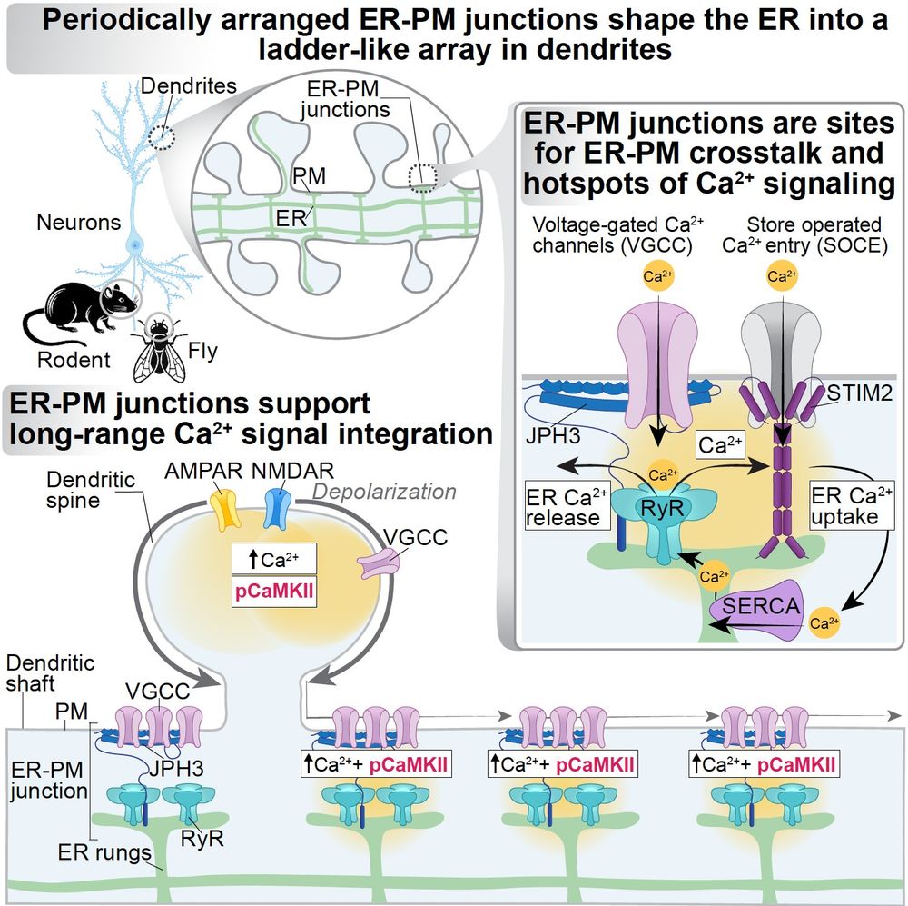
Neuroscientist and cell biologist dedicated to unraveling how organelles adapt their structure and function to sustain neuronal signaling and metabolism. Postdoc in the JLS lab @ HHMI Janelia
Check out the stunning movie by @xianglejerry.bsky.social from @wumin-lab.bsky.social, the winner of the 2024 ASCB Video Contest! Traveling waves of FBP17-EGFP at the cortex of immune mast cells #ASCB2024 @ascbiology.bsky.social
December 27, 2024 at 7:45 PM
Check out the stunning movie by @xianglejerry.bsky.social from @wumin-lab.bsky.social, the winner of the 2024 ASCB Video Contest! Traveling waves of FBP17-EGFP at the cortex of immune mast cells #ASCB2024 @ascbiology.bsky.social
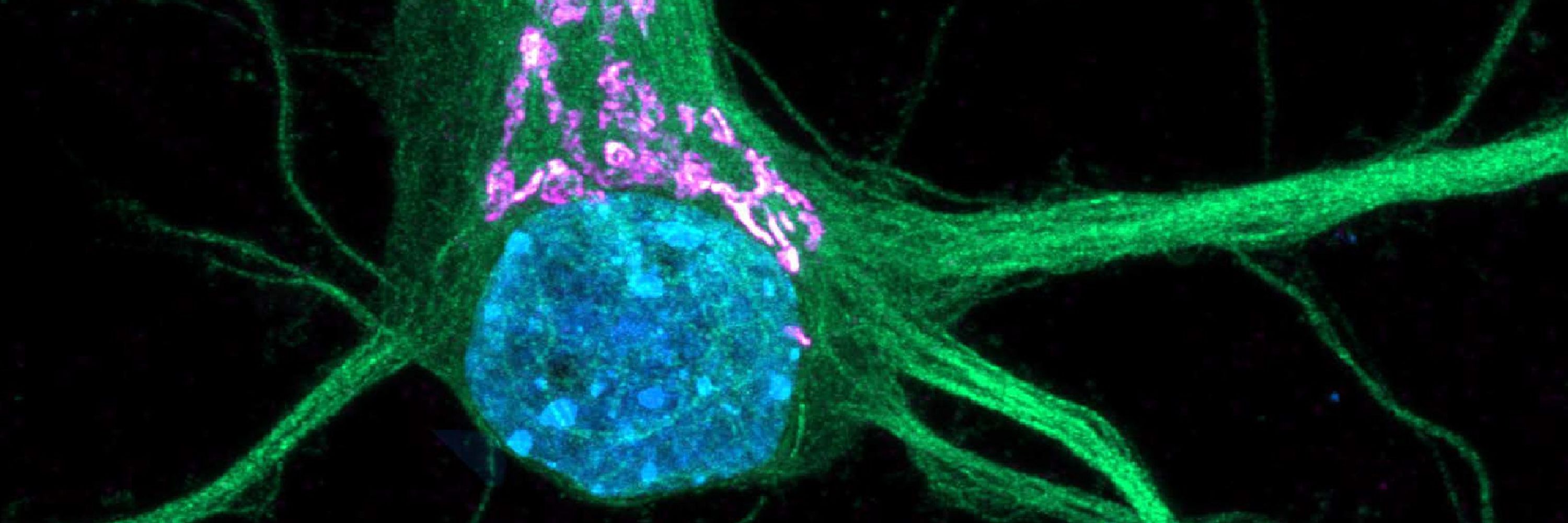
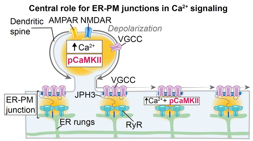
11/Can Ca²⁺ release at ER-PM junctions activate Ca²⁺-dependent intracellular signaling? Upon stimulation, pCaMKII accumulates in spine's heads and JPH3 contacts along dendrites, suggesting that ER-PM junctions serve as preferential sites for Ca²⁺-dependent intracellular signaling along dendrites.

December 20, 2024 at 9:13 PM
11/Can Ca²⁺ release at ER-PM junctions activate Ca²⁺-dependent intracellular signaling? Upon stimulation, pCaMKII accumulates in spine's heads and JPH3 contacts along dendrites, suggesting that ER-PM junctions serve as preferential sites for Ca²⁺-dependent intracellular signaling along dendrites.
10/ One of the most exciting aspects was that local spine activation triggers Ca²⁺ release from the ER via RyRs at these ER-PM contact sites. Contact sites support Ca²⁺ signal propagation over distances 100x greater than spine’s head, revealing a mechanism for amplifying and distributing signals.

December 20, 2024 at 9:13 PM
10/ One of the most exciting aspects was that local spine activation triggers Ca²⁺ release from the ER via RyRs at these ER-PM contact sites. Contact sites support Ca²⁺ signal propagation over distances 100x greater than spine’s head, revealing a mechanism for amplifying and distributing signals.
9/ These junctions are populated with Ca²⁺ tuners and responders. Among them, on the ER membrane, we found that the Ca²⁺-activated Ca²⁺ release channel, RyR, is enriched at JPH3 junctions, where it is maintained in nanometer proximity to plasma membrane Ca²⁺ sources.

December 20, 2024 at 9:13 PM
9/ These junctions are populated with Ca²⁺ tuners and responders. Among them, on the ER membrane, we found that the Ca²⁺-activated Ca²⁺ release channel, RyR, is enriched at JPH3 junctions, where it is maintained in nanometer proximity to plasma membrane Ca²⁺ sources.
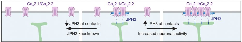
7/ We found that JPH3 plays a critical role in controlling the positioning and accumulation of voltage-gated ion channels at ER-PM junctions. These data reveal a new fundamental role for the ER in regulating the positioning of voltage-gated ion channels at the plasma membrane of dendrites.
December 20, 2024 at 9:13 PM
7/ We found that JPH3 plays a critical role in controlling the positioning and accumulation of voltage-gated ion channels at ER-PM junctions. These data reveal a new fundamental role for the ER in regulating the positioning of voltage-gated ion channels at the plasma membrane of dendrites.
6/ These junctions are populated by the ER-PM tethering protein JPH3.
December 20, 2024 at 9:13 PM
6/ These junctions are populated by the ER-PM tethering protein JPH3.
5/ and in neurons of the fly brain.
December 20, 2024 at 9:13 PM
5/ and in neurons of the fly brain.
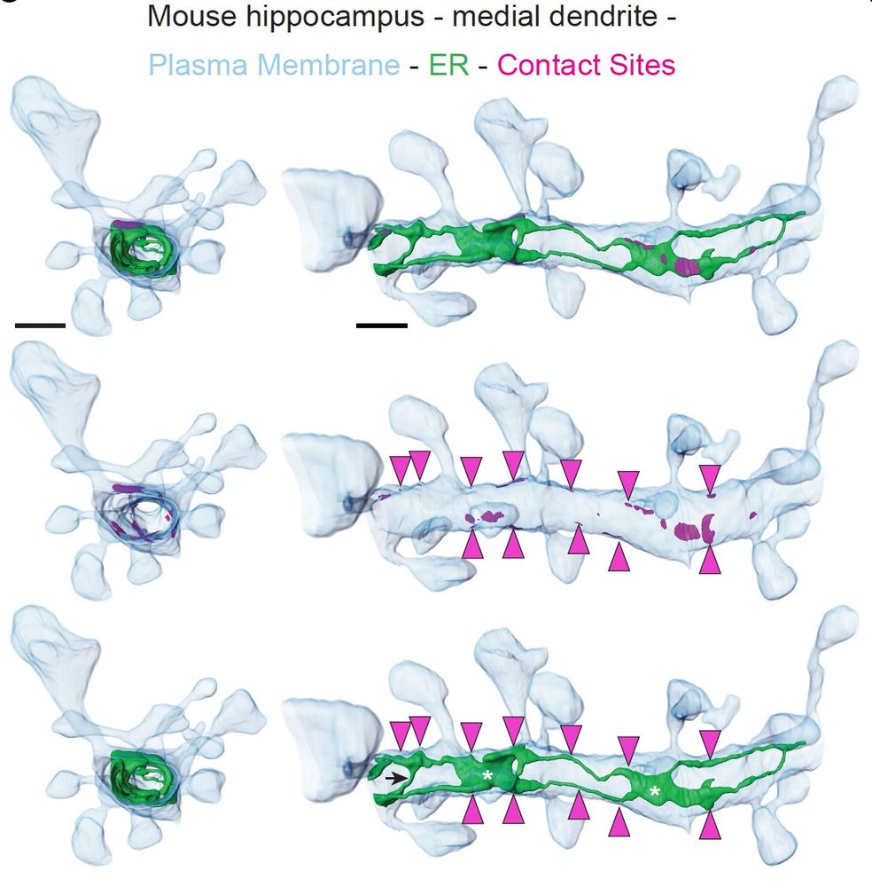
4/ These transverse ER elements protrude and form regular connections with the plasma membrane. Using volumetric EM reconstructions, we confirmed this in vivo in the mouse hippocampus.
December 20, 2024 at 9:13 PM
4/ These transverse ER elements protrude and form regular connections with the plasma membrane. Using volumetric EM reconstructions, we confirmed this in vivo in the mouse hippocampus.
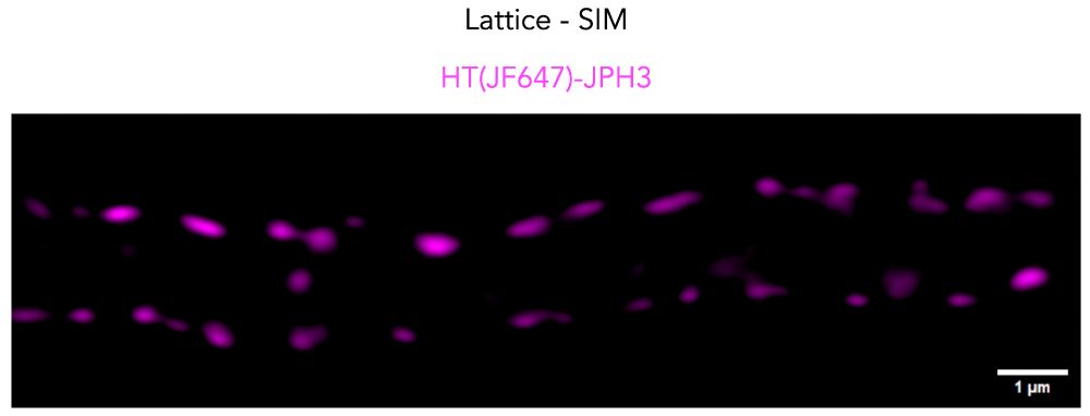
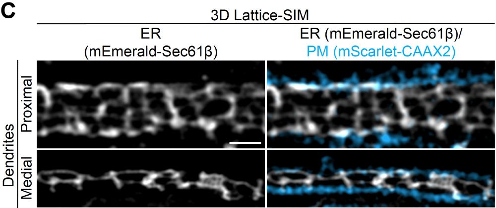
3/ Using live 3D lattice SIM imaging, we observed that along proximal and medial dendrites, longitudinal ER elements are connected by very regularly interspersed.
transverse ER elements.
transverse ER elements.
December 20, 2024 at 9:13 PM
3/ Using live 3D lattice SIM imaging, we observed that along proximal and medial dendrites, longitudinal ER elements are connected by very regularly interspersed.
transverse ER elements.
transverse ER elements.
2/We reveal how a unique organization of ER-PM contact sites shapes the ER in dendrites into a ladder-like structure. These periodic ER-PM contacts act as local, spatial, and temporal amplifiers for Ca²⁺-dependent signaling.
December 20, 2024 at 9:13 PM
2/We reveal how a unique organization of ER-PM contact sites shapes the ER in dendrites into a ladder-like structure. These periodic ER-PM contacts act as local, spatial, and temporal amplifiers for Ca²⁺-dependent signaling.
I am super excited to share our article addressing a longstanding question in neurobiology: how Ca²⁺ signaling cascades initiated at dendritic spines operate at a distance. Check it out here: authors.elsevier.com/sd/article/S... @cp-cell.bsky.social.
December 20, 2024 at 9:13 PM
I am super excited to share our article addressing a longstanding question in neurobiology: how Ca²⁺ signaling cascades initiated at dendritic spines operate at a distance. Check it out here: authors.elsevier.com/sd/article/S... @cp-cell.bsky.social.
Kicking off here with some old but gold movies showing ER-lysosome interactions triggered by blue-light excitation of the light-sensitive protein eMags. A shining example of how light can control cellular dynamics! 💡🧫✨
November 30, 2024 at 2:39 AM
Kicking off here with some old but gold movies showing ER-lysosome interactions triggered by blue-light excitation of the light-sensitive protein eMags. A shining example of how light can control cellular dynamics! 💡🧫✨