

Alexandre Dumoulin
@axonsalex.bsky.social
Developmental Neurobiologist at UZH | Microscopy Enthusiast. Using the chicken embryo to decipher the molecular mechanisms of neural circuit formation. Fascinated by axonal growth cones, primary cilia, and extracellular vesicles.
Thrilled to collaborate with @zhixingchen2.bsky.social Lab @pku1898.bsky.social !
We tested new PK Mem dyes—gentle, photostable tools to label the plasma membrane in live neurons, making it easier to track growth cone motility & axonal transport. #liveimaging #neuroscience
tiny.cc/3qhp001
We tested new PK Mem dyes—gentle, photostable tools to label the plasma membrane in live neurons, making it easier to track growth cone motility & axonal transport. #liveimaging #neuroscience
tiny.cc/3qhp001
July 16, 2025 at 6:44 AM
Thrilled to collaborate with @zhixingchen2.bsky.social Lab @pku1898.bsky.social !
We tested new PK Mem dyes—gentle, photostable tools to label the plasma membrane in live neurons, making it easier to track growth cone motility & axonal transport. #liveimaging #neuroscience
tiny.cc/3qhp001
We tested new PK Mem dyes—gentle, photostable tools to label the plasma membrane in live neurons, making it easier to track growth cone motility & axonal transport. #liveimaging #neuroscience
tiny.cc/3qhp001
it is a filopodial (thin protusions) party in there! 🕺🧐
🧪 #liveimaging
🧪 #liveimaging
April 30, 2025 at 7:54 AM
it is a filopodial (thin protusions) party in there! 🕺🧐
🧪 #liveimaging
🧪 #liveimaging
This time-lapse captures 17 hours of axonal growth from a chicken dorsal root ganglion explant, visualized through the actin cytoskeleton using live confocal imaging.
I just submitted this video to the Nikon Small World in Motion competition. Today is the last day to upload yours! 😉
🧪
I just submitted this video to the Nikon Small World in Motion competition. Today is the last day to upload yours! 😉
🧪
April 30, 2025 at 7:16 AM
This time-lapse captures 17 hours of axonal growth from a chicken dorsal root ganglion explant, visualized through the actin cytoskeleton using live confocal imaging.
I just submitted this video to the Nikon Small World in Motion competition. Today is the last day to upload yours! 😉
🧪
I just submitted this video to the Nikon Small World in Motion competition. Today is the last day to upload yours! 😉
🧪
For this #FluorescenceFriday, let's watch an axonal growth cone (for a change) displaying its most beautiful filopodia (this time)! 🤩 They are so dynamic! 1 image taken every 500 ms for 5 min played at 15 fps. 🧪
December 20, 2024 at 2:25 PM
For this #FluorescenceFriday, let's watch an axonal growth cone (for a change) displaying its most beautiful filopodia (this time)! 🤩 They are so dynamic! 1 image taken every 500 ms for 5 min played at 15 fps. 🧪
This axonal growth cone from a culture of spinal neurons decided to display its most beautiful lamellipodia for this #FluorescenceFriday! 🤩 #neuroscience #liveimaging 🧪
1 image taken every second for 10 min, played at 15 images/s
1 image taken every second for 10 min, played at 15 images/s
December 13, 2024 at 8:43 AM
This axonal growth cone from a culture of spinal neurons decided to display its most beautiful lamellipodia for this #FluorescenceFriday! 🤩 #neuroscience #liveimaging 🧪
1 image taken every second for 10 min, played at 15 images/s
1 image taken every second for 10 min, played at 15 images/s
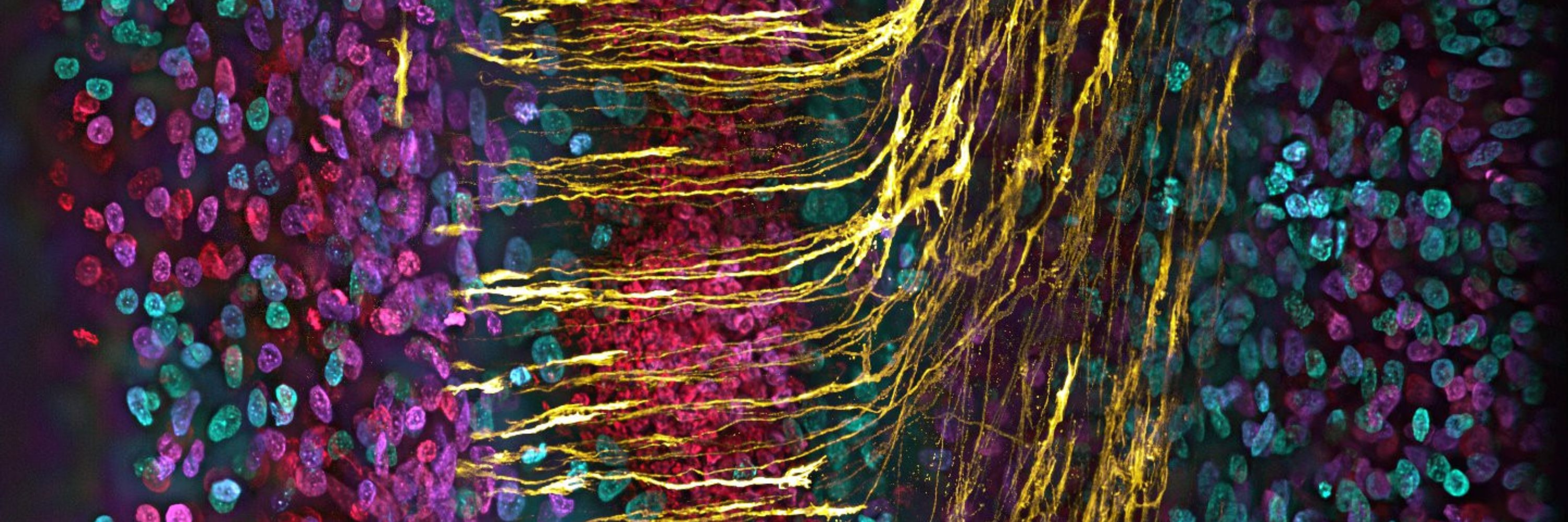
🚀 It's #FluorescenceFriday, and commissural axons are coming at you!
🧪🔬 15-hour time-lapse of chick commissural axons navigating and crossing the CNS midline (and reverse).
One stack taken every 10 minutes. Pixels were color-coded in the z-axis.
#LiveImaging #Neuroscience #Science #Microscopy 🧠✨
🧪🔬 15-hour time-lapse of chick commissural axons navigating and crossing the CNS midline (and reverse).
One stack taken every 10 minutes. Pixels were color-coded in the z-axis.
#LiveImaging #Neuroscience #Science #Microscopy 🧠✨
November 29, 2024 at 7:20 AM
🚀 It's #FluorescenceFriday, and commissural axons are coming at you!
🧪🔬 15-hour time-lapse of chick commissural axons navigating and crossing the CNS midline (and reverse).
One stack taken every 10 minutes. Pixels were color-coded in the z-axis.
#LiveImaging #Neuroscience #Science #Microscopy 🧠✨
🧪🔬 15-hour time-lapse of chick commissural axons navigating and crossing the CNS midline (and reverse).
One stack taken every 10 minutes. Pixels were color-coded in the z-axis.
#LiveImaging #Neuroscience #Science #Microscopy 🧠✨
Starting the week strong! Cheering myself up for the next challenges with 15 min of axonal growth cone dynamic madness. F-actin (cyan) and membrane (magenta) show their dance. One image taken every 10 s.🧠🔬
#MicroscopyMonday #LiveImaging #Neuroscience #Microscopy
#MicroscopyMonday #LiveImaging #Neuroscience #Microscopy
November 25, 2024 at 1:05 PM
Starting the week strong! Cheering myself up for the next challenges with 15 min of axonal growth cone dynamic madness. F-actin (cyan) and membrane (magenta) show their dance. One image taken every 10 s.🧠🔬
#MicroscopyMonday #LiveImaging #Neuroscience #Microscopy
#MicroscopyMonday #LiveImaging #Neuroscience #Microscopy
Leave me alone it's #Fluorescencefriday !➡️ two axonal growth cones growing together until one got fed up of the other... #neuroscience #microscopy #liveimaging
November 22, 2024 at 4:56 PM
Leave me alone it's #Fluorescencefriday !➡️ two axonal growth cones growing together until one got fed up of the other... #neuroscience #microscopy #liveimaging
Let's unleash the axonal growth cones for this #Fluorescencefriday ! #neuroscience #Liveimaging #microscopy #Axonguidance
November 15, 2024 at 7:19 AM
Let's unleash the axonal growth cones for this #Fluorescencefriday ! #neuroscience #Liveimaging #microscopy #Axonguidance
November 13, 2024 at 12:16 PM