


Daryan Chitsaz
@dchits.bsky.social
Photographer of brain cells. Researcher at the Montreal Neurological Hospital. I like glia, technology, and political shitposts.
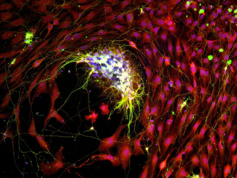
In my MSc I co-cultured motor neurons (yellow) with muscle cells (red) in a separate compartment. One time the barrier between them broke and the chunk of neural tissue was swept up in a wave of proliferating myoblasts. The result reminds me of a boat in a tumultuous ocean storm. #fluorescenceFriday
April 11, 2025 at 3:04 PM
In my MSc I co-cultured motor neurons (yellow) with muscle cells (red) in a separate compartment. One time the barrier between them broke and the chunk of neural tissue was swept up in a wave of proliferating myoblasts. The result reminds me of a boat in a tumultuous ocean storm. #fluorescenceFriday
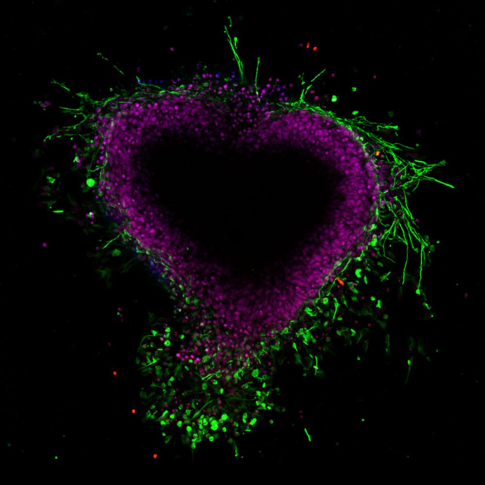
8 years ago as a bumbling RA I tried to impress the cute French intern in the neighbouring lab by sculpting a spinal cord explant into a heart. The neurons did not fare particularly well and labelling leaves something to be desired, but we're still happily together 😍 #fluorescenceFriday #valentines
February 14, 2025 at 2:07 PM
8 years ago as a bumbling RA I tried to impress the cute French intern in the neighbouring lab by sculpting a spinal cord explant into a heart. The neurons did not fare particularly well and labelling leaves something to be desired, but we're still happily together 😍 #fluorescenceFriday #valentines
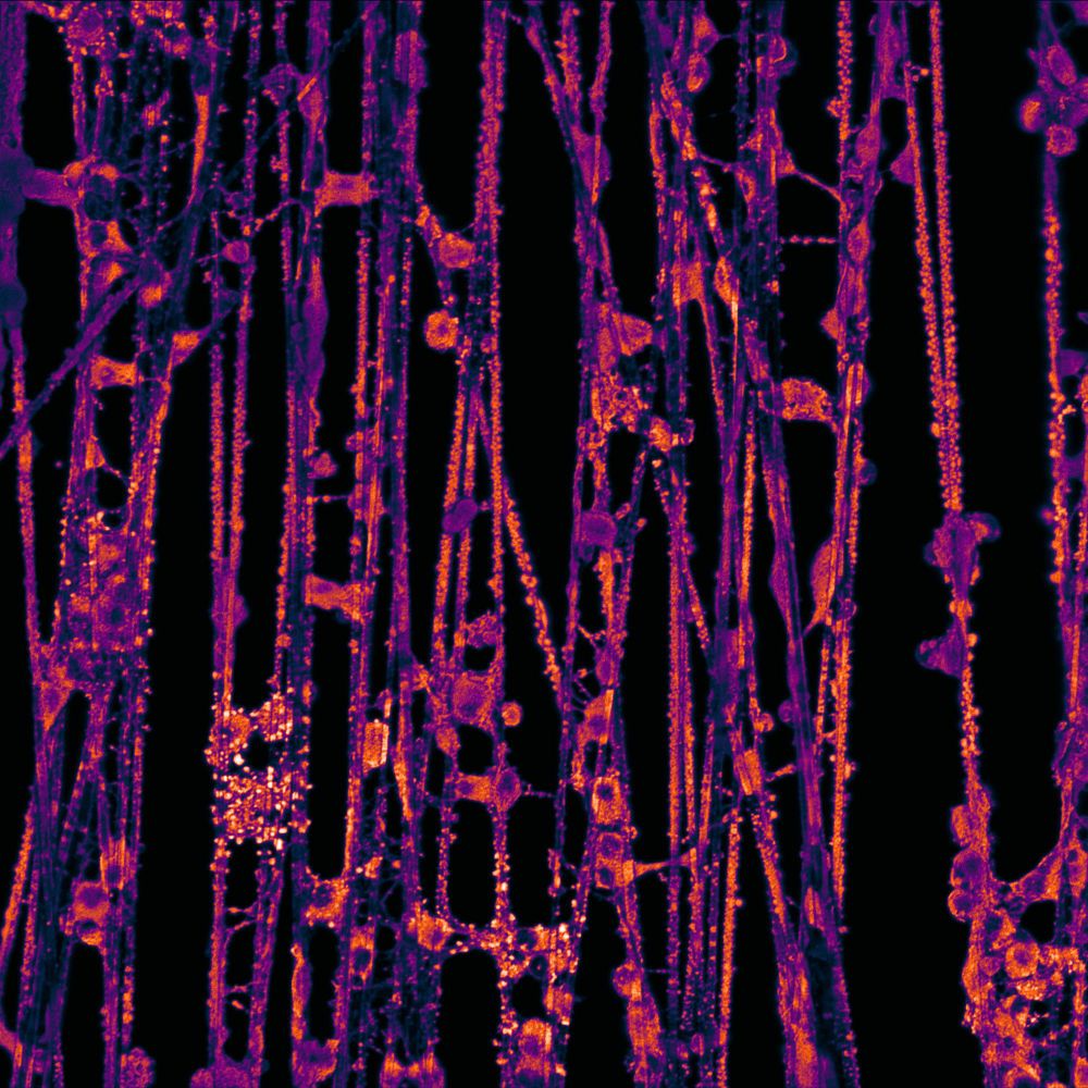
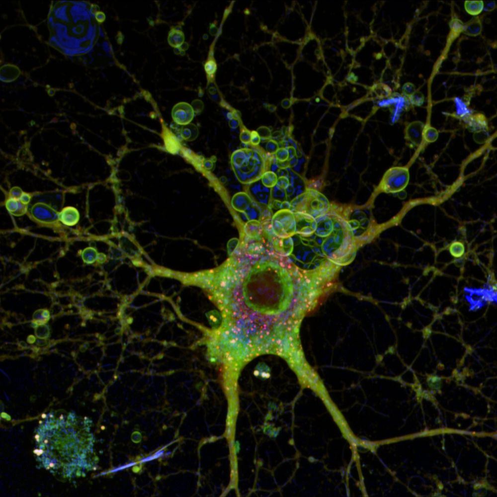
Oligodendrocytes normally make myelin by ensheathing neuronal axons, but these cells are so hell-bent on myelinating that they will wrap up anything, even tiny plastic fibers strung across a cell culture dish. These microfiber cultures let us rapidly screen for myelin-promoting drugs, and look cool.
February 11, 2025 at 5:02 PM
Oligodendrocytes normally make myelin by ensheathing neuronal axons, but these cells are so hell-bent on myelinating that they will wrap up anything, even tiny plastic fibers strung across a cell culture dish. These microfiber cultures let us rapidly screen for myelin-promoting drugs, and look cool.
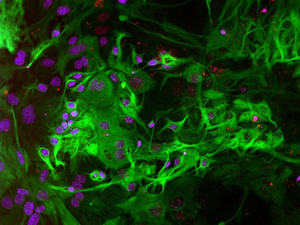
Happy #fluorescenceFriday! This pic shows a gaggle of astrocytes with GFAP labelled, a major cytoskeletal protein specific to these cells. Astrocytes keep your neurons happy and well-fed, and have many other roles including regulating brain immune responses and maintaining the blood-brain barrier.
January 31, 2025 at 6:47 PM
Happy #fluorescenceFriday! This pic shows a gaggle of astrocytes with GFAP labelled, a major cytoskeletal protein specific to these cells. Astrocytes keep your neurons happy and well-fed, and have many other roles including regulating brain immune responses and maintaining the blood-brain barrier.
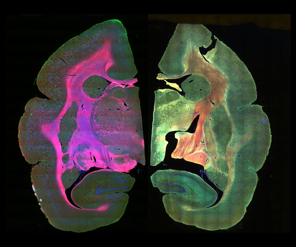
For #fluorescenceFriday I'm celebrating a paper that got accepted this week on immunofluorescent brain slice labelling in Marmosets! These new-world primates are increasingly being used as research models for Austism spectrum disorder, but notably have not yet been reported Sieg Heil-ing.
January 24, 2025 at 5:28 PM
For #fluorescenceFriday I'm celebrating a paper that got accepted this week on immunofluorescent brain slice labelling in Marmosets! These new-world primates are increasingly being used as research models for Austism spectrum disorder, but notably have not yet been reported Sieg Heil-ing.
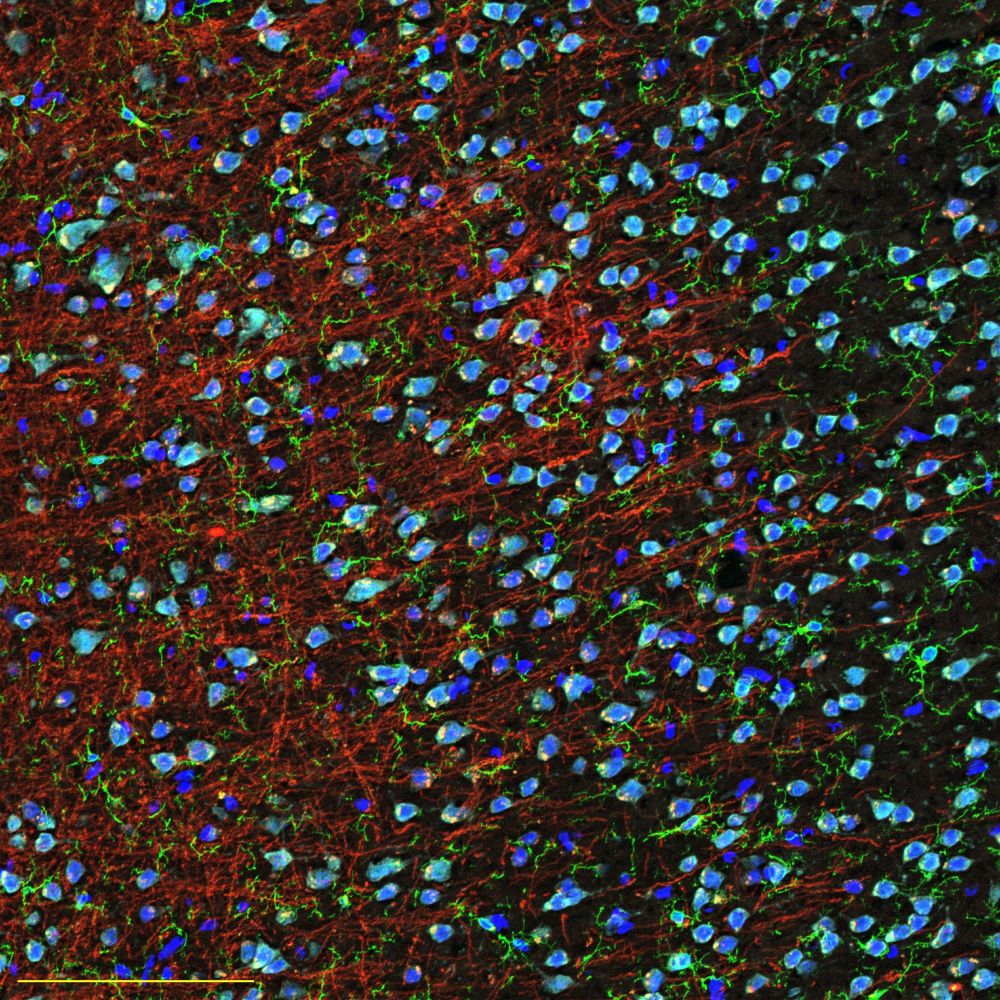
This chaotic scene shows microglia (green), nimble little custodian-like brain cells, navigating through a dense canopy of myelin (red). Microglia support their communities, dissolve toxic particles, and don't threaten the sovereignty of friendly nations. Be more like microglia. #fluorescenceFridays
January 17, 2025 at 8:08 PM
This chaotic scene shows microglia (green), nimble little custodian-like brain cells, navigating through a dense canopy of myelin (red). Microglia support their communities, dissolve toxic particles, and don't threaten the sovereignty of friendly nations. Be more like microglia. #fluorescenceFridays
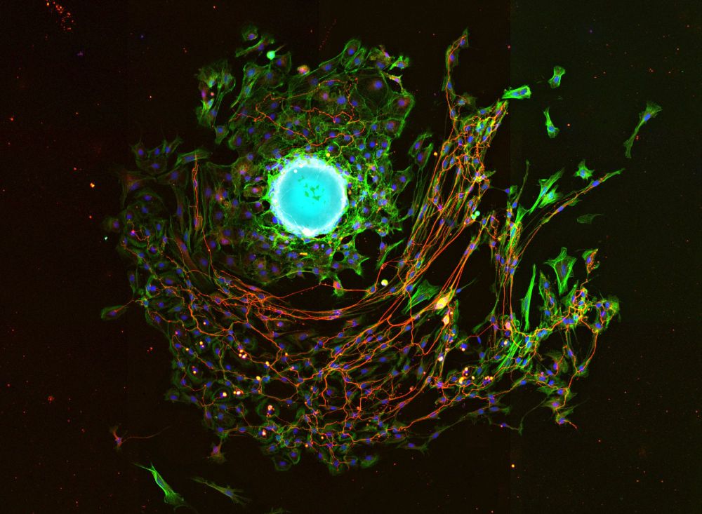
Happy New Year y'all, here's a firework-ish pic of neurons "exploding" from a chunk of ganglia. Fiboblasts (green) bursting out of the left side provide an attractive substrate for sensory neuron axons (red), while other axons at the bottom grow in silly looping circles. #FluorescenceFriday #SciArt
January 3, 2025 at 8:18 PM
Happy New Year y'all, here's a firework-ish pic of neurons "exploding" from a chunk of ganglia. Fiboblasts (green) bursting out of the left side provide an attractive substrate for sensory neuron axons (red), while other axons at the bottom grow in silly looping circles. #FluorescenceFriday #SciArt
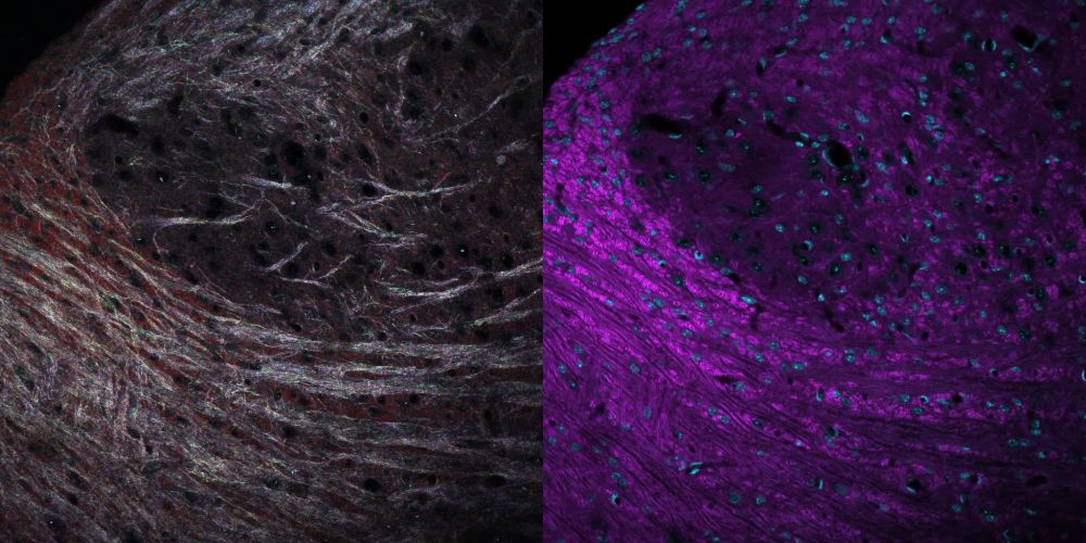
Myelin consists of many miniscule layers of smooth membrane, making it highly reflective. This lets you image it with #microscopy without any staining - left shows reflections of RGB lasers from brainstem myelin lit up like a disco ball, right is the same region with a fluorescent dye. #neuroskyence
December 27, 2024 at 3:10 AM
Myelin consists of many miniscule layers of smooth membrane, making it highly reflective. This lets you image it with #microscopy without any staining - left shows reflections of RGB lasers from brainstem myelin lit up like a disco ball, right is the same region with a fluorescent dye. #neuroskyence
After a long day of live cell imaging this impressive specimen of an oligodendrocyte is heading for the sweet embrace of apoptosis. Happy weekend y'all #FluorescenceFriday #microscopy #cellbiology
December 13, 2024 at 7:24 PM
After a long day of live cell imaging this impressive specimen of an oligodendrocyte is heading for the sweet embrace of apoptosis. Happy weekend y'all #FluorescenceFriday #microscopy #cellbiology
Explanted neural tissue can make for a dramatic image. This 2-day old chunk of dorsal root ganglia contains the sensory neurons (red) that allow you to feel. They don't particularly like growing on the glass plate, preferring to hop along hardy epithelial cells (green). #microscopy #neuroskyence
December 10, 2024 at 1:58 PM
Explanted neural tissue can make for a dramatic image. This 2-day old chunk of dorsal root ganglia contains the sensory neurons (red) that allow you to feel. They don't particularly like growing on the glass plate, preferring to hop along hardy epithelial cells (green). #microscopy #neuroskyence
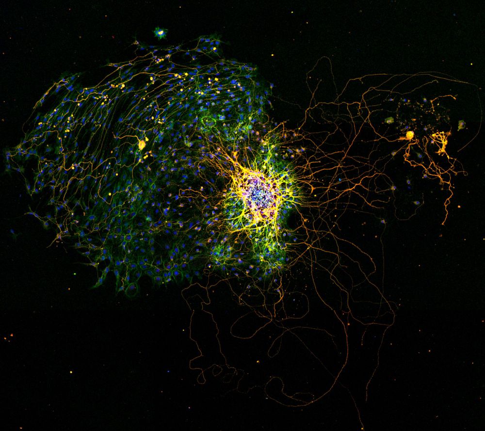
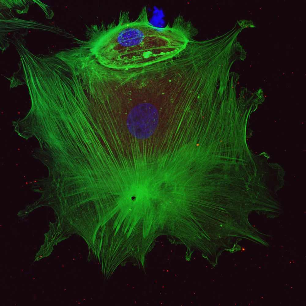
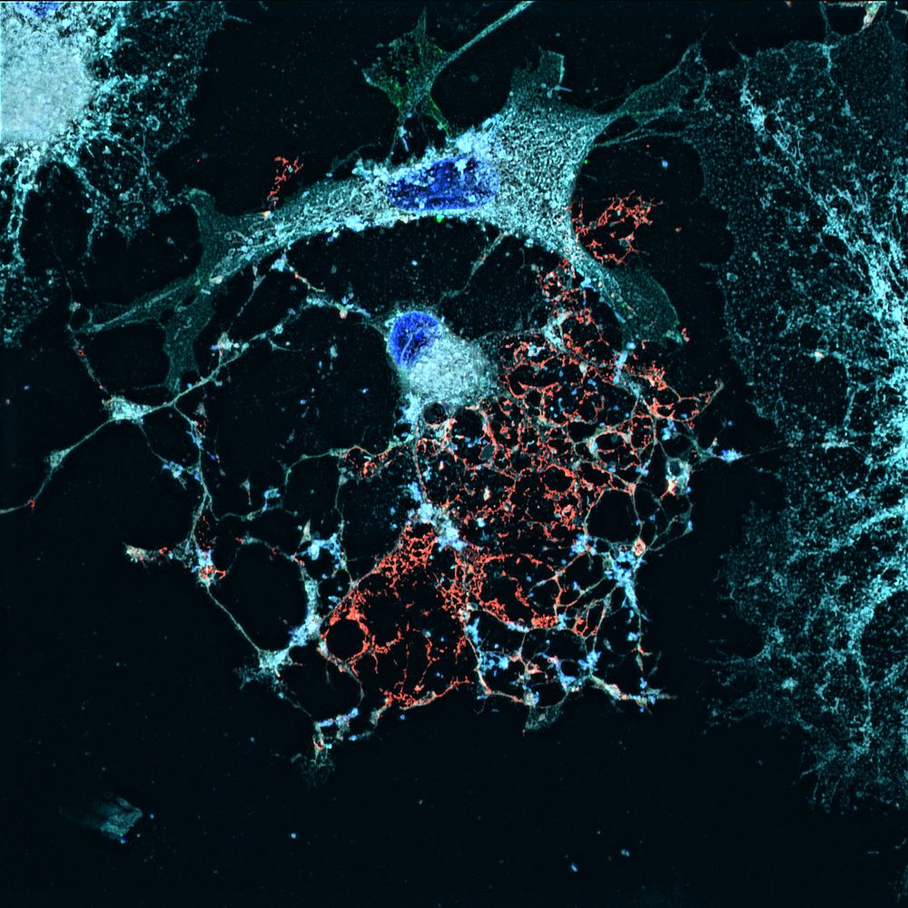
For #fluorescenceFriday sharing some sweet-ass actin: "birth of an endothelial cell" #cellbiology #microscopy
December 6, 2024 at 2:09 PM
For #fluorescenceFriday sharing some sweet-ass actin: "birth of an endothelial cell" #cellbiology #microscopy
phd oral defense in about 24 hours, feeling like this stressed out little glial cell being treated with LPS #academiasky
December 3, 2024 at 12:43 PM
phd oral defense in about 24 hours, feeling like this stressed out little glial cell being treated with LPS #academiasky
Here's a weird type of microscopy - interference reflection microscopy (IRM)! Oligodendrocyte membranes are so smooth and reflective, you can see them without needing a dye by just bouncing laser light off of them (shown in blue, with cell body and processes in red). #neuroskyence
#microscopyMonday
#microscopyMonday
December 2, 2024 at 1:21 PM
Here's a weird type of microscopy - interference reflection microscopy (IRM)! Oligodendrocyte membranes are so smooth and reflective, you can see them without needing a dye by just bouncing laser light off of them (shown in blue, with cell body and processes in red). #neuroskyence
#microscopyMonday
#microscopyMonday
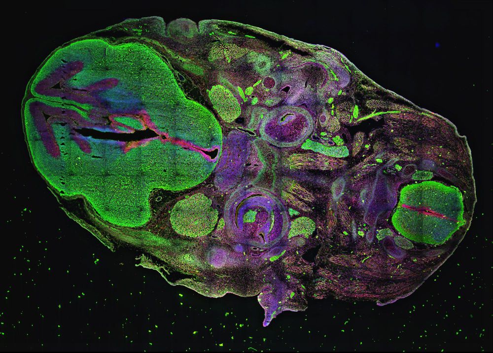
This slightly-botched tissue slice shows embryonic nervous system development. A blue DNA dye labels all cell nuclei, while neurons are green and sites where new ones are being produced are red. This pic was stitched from 100+ images, which is where that nasty grid comes from. #sciArt #neuroscience
November 29, 2024 at 11:49 PM
This slightly-botched tissue slice shows embryonic nervous system development. A blue DNA dye labels all cell nuclei, while neurons are green and sites where new ones are being produced are red. This pic was stitched from 100+ images, which is where that nasty grid comes from. #sciArt #neuroscience
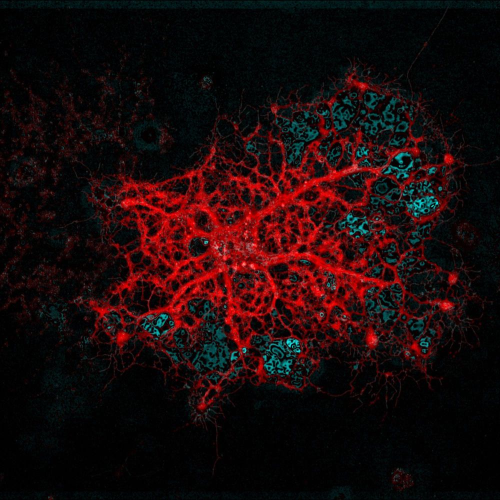
These are a couple of glia (non-neuron brain cells). The lattice-like red stuff comes from a specialized cell that produces myelin, a substance that wraps around neurons to support them. This oligodendrocyte is hoping to find an axon to wrap but alas, there is none to be found. #SciArt #neuroscience
November 29, 2024 at 11:46 PM
These are a couple of glia (non-neuron brain cells). The lattice-like red stuff comes from a specialized cell that produces myelin, a substance that wraps around neurons to support them. This oligodendrocyte is hoping to find an axon to wrap but alas, there is none to be found. #SciArt #neuroscience
I'm nearing the end of my neuroscience PhD and want to share some pics I took along the way.
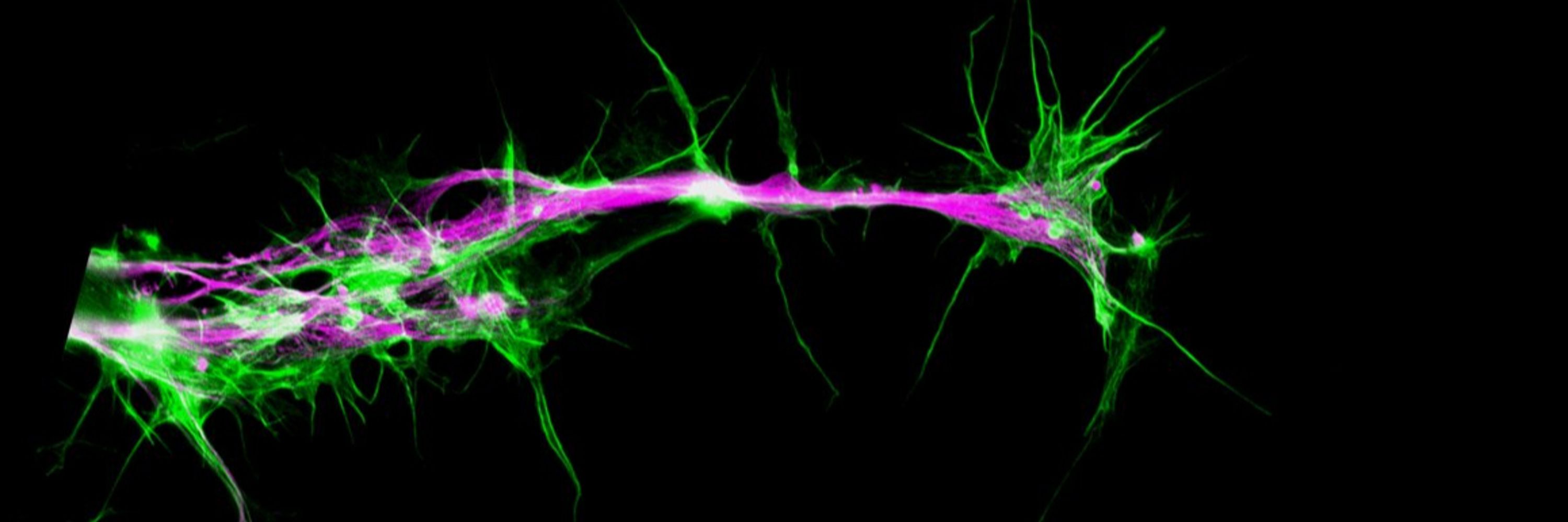
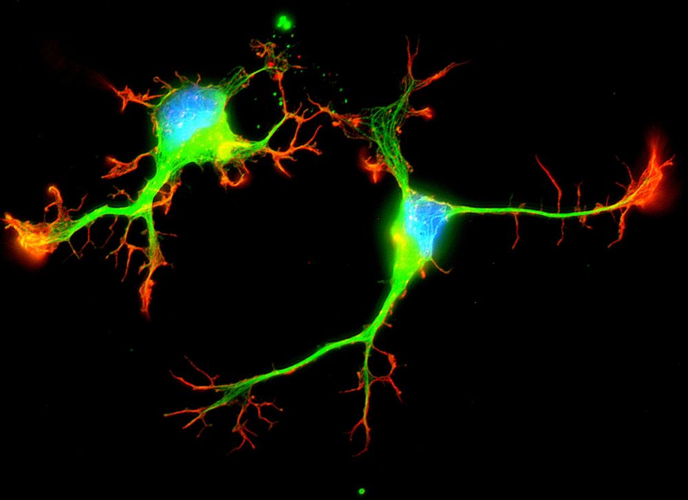
Here are 2 cultured sensory neurons, the ones that carry information from your skin to spinal cord. These little guys only reach ~1/10th of a millimeter now, but can span several meters in large mammals.
Here are 2 cultured sensory neurons, the ones that carry information from your skin to spinal cord. These little guys only reach ~1/10th of a millimeter now, but can span several meters in large mammals.
November 29, 2024 at 11:13 PM
I'm nearing the end of my neuroscience PhD and want to share some pics I took along the way.
Here are 2 cultured sensory neurons, the ones that carry information from your skin to spinal cord. These little guys only reach ~1/10th of a millimeter now, but can span several meters in large mammals.
Here are 2 cultured sensory neurons, the ones that carry information from your skin to spinal cord. These little guys only reach ~1/10th of a millimeter now, but can span several meters in large mammals.