


Vitor Lopes dos Santos
@vitorlds.bsky.social
The embedding enables a cool experiment: start with a tetrode in CA1 pyr, lower it gradually, and project the signal at each step.
The tetrode follows the predicted path, and the gamma profiles match the silicon probe data beautifully at each step.
See one going from CA1 pyr to DG midmol below!
The tetrode follows the predicted path, and the gamma profiles match the silicon probe data beautifully at each step.
See one going from CA1 pyr to DG midmol below!

June 6, 2025 at 11:38 AM
The embedding enables a cool experiment: start with a tetrode in CA1 pyr, lower it gradually, and project the signal at each step.
The tetrode follows the predicted path, and the gamma profiles match the silicon probe data beautifully at each step.
See one going from CA1 pyr to DG midmol below!
The tetrode follows the predicted path, and the gamma profiles match the silicon probe data beautifully at each step.
See one going from CA1 pyr to DG midmol below!
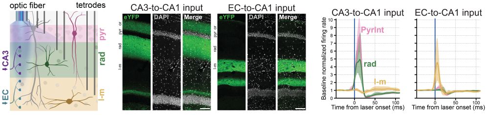
Using optogenetics, we tested how CA1 interneurons across layers respond to upstream input.
CA3 terminal stimulation strongly activated pyramidale (PyrInt) and radiatum (rad) interneurons, while EC terminal stimulation strongly drove LM interneurons.
(10/13)
CA3 terminal stimulation strongly activated pyramidale (PyrInt) and radiatum (rad) interneurons, while EC terminal stimulation strongly drove LM interneurons.
(10/13)
June 5, 2025 at 5:10 PM
Using optogenetics, we tested how CA1 interneurons across layers respond to upstream input.
CA3 terminal stimulation strongly activated pyramidale (PyrInt) and radiatum (rad) interneurons, while EC terminal stimulation strongly drove LM interneurons.
(10/13)
CA3 terminal stimulation strongly activated pyramidale (PyrInt) and radiatum (rad) interneurons, while EC terminal stimulation strongly drove LM interneurons.
(10/13)
Using embedding-guided tetrodes, Demi recorded interneurons in sparse CA1 layers: rad and LM (btw great dataset, stay tuned for future work on this!).
Firing behavior was layer-specific; e.g., LM interneurons were barely active in ripples.
Preferred theta phase also varied across layers.
(9/13)
Firing behavior was layer-specific; e.g., LM interneurons were barely active in ripples.
Preferred theta phase also varied across layers.
(9/13)

June 5, 2025 at 5:10 PM
Using embedding-guided tetrodes, Demi recorded interneurons in sparse CA1 layers: rad and LM (btw great dataset, stay tuned for future work on this!).
Firing behavior was layer-specific; e.g., LM interneurons were barely active in ripples.
Preferred theta phase also varied across layers.
(9/13)
Firing behavior was layer-specific; e.g., LM interneurons were barely active in ripples.
Preferred theta phase also varied across layers.
(9/13)
I love this figure
By projecting tetrode LFPs from several mice onto the embedding (estimating each tetrode’s “depth”), we recover gradually changing theta and ripple waveforms, as if recorded with a single linear probe.
No need for alignment or histology. Just the signals and the embedding.
(8/13)
By projecting tetrode LFPs from several mice onto the embedding (estimating each tetrode’s “depth”), we recover gradually changing theta and ripple waveforms, as if recorded with a single linear probe.
No need for alignment or histology. Just the signals and the embedding.
(8/13)

June 5, 2025 at 5:10 PM
I love this figure
By projecting tetrode LFPs from several mice onto the embedding (estimating each tetrode’s “depth”), we recover gradually changing theta and ripple waveforms, as if recorded with a single linear probe.
No need for alignment or histology. Just the signals and the embedding.
(8/13)
By projecting tetrode LFPs from several mice onto the embedding (estimating each tetrode’s “depth”), we recover gradually changing theta and ripple waveforms, as if recorded with a single linear probe.
No need for alignment or histology. Just the signals and the embedding.
(8/13)
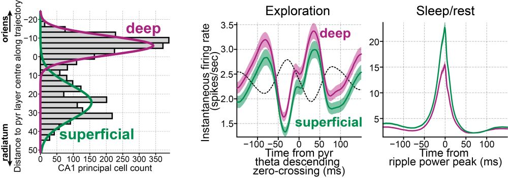
Zooming in, the embedding also separates deep vs superficial CA1 pyramidal cells.
These groups differ in firing rates (awake and sleep), theta modulation, and coupling to gamma and ripple.
This let us replicate findings from fancy silicon probes using tetrodes, and extend them further.
(7/13)
These groups differ in firing rates (awake and sleep), theta modulation, and coupling to gamma and ripple.
This let us replicate findings from fancy silicon probes using tetrodes, and extend them further.
(7/13)
June 5, 2025 at 5:10 PM
Zooming in, the embedding also separates deep vs superficial CA1 pyramidal cells.
These groups differ in firing rates (awake and sleep), theta modulation, and coupling to gamma and ripple.
This let us replicate findings from fancy silicon probes using tetrodes, and extend them further.
(7/13)
These groups differ in firing rates (awake and sleep), theta modulation, and coupling to gamma and ripple.
This let us replicate findings from fancy silicon probes using tetrodes, and extend them further.
(7/13)
From our spike analysis:
In pyr fast gamma, principal cells fire before interneurons. In rad fast gamma, this flips!
Same freq range, but ≠ mechanism.
+ pyr fast gamma and ripples may arise from the same oscillator: called 'fast gamma' when theta-driven, 'ripples' when driven by SW inputs.
(6/13)
In pyr fast gamma, principal cells fire before interneurons. In rad fast gamma, this flips!
Same freq range, but ≠ mechanism.
+ pyr fast gamma and ripples may arise from the same oscillator: called 'fast gamma' when theta-driven, 'ripples' when driven by SW inputs.
(6/13)

June 5, 2025 at 5:10 PM
From our spike analysis:
In pyr fast gamma, principal cells fire before interneurons. In rad fast gamma, this flips!
Same freq range, but ≠ mechanism.
+ pyr fast gamma and ripples may arise from the same oscillator: called 'fast gamma' when theta-driven, 'ripples' when driven by SW inputs.
(6/13)
In pyr fast gamma, principal cells fire before interneurons. In rad fast gamma, this flips!
Same freq range, but ≠ mechanism.
+ pyr fast gamma and ripples may arise from the same oscillator: called 'fast gamma' when theta-driven, 'ripples' when driven by SW inputs.
(6/13)
Histological validation: lower tetrodes along the CA1–DG axis until they reach a target layer, then confirm final position with histology.
It’s really robust! Ephys patterns do predict anatomical layers.
See two tetrodes going to DG molecular layer below (more examples in the paper!).
(5/13)
It’s really robust! Ephys patterns do predict anatomical layers.
See two tetrodes going to DG molecular layer below (more examples in the paper!).
(5/13)

June 5, 2025 at 5:10 PM
Histological validation: lower tetrodes along the CA1–DG axis until they reach a target layer, then confirm final position with histology.
It’s really robust! Ephys patterns do predict anatomical layers.
See two tetrodes going to DG molecular layer below (more examples in the paper!).
(5/13)
It’s really robust! Ephys patterns do predict anatomical layers.
See two tetrodes going to DG molecular layer below (more examples in the paper!).
(5/13)
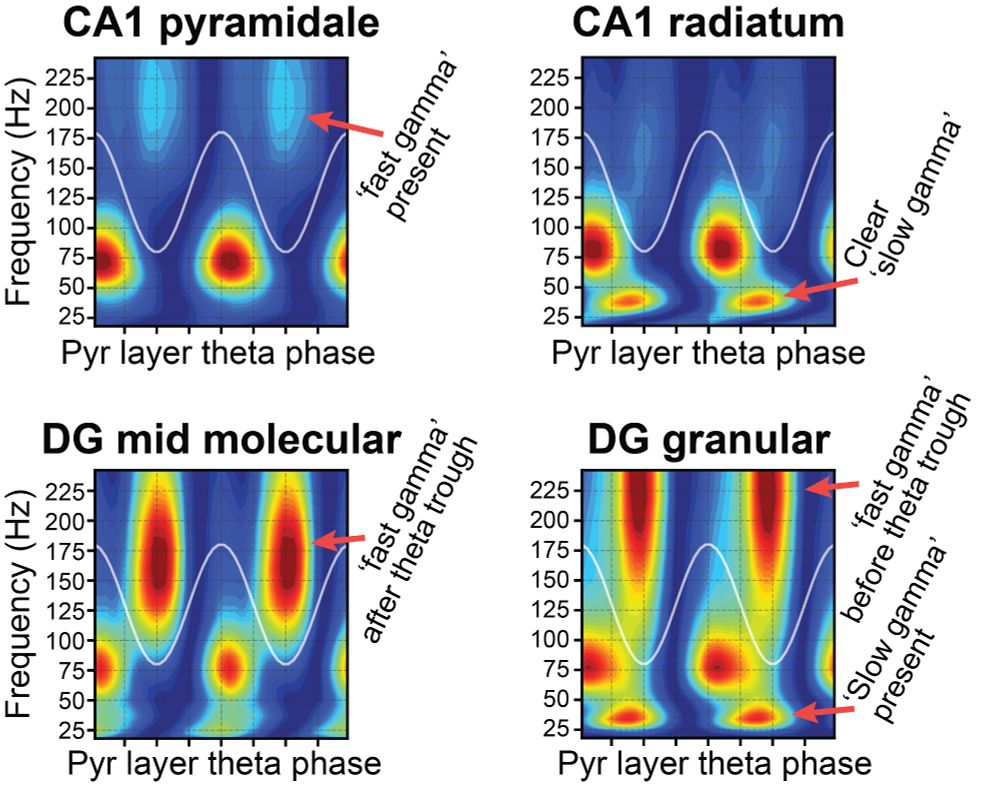
We characterize multiple coexisting gamma rhythms organized anatomically and by theta phase.
This spatiotemporal structure was robust across mice in our silicon probe dataset.
See examples below and check the paper for all the details (including a few novel gamma rhythms)!
(3/13)
This spatiotemporal structure was robust across mice in our silicon probe dataset.
See examples below and check the paper for all the details (including a few novel gamma rhythms)!
(3/13)
June 5, 2025 at 5:10 PM
We characterize multiple coexisting gamma rhythms organized anatomically and by theta phase.
This spatiotemporal structure was robust across mice in our silicon probe dataset.
See examples below and check the paper for all the details (including a few novel gamma rhythms)!
(3/13)
This spatiotemporal structure was robust across mice in our silicon probe dataset.
See examples below and check the paper for all the details (including a few novel gamma rhythms)!
(3/13)
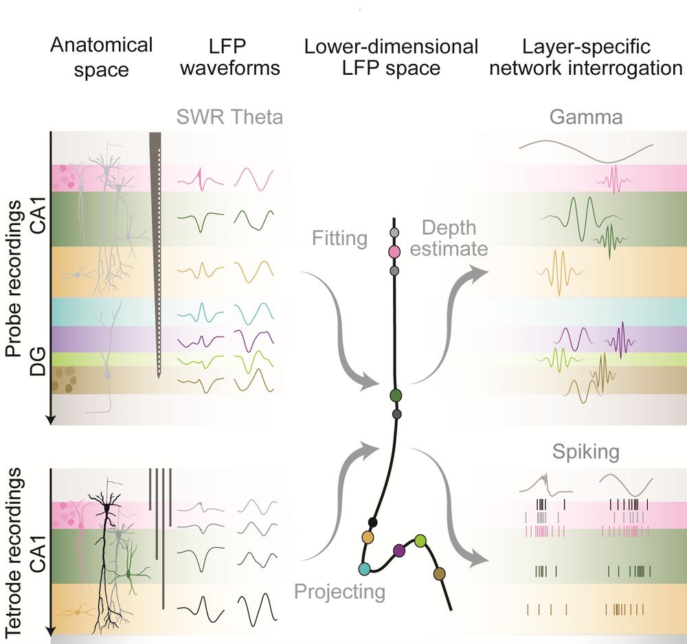
We start by building an embedding that transforms LFP signals (theta and SWR features) from a single channel into a 2D coordinate, which predicts its anatomical layer along the CA1–DG axis.
In essence, the embedding links anatomical position to electrophysiological features.
(2/13)
In essence, the embedding links anatomical position to electrophysiological features.
(2/13)

June 5, 2025 at 5:10 PM
We start by building an embedding that transforms LFP signals (theta and SWR features) from a single channel into a 2D coordinate, which predicts its anatomical layer along the CA1–DG axis.
In essence, the embedding links anatomical position to electrophysiological features.
(2/13)
In essence, the embedding links anatomical position to electrophysiological features.
(2/13)
I'm pleased to share our new work, “Spatio-temporal organization of network activity patterns in the hippocampus”, out in @cp-cellreports.bsky.social !
With Demi Brizee & David Dupret, we track how oscillations and spiking behaviour map onto hippocampal layers using an LFP-based embedding.
(1/13)
With Demi Brizee & David Dupret, we track how oscillations and spiking behaviour map onto hippocampal layers using an LFP-based embedding.
(1/13)
June 5, 2025 at 5:10 PM
I'm pleased to share our new work, “Spatio-temporal organization of network activity patterns in the hippocampus”, out in @cp-cellreports.bsky.social !
With Demi Brizee & David Dupret, we track how oscillations and spiking behaviour map onto hippocampal layers using an LFP-based embedding.
(1/13)
With Demi Brizee & David Dupret, we track how oscillations and spiking behaviour map onto hippocampal layers using an LFP-based embedding.
(1/13)