


Tom Cumming
@tomcumming.bsky.social
PhD student at Institut Pasteur studying commitment to cell death downstream of the caspases in epithelia.
Thanks also to Florence Levillayer, Léo Valon, Anne Loap, Vaishnav Manoj, Georgy Sapozhnikov, Gaëlle Letort and Alexis Matamoro-Vidal for their contributions, and finally my greatest thanks to Romain @levayerr.bsky.social for supervising the project. We hope you enjoy reading it!
May 21, 2025 at 8:52 AM
Thanks also to Florence Levillayer, Léo Valon, Anne Loap, Vaishnav Manoj, Georgy Sapozhnikov, Gaëlle Letort and Alexis Matamoro-Vidal for their contributions, and finally my greatest thanks to Romain @levayerr.bsky.social for supervising the project. We hope you enjoy reading it!
Huge thanks to all @levayerr.bsky.social lab members who contributed to this work, particularly the fantastic @avillars.bsky.social who pioneered the project and the analysis during his PhD, and bioinformatics expert Anđela Davidović who handled the large segmented datasets and ML-based prediction.
May 21, 2025 at 8:52 AM
Huge thanks to all @levayerr.bsky.social lab members who contributed to this work, particularly the fantastic @avillars.bsky.social who pioneered the project and the analysis during his PhD, and bioinformatics expert Anđela Davidović who handled the large segmented datasets and ML-based prediction.
Altogether, we present the first systematic and quantitative analysis of the relationship between caspase activity and death in vivo. Caspase threshold for death is variable between cells, spatially patterned, and informed by past exposure which can be leveraged to predict dying cells.
May 21, 2025 at 8:52 AM
Altogether, we present the first systematic and quantitative analysis of the relationship between caspase activity and death in vivo. Caspase threshold for death is variable between cells, spatially patterned, and informed by past exposure which can be leveraged to predict dying cells.
Finally, we triggered mild optoDronc activation in clones and found that their death rate 1.5-5hr later was 3.5-fold that of their WT neighbours, indicating that sublethal caspase activity can bias clone elimination in vivo.
May 21, 2025 at 8:52 AM
Finally, we triggered mild optoDronc activation in clones and found that their death rate 1.5-5hr later was 3.5-fold that of their WT neighbours, indicating that sublethal caspase activity can bias clone elimination in vivo.
Furthermore, we trained a time series forest machine learning model to predict the dying cell from a neighbouring pair (one dying cell and one surviving neighbour), and found that we could reach a strong predictive value using relative caspase values up to 3 hours prior to engagement in extrusion.

May 21, 2025 at 8:52 AM
Furthermore, we trained a time series forest machine learning model to predict the dying cell from a neighbouring pair (one dying cell and one surviving neighbour), and found that we could reach a strong predictive value using relative caspase values up to 3 hours prior to engagement in extrusion.
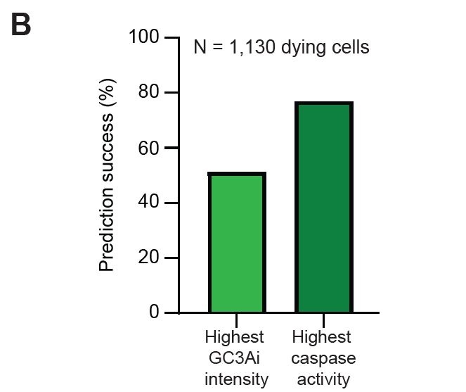
Among 1,130 groups of dying cells, the first dying cell had the highest cumulative caspase activity in 53% of cases, and the highest instantaneous caspase activity in 77% of cases. This suggested that relative caspase activity and past exposure to caspase can bias elimination between neighbours.
May 21, 2025 at 8:52 AM
Among 1,130 groups of dying cells, the first dying cell had the highest cumulative caspase activity in 53% of cases, and the highest instantaneous caspase activity in 77% of cases. This suggested that relative caspase activity and past exposure to caspase can bias elimination between neighbours.
Our lab previously showed that dying cells protect their neighbours via ERK, so locally the decision to die is not fully cell-autonomous and relies on which cell dies first in a group. We therefore checked whether relative differences in past/current caspase activity can bias clone elimination.
May 21, 2025 at 8:52 AM
Our lab previously showed that dying cells protect their neighbours via ERK, so locally the decision to die is not fully cell-autonomous and relies on which cell dies first in a group. We therefore checked whether relative differences in past/current caspase activity can bias clone elimination.
Combining optoDronc with GC3Ai we saw that cumulative caspase activity at the point of further activation is strongly predictive of cell fate at the single cell level, as the higher a cell's GC3Ai at t0 the more likely it was to die following the optoDronc pulse.

May 21, 2025 at 8:52 AM
Combining optoDronc with GC3Ai we saw that cumulative caspase activity at the point of further activation is strongly predictive of cell fate at the single cell level, as the higher a cell's GC3Ai at t0 the more likely it was to die following the optoDronc pulse.
Interestingly, when we block prior caspase with RNAi and trigger optoDronc, there is still a bias towards increased death at the midline and anterior side of the tissue, indicating other unknown spatially patterned factors modulating sensitivity to caspase independently of past activation.
May 21, 2025 at 8:52 AM
Interestingly, when we block prior caspase with RNAi and trigger optoDronc, there is still a bias towards increased death at the midline and anterior side of the tissue, indicating other unknown spatially patterned factors modulating sensitivity to caspase independently of past activation.
So could our map of caspase sensitivity be explained by the pattern of cumulative caspase activation? Indeed, the maps of deaths triggered by optoDronc in early and late pupae closely matched the underlying GC3Ai pattern, and abolishing prior caspase activity led to a more homogenous extrusion map.

May 21, 2025 at 8:52 AM
So could our map of caspase sensitivity be explained by the pattern of cumulative caspase activation? Indeed, the maps of deaths triggered by optoDronc in early and late pupae closely matched the underlying GC3Ai pattern, and abolishing prior caspase activity led to a more homogenous extrusion map.
We found this sublethal pulse of caspase was sufficient to prime cells to die over a short timescale, with cells considerably more likely to die in the region that experienced prior caspase activation 1-3 hours in the past.
May 21, 2025 at 8:52 AM
We found this sublethal pulse of caspase was sufficient to prime cells to die over a short timescale, with cells considerably more likely to die in the region that experienced prior caspase activation 1-3 hours in the past.
To test if such priming occurs, we used optoDronc to subject one lateral domain of the tissue to two pulses of mild caspase activation separated by a number of hours (blue box), and measured the number of deaths against the control contralateral domain only subjected to the latter pulse (red box).
May 21, 2025 at 8:52 AM
To test if such priming occurs, we used optoDronc to subject one lateral domain of the tissue to two pulses of mild caspase activation separated by a number of hours (blue box), and measured the number of deaths against the control contralateral domain only subjected to the latter pulse (red box).
What, then, sets the caspase threshold for a single cell? Analysing over 1,000 dying cells we found a correlation indicating that cells with high caspase a few hours before death tended to commit to extrusion at lower caspase. This suggested sublethal caspase could prime cells for later death.
May 21, 2025 at 8:52 AM
What, then, sets the caspase threshold for a single cell? Analysing over 1,000 dying cells we found a correlation indicating that cells with high caspase a few hours before death tended to commit to extrusion at lower caspase. This suggested sublethal caspase could prime cells for later death.
Compiling these maps of optoDronc-induced deaths, we saw clear hotspots of caspase sensitivity in the midline, posterior and anterior edges of the tissue. This indicated that engagement in apoptosis downstream of effector caspases can be developmentally regulated.

May 21, 2025 at 8:52 AM
Compiling these maps of optoDronc-induced deaths, we saw clear hotspots of caspase sensitivity in the midline, posterior and anterior edges of the tissue. This indicated that engagement in apoptosis downstream of effector caspases can be developmentally regulated.
To test the physiological relevance of this cell-to-cell heterogeneity in caspase threshold, we used optoDronc, an optogenetic caspase, to trigger mild caspase activation everywhere in the notum and track where cells were most and least likely to die.
May 21, 2025 at 8:52 AM
To test the physiological relevance of this cell-to-cell heterogeneity in caspase threshold, we used optoDronc, an optogenetic caspase, to trigger mild caspase activation everywhere in the notum and track where cells were most and least likely to die.
Cumulative caspase activity was very poorly predictive of engagement in apoptosis, while probability of death scaled quasi-linearly with instantaneous caspase activity. Nonetheless, no universal caspase threshold was found, and cells committed to die across a wide range of caspase levels.

May 21, 2025 at 8:52 AM
Cumulative caspase activity was very poorly predictive of engagement in apoptosis, while probability of death scaled quasi-linearly with instantaneous caspase activity. Nonetheless, no universal caspase threshold was found, and cells committed to die across a wide range of caspase levels.
By detecting the point at which individual tracked cells irreversibly commit to apoptotic extrusion, we searched for a shared threshold of cumulative (GC3Ai) or instantaneous (δGC3Ai) caspase activity that defines this point of commitment.
May 21, 2025 at 8:52 AM
By detecting the point at which individual tracked cells irreversibly commit to apoptotic extrusion, we searched for a shared threshold of cumulative (GC3Ai) or instantaneous (δGC3Ai) caspase activity that defines this point of commitment.
The bifurcation between survival and death was generally assumed to be driven by a threshold of caspase activity, but this was never tested quantitatively in vivo. We tracked over 15,000 cells expressing a caspase activity sensor in a developing epithelium to probe the existence of such a threshold.
May 21, 2025 at 8:52 AM
The bifurcation between survival and death was generally assumed to be driven by a threshold of caspase activity, but this was never tested quantitatively in vivo. We tracked over 15,000 cells expressing a caspase activity sensor in a developing epithelium to probe the existence of such a threshold.
Effector caspase activation was previously thought to trigger definitive commitment to cell death. However, the discovery of widespread survival from this event in various model systems and diverse non-apoptotic functions of caspases indicate a decision-making step downstream of caspase activation.
May 21, 2025 at 8:52 AM
Effector caspase activation was previously thought to trigger definitive commitment to cell death. However, the discovery of widespread survival from this event in various model systems and diverse non-apoptotic functions of caspases indicate a decision-making step downstream of caspase activation.