


Swathivsekhar
@swathivsekhar.bsky.social
Post-doctoral researcher at Tata Institute of Fundamental Research Hyderabad.
Veetil Lab, chemical biology.
Veetil Lab, chemical biology.
Reposted by Swathivsekhar

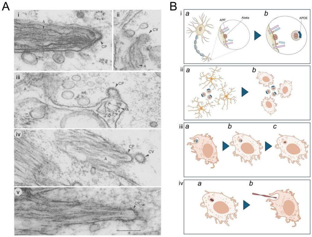
'Evidence suggesting that #microglia make #amyloid from neuronally expressed APP: a hypothesis'
John Hardy & Patrick Lewis
molecularneurodegeneration.biomedcentral.com/articles/10....
John Hardy & Patrick Lewis
molecularneurodegeneration.biomedcentral.com/articles/10....
Evidence suggesting that microglia make amyloid from neuronally expressed APP: a hypothesis - Molecular Neurodegeneration
While APP is largely neuonally expressed, Aβ amyloid is largely produced by microglia as the clearance mechanisms for damaged membranes becomes overwhelmed.
molecularneurodegeneration.biomedcentral.com
May 9, 2025 at 9:39 PM
'Evidence suggesting that #microglia make #amyloid from neuronally expressed APP: a hypothesis'
John Hardy & Patrick Lewis
molecularneurodegeneration.biomedcentral.com/articles/10....
John Hardy & Patrick Lewis
molecularneurodegeneration.biomedcentral.com/articles/10....
Rapid extension and withdrawal of microglial processes surveying CRH neurons.

🧠 🧪I am still awed by this live imaging of microglia (green) surveying #stress-sensitive CRH cells (red) in #hypothalamus and lopping off extraneous synapses. By the amazing @jessicalbolton.bsky.social #FluorescentFriday
May 4, 2025 at 4:01 AM
Rapid extension and withdrawal of microglial processes surveying CRH neurons.
This is microglial cells from mouse brain visualized using the modular fluorescent probe developed in our lab. Check out the first preprint from our lab for imaging and manipulation of these immune cells.
doi.org/10.1101/2025...
doi.org/10.1101/2025...
Here is a first look at microglia (red) from a mouse brain using MITIGATE.
April 7, 2025 at 7:29 PM
This is microglial cells from mouse brain visualized using the modular fluorescent probe developed in our lab. Check out the first preprint from our lab for imaging and manipulation of these immune cells.
doi.org/10.1101/2025...
doi.org/10.1101/2025...
Reposted by Swathivsekhar

Today, we report the first independent study from our lab describing a new technology for live imaging and manipulation of homeostatic microglia (brain-resident macrophages) in the brain.
www.biorxiv.org/content/10.1...
www.biorxiv.org/content/10.1...

GPCR-targeted imaging and manipulation of homeostatic microglia in living systems
Microglia, the resident innate immune cells of the brain, are known to perform key roles such as synaptic pruning, apoptotic debris removal, and pathogen defense in the central nervous system. Microglial mutations are directly linked to many neurodevelopmental (e.g., schizophrenia) and neurodegenerative (e.g., Alzheimer's disease) disorders, indicating the diagnostic and therapeutic potential of microglia for treating these conditions. Currently, we lack robust molecular tools to specifically image and manipulate microglia in vivo, which presents a major hurdle in our understanding of the brain-wide functions of these cells during the early onset of brain diseases. Here, we describe a molecular technology for imaging and manipulation of homeostatic microglia in live organisms (e.g., in zebrafish and mice) by covalently targeting the purinergic receptor, P2RY12. Using this technology, we imaged microglia-pathogen interactions in the larval zebrafish brain and revealed various morphological states of microglia in the adult mouse brain. We further expanded the microglia labelling approach to single-microglia tracking and microglial surfaceome mapping using photoactivatable fluorophores and photoproximity labelling, respectively. We anticipate the use of this universal tool for studying microglial biology across species to reveal the dynamics and polarization of resting microglia into a reactive state found in many neurodegenerative diseases. ### Competing Interest Statement The authors have declared no competing interest.
www.biorxiv.org
April 6, 2025 at 6:50 PM
Today, we report the first independent study from our lab describing a new technology for live imaging and manipulation of homeostatic microglia (brain-resident macrophages) in the brain.
www.biorxiv.org/content/10.1...
www.biorxiv.org/content/10.1...