


Søren Grubb
@sorengrubb.bsky.social
Biologist👩🔬, physiologist🔬, electrophysiologist⚡, cardiovascular scientist❤️, neuroscientist🧠 #precapillarysphincters #microvasculature #meninges #neurovascularcoupling #pericytes #MICrONS #microglia #macrophages #wasteclearance #aging
Also thanks to @helenelab.bsky.social for access to the CA1 dataset!
November 20, 2025 at 9:48 PM
Also thanks to @helenelab.bsky.social for access to the CA1 dataset!
We would like to acknowledge the amazing #OpenScience #ElectronMicroscopy datasets by @alleninstitute.org , @hhmijanelia.bsky.social and other sources!
This work is a collaboration between me at @ucph.bsky.social, @amreenmughal.bsky.social and Vanshika at #NIH, and Carolyn and Jennifer at #Janelia
This work is a collaboration between me at @ucph.bsky.social, @amreenmughal.bsky.social and Vanshika at #NIH, and Carolyn and Jennifer at #Janelia

a man in a suit and tie is giving a thank you sign .
ALT: a man in a suit and tie is giving a thank you sign .
media.tenor.com
November 20, 2025 at 9:04 PM
We would like to acknowledge the amazing #OpenScience #ElectronMicroscopy datasets by @alleninstitute.org , @hhmijanelia.bsky.social and other sources!
This work is a collaboration between me at @ucph.bsky.social, @amreenmughal.bsky.social and Vanshika at #NIH, and Carolyn and Jennifer at #Janelia
This work is a collaboration between me at @ucph.bsky.social, @amreenmughal.bsky.social and Vanshika at #NIH, and Carolyn and Jennifer at #Janelia
🧩 Summary:
✅ #Pericytes have #PrimaryCilia with an arterio-venular gradient🏳️🌈
✅ 🚶♀️🧠 ECs have primary #cilia, 🐁🧠 ECs don’t
✅ EC polarity aligns opposite to #BloodFlow 🧭
❔Lots of open questions about cilia’s role in #NeurovascularCoupling!
Read the full preprint: 🔗 www.biorxiv.org/content/10.1...
🧪🧵8/8
✅ #Pericytes have #PrimaryCilia with an arterio-venular gradient🏳️🌈
✅ 🚶♀️🧠 ECs have primary #cilia, 🐁🧠 ECs don’t
✅ EC polarity aligns opposite to #BloodFlow 🧭
❔Lots of open questions about cilia’s role in #NeurovascularCoupling!
Read the full preprint: 🔗 www.biorxiv.org/content/10.1...
🧪🧵8/8

November 20, 2025 at 9:04 PM
🧩 Summary:
✅ #Pericytes have #PrimaryCilia with an arterio-venular gradient🏳️🌈
✅ 🚶♀️🧠 ECs have primary #cilia, 🐁🧠 ECs don’t
✅ EC polarity aligns opposite to #BloodFlow 🧭
❔Lots of open questions about cilia’s role in #NeurovascularCoupling!
Read the full preprint: 🔗 www.biorxiv.org/content/10.1...
🧪🧵8/8
✅ #Pericytes have #PrimaryCilia with an arterio-venular gradient🏳️🌈
✅ 🚶♀️🧠 ECs have primary #cilia, 🐁🧠 ECs don’t
✅ EC polarity aligns opposite to #BloodFlow 🧭
❔Lots of open questions about cilia’s role in #NeurovascularCoupling!
Read the full preprint: 🔗 www.biorxiv.org/content/10.1...
🧪🧵8/8
🧭 #EndothelialCells are polarized against #BloodFlow direction ⤴️. #Centrioles are consistently upstream of the #nucleus. This polarity persists even in fixed tissue and could serve as a structural marker for flow direction. 📌
🧪🧵7/8
🧪🧵7/8
November 20, 2025 at 9:04 PM
🧭 #EndothelialCells are polarized against #BloodFlow direction ⤴️. #Centrioles are consistently upstream of the #nucleus. This polarity persists even in fixed tissue and could serve as a structural marker for flow direction. 📌
🧪🧵7/8
🧪🧵7/8
😮Surprise: 🚶♀️🧠 #EndothelialCells (EC) have #PrimaryCilia facing the vessel lumen, but 🐁🧠 ECs don’t. ~18% of 🚶♀️🧠 ECs are ciliated, mostly in arteriolar-end capillaries. What do these #cilia sense in the 🩸?
🧪🧵6/8
🧪🧵6/8

November 20, 2025 at 9:04 PM
😮Surprise: 🚶♀️🧠 #EndothelialCells (EC) have #PrimaryCilia facing the vessel lumen, but 🐁🧠 ECs don’t. ~18% of 🚶♀️🧠 ECs are ciliated, mostly in arteriolar-end capillaries. What do these #cilia sense in the 🩸?
🧪🧵6/8
🧪🧵6/8
In 🐁, some #pericyte #PrimaryCilia penetrate #astrocytic #endfeet and reach the #neuropil - sometimes near #synapses! Could these #cilia act as sensory “antennae” 📡for #NeurovascularCoupling?
🧪🧵5/8
🧪🧵5/8
November 20, 2025 at 9:04 PM
In 🐁, some #pericyte #PrimaryCilia penetrate #astrocytic #endfeet and reach the #neuropil - sometimes near #synapses! Could these #cilia act as sensory “antennae” 📡for #NeurovascularCoupling?
🧪🧵5/8
🧪🧵5/8
In 🐁🧠, #ultrastructure shows that #pericyte #cilia are often ensheathed by #astrocytic #endfeet. In 🚶♀️🧠, #astrocytes are swollen and cilia stay confined within the basal lamina. What are they sensing from the astrocytes?
🧪🧵4/8
🧪🧵4/8
November 20, 2025 at 9:04 PM
In 🐁🧠, #ultrastructure shows that #pericyte #cilia are often ensheathed by #astrocytic #endfeet. In 🚶♀️🧠, #astrocytes are swollen and cilia stay confined within the basal lamina. What are they sensing from the astrocytes?
🧪🧵4/8
🧪🧵4/8
🗺️We mapped #MuralCells — including #pericytes and #smoothmuscle — as vectors➡️ to visualize where #PrimaryCilia🌟 occur across the 🐁🧠 #microvasculature.
The result? A striking pattern: #cilia are abundant on #capillaries and #venules, but almost absent on #arterioles.
🧪🧵3/8
The result? A striking pattern: #cilia are abundant on #capillaries and #venules, but almost absent on #arterioles.
🧪🧵3/8
November 20, 2025 at 9:04 PM
🗺️We mapped #MuralCells — including #pericytes and #smoothmuscle — as vectors➡️ to visualize where #PrimaryCilia🌟 occur across the 🐁🧠 #microvasculature.
The result? A striking pattern: #cilia are abundant on #capillaries and #venules, but almost absent on #arterioles.
🧪🧵3/8
The result? A striking pattern: #cilia are abundant on #capillaries and #venules, but almost absent on #arterioles.
🧪🧵3/8
📊 #Pericytes are frequently ciliated in both 🐁 and 🚶♀️🧠—but mostly near #venules and venous-end #capillaries.
Venular mural cells: up to 90% ciliated in 🐁 datasets!
Arteriolar mural cells: almost none.
Why this gradient? Flow, pressure, permeability?
🧪🧵2/8
Venular mural cells: up to 90% ciliated in 🐁 datasets!
Arteriolar mural cells: almost none.
Why this gradient? Flow, pressure, permeability?
🧪🧵2/8

November 20, 2025 at 9:04 PM
📊 #Pericytes are frequently ciliated in both 🐁 and 🚶♀️🧠—but mostly near #venules and venous-end #capillaries.
Venular mural cells: up to 90% ciliated in 🐁 datasets!
Arteriolar mural cells: almost none.
Why this gradient? Flow, pressure, permeability?
🧪🧵2/8
Venular mural cells: up to 90% ciliated in 🐁 datasets!
Arteriolar mural cells: almost none.
Why this gradient? Flow, pressure, permeability?
🧪🧵2/8
That reminds me of Thor fighting “midgårdsormen” the world serpent 🤣

September 27, 2025 at 8:31 AM
That reminds me of Thor fighting “midgårdsormen” the world serpent 🤣
🔗 Full Open Access manuscript: link.springer.com/article/10.1...
👇 Curious to hear more about #PrecapillarySphincters?
Check out these threads about the subject:
Characterization: x.com/SorenGrubb/s...
Ultrastructure: x.com/SorenGrubb/s...
👇 Curious to hear more about #PrecapillarySphincters?
Check out these threads about the subject:
Characterization: x.com/SorenGrubb/s...
Ultrastructure: x.com/SorenGrubb/s...
Brain precapillary sphincters modulate myogenic tone in adult and aged mice - GeroScience
Brain precapillary sphincters, which are surrounded by contractile pericytes and are located at the junction of penetrating arterioles and first-order capillaries, can increase their diameter by ~ 30%...
link.springer.com
June 10, 2025 at 7:38 PM
🔗 Full Open Access manuscript: link.springer.com/article/10.1...
👇 Curious to hear more about #PrecapillarySphincters?
Check out these threads about the subject:
Characterization: x.com/SorenGrubb/s...
Ultrastructure: x.com/SorenGrubb/s...
👇 Curious to hear more about #PrecapillarySphincters?
Check out these threads about the subject:
Characterization: x.com/SorenGrubb/s...
Ultrastructure: x.com/SorenGrubb/s...
In older mice, where #MyogenicTone is weaker, #PrecapillarySphincters become even more critical barriers against #BloodPressure surges—compensating for #VascularStiffening and remodeling.
June 10, 2025 at 7:38 PM
In older mice, where #MyogenicTone is weaker, #PrecapillarySphincters become even more critical barriers against #BloodPressure surges—compensating for #VascularStiffening and remodeling.
These changes suggest that #PrecapillarySphincters protect the capillaries 🛡️ by acting as a dynamic #resistance that regulates #capillary #BloodFlow and #BloodPressure.
June 10, 2025 at 7:38 PM
These changes suggest that #PrecapillarySphincters protect the capillaries 🛡️ by acting as a dynamic #resistance that regulates #capillary #BloodFlow and #BloodPressure.
Using #InVivo #VolumeImaging, we found #age related #vascular changes:
• ↑ #Tortuosity of #PenetratingArterioles
• ↓ #Arteriolar-capillary density
• ↑ #Capillaries at the venous end + #aneurysm-like bulbs downstream of #PrecapillarySphincters.
• ↑ #Tortuosity of #PenetratingArterioles
• ↓ #Arteriolar-capillary density
• ↑ #Capillaries at the venous end + #aneurysm-like bulbs downstream of #PrecapillarySphincters.
June 10, 2025 at 7:38 PM
Using #InVivo #VolumeImaging, we found #age related #vascular changes:
• ↑ #Tortuosity of #PenetratingArterioles
• ↓ #Arteriolar-capillary density
• ↑ #Capillaries at the venous end + #aneurysm-like bulbs downstream of #PrecapillarySphincters.
• ↑ #Tortuosity of #PenetratingArterioles
• ↓ #Arteriolar-capillary density
• ↑ #Capillaries at the venous end + #aneurysm-like bulbs downstream of #PrecapillarySphincters.
By recording #cortical #angiograms during i.v. injection of FITC-dextran 🟢 in #adult and #old #NG2DsRed mice, we time-colored the #arterioles 🔴 and #venules 🔵. We found that #OldMice have less #collaterals and more #tortuous #PenetratingArterioles.
June 10, 2025 at 7:38 PM
By recording #cortical #angiograms during i.v. injection of FITC-dextran 🟢 in #adult and #old #NG2DsRed mice, we time-colored the #arterioles 🔴 and #venules 🔵. We found that #OldMice have less #collaterals and more #tortuous #PenetratingArterioles.
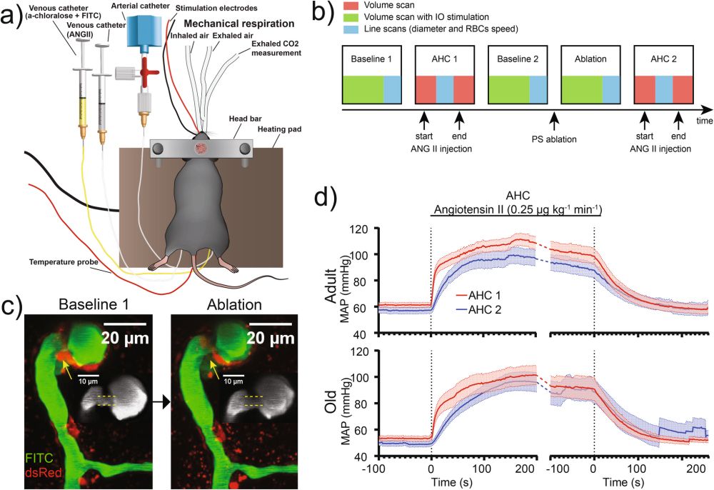
After #LaserAblating pericytes around #PrecapillarySphincters, we observed impaired #MyogenicResponse and #NeurovascularCoupling—demonstrating #PrecapillarySphincter pericyte contractility is essential.

June 10, 2025 at 7:38 PM
After #LaserAblating pericytes around #PrecapillarySphincters, we observed impaired #MyogenicResponse and #NeurovascularCoupling—demonstrating #PrecapillarySphincter pericyte contractility is essential.
#OldMice also showed decreased pulsations of microvessel diameters measured by transverse line scans, likely due to #VascularStiffening.

June 10, 2025 at 7:38 PM
#OldMice also showed decreased pulsations of microvessel diameters measured by transverse line scans, likely due to #VascularStiffening.
In aged mice, the #MyogenicResponse was significantly blunted. This was shown by an increased #CorrelationCoefficient (CC) between #VascularDiameter and #BloodPressure

June 10, 2025 at 7:38 PM
In aged mice, the #MyogenicResponse was significantly blunted. This was shown by an increased #CorrelationCoefficient (CC) between #VascularDiameter and #BloodPressure