


Stefan Mereiter
@smereiter.bsky.social
Glycobiologist & Cancer Researcher working at IMBA and MedUni Vienna, Austria. Exploring the structural-functional glycoproteome and cancer immunoregulation.
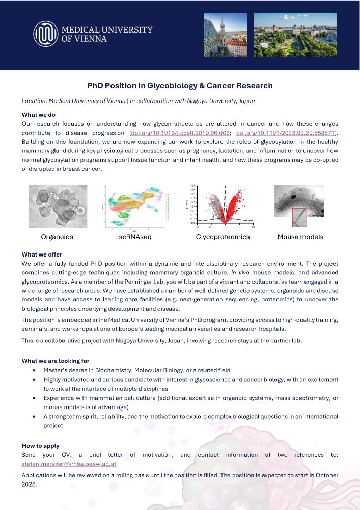
🚨 PhD position open! 🚨
Join our team at the Medical University of Vienna to study glycan regulation in mammary organoids, milk, and breast cancer.
Details in the image – please share! 🧬✨
Join our team at the Medical University of Vienna to study glycan regulation in mammary organoids, milk, and breast cancer.
Details in the image – please share! 🧬✨
May 21, 2025 at 1:16 PM
🚨 PhD position open! 🚨
Join our team at the Medical University of Vienna to study glycan regulation in mammary organoids, milk, and breast cancer.
Details in the image – please share! 🧬✨
Join our team at the Medical University of Vienna to study glycan regulation in mammary organoids, milk, and breast cancer.
Details in the image – please share! 🧬✨
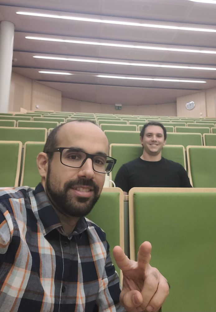
Feeling honored to work in a lab with two (!) of 2024’s most cited scientists: mentor Josef Penninger 🧠 and lab colleague Max Kellner 📚 (@clarivate). Max (spot him sitting behind me in yesterday’s lab meeting) is a PhD student 🤯
November 21, 2024 at 9:46 AM
Feeling honored to work in a lab with two (!) of 2024’s most cited scientists: mentor Josef Penninger 🧠 and lab colleague Max Kellner 📚 (@clarivate). Max (spot him sitting behind me in yesterday’s lab meeting) is a PhD student 🤯
12/
Great collaboration between BOKU University, IMBA, MedUni Vienna, and Michigan University! Thank you, J. Helm and the amazing Stadlmann Lab for taking me on this ride!! Together, we’re uncovering new insights into glycans. 🟦 🟡 🔺 🟢 🔶 🟨
Great collaboration between BOKU University, IMBA, MedUni Vienna, and Michigan University! Thank you, J. Helm and the amazing Stadlmann Lab for taking me on this ride!! Together, we’re uncovering new insights into glycans. 🟦 🟡 🔺 🟢 🔶 🟨
November 19, 2024 at 9:50 AM
12/
Great collaboration between BOKU University, IMBA, MedUni Vienna, and Michigan University! Thank you, J. Helm and the amazing Stadlmann Lab for taking me on this ride!! Together, we’re uncovering new insights into glycans. 🟦 🟡 🔺 🟢 🔶 🟨
Great collaboration between BOKU University, IMBA, MedUni Vienna, and Michigan University! Thank you, J. Helm and the amazing Stadlmann Lab for taking me on this ride!! Together, we’re uncovering new insights into glycans. 🟦 🟡 🔺 🟢 🔶 🟨
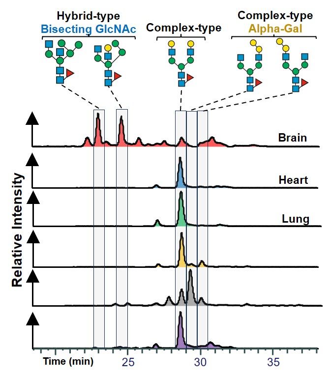
10/
For those ready to dive deeper: We haven’t even started with the chromatography data! PGC allows us to differentiate N-glycans with the same mass but distinct structures, adding more layers of tissue-specific complexity.
For those ready to dive deeper: We haven’t even started with the chromatography data! PGC allows us to differentiate N-glycans with the same mass but distinct structures, adding more layers of tissue-specific complexity.
November 19, 2024 at 9:50 AM
10/
For those ready to dive deeper: We haven’t even started with the chromatography data! PGC allows us to differentiate N-glycans with the same mass but distinct structures, adding more layers of tissue-specific complexity.
For those ready to dive deeper: We haven’t even started with the chromatography data! PGC allows us to differentiate N-glycans with the same mass but distinct structures, adding more layers of tissue-specific complexity.
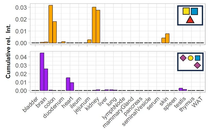
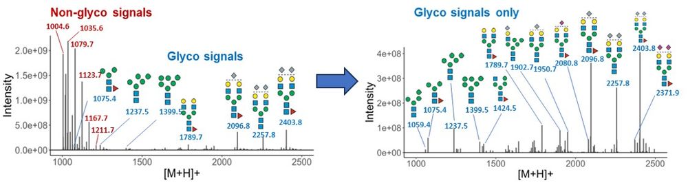
9/
But wait—there was more! After filtering non-glycan spectra and expected glycans, we found spectra representing unexpected glycan structures and mapped them across all tissues! 🔍
But wait—there was more! After filtering non-glycan spectra and expected glycans, we found spectra representing unexpected glycan structures and mapped them across all tissues! 🔍
November 19, 2024 at 9:50 AM
9/
But wait—there was more! After filtering non-glycan spectra and expected glycans, we found spectra representing unexpected glycan structures and mapped them across all tissues! 🔍
But wait—there was more! After filtering non-glycan spectra and expected glycans, we found spectra representing unexpected glycan structures and mapped them across all tissues! 🔍
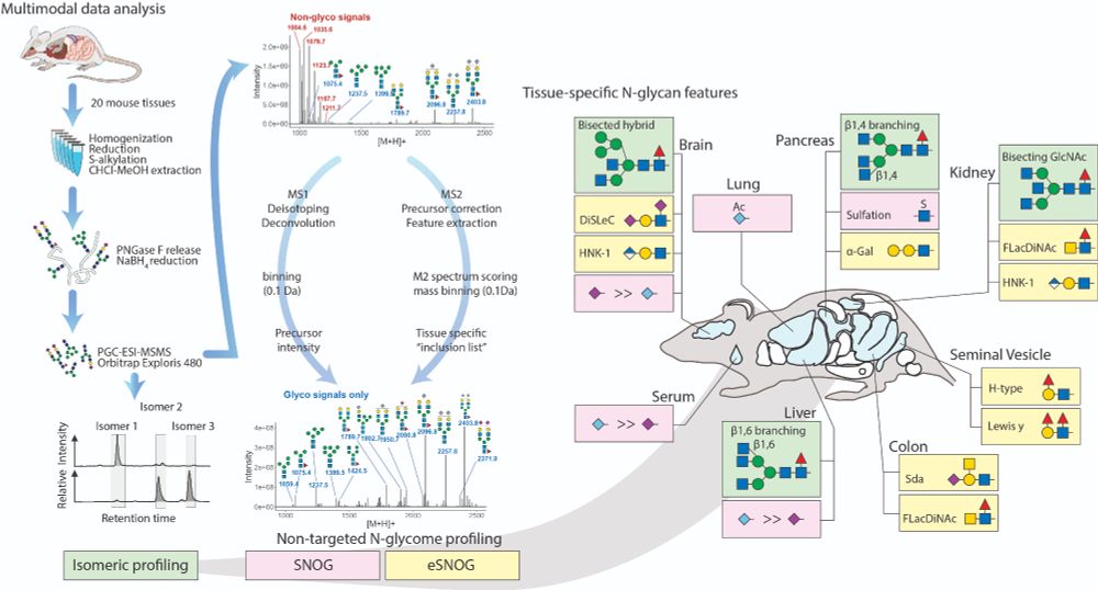
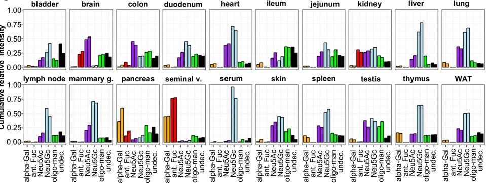
8/
Next, we extended SNOG with eSNOG, extracting spectra with special fragment ions to compare glycan structural features like α-Gal, antennary Fuc, Neu5Ac, Neu5Gc, and oligo-Man across tissues.
Next, we extended SNOG with eSNOG, extracting spectra with special fragment ions to compare glycan structural features like α-Gal, antennary Fuc, Neu5Ac, Neu5Gc, and oligo-Man across tissues.
November 19, 2024 at 9:50 AM
8/
Next, we extended SNOG with eSNOG, extracting spectra with special fragment ions to compare glycan structural features like α-Gal, antennary Fuc, Neu5Ac, Neu5Gc, and oligo-Man across tissues.
Next, we extended SNOG with eSNOG, extracting spectra with special fragment ions to compare glycan structural features like α-Gal, antennary Fuc, Neu5Ac, Neu5Gc, and oligo-Man across tissues.
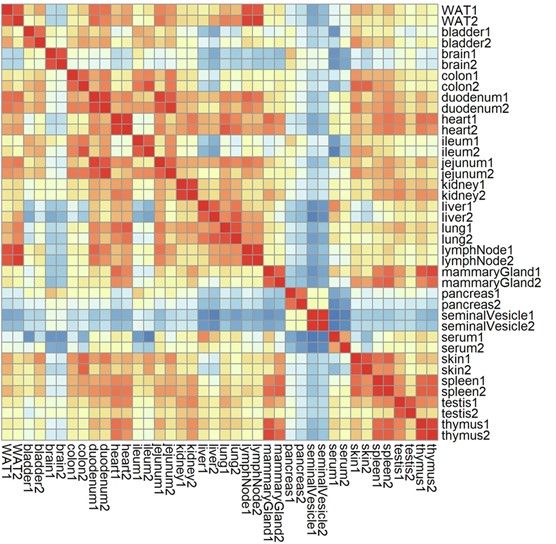
7/
With SNOG-filtered data, we saw clear, tissue-specific glycan clusters emerge, with high technical reproducibility across replicates. This filtering step was a game-changer!
With SNOG-filtered data, we saw clear, tissue-specific glycan clusters emerge, with high technical reproducibility across replicates. This filtering step was a game-changer!
November 19, 2024 at 9:50 AM
7/
With SNOG-filtered data, we saw clear, tissue-specific glycan clusters emerge, with high technical reproducibility across replicates. This filtering step was a game-changer!
With SNOG-filtered data, we saw clear, tissue-specific glycan clusters emerge, with high technical reproducibility across replicates. This filtering step was a game-changer!
6/
First up: SNOG-filtering. This automated step removes contaminants (up to 75% in some tissues!) so we can focus on genuine N-glycan signals.
First up: SNOG-filtering. This automated step removes contaminants (up to 75% in some tissues!) so we can focus on genuine N-glycan signals.
November 19, 2024 at 9:50 AM
6/
First up: SNOG-filtering. This automated step removes contaminants (up to 75% in some tissues!) so we can focus on genuine N-glycan signals.
First up: SNOG-filtering. This automated step removes contaminants (up to 75% in some tissues!) so we can focus on genuine N-glycan signals.
5/
Our approach had to be scalable, non-targeted (no pre-built mass lists, please!), and data-driven. Automated spectral ID would be the icing on the cake! 🧁 We combined PGC analysis with multiple profiling strategies for semi/quantitative N-glycomics.
Our approach had to be scalable, non-targeted (no pre-built mass lists, please!), and data-driven. Automated spectral ID would be the icing on the cake! 🧁 We combined PGC analysis with multiple profiling strategies for semi/quantitative N-glycomics.

November 19, 2024 at 9:50 AM
5/
Our approach had to be scalable, non-targeted (no pre-built mass lists, please!), and data-driven. Automated spectral ID would be the icing on the cake! 🧁 We combined PGC analysis with multiple profiling strategies for semi/quantitative N-glycomics.
Our approach had to be scalable, non-targeted (no pre-built mass lists, please!), and data-driven. Automated spectral ID would be the icing on the cake! 🧁 We combined PGC analysis with multiple profiling strategies for semi/quantitative N-glycomics.
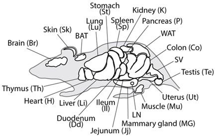
3/
So, we analyzed N-glycans from 20 mouse tissues, generating a comprehensive, structure-resolved N-glycome Atlas. Using PGC columns for isomer separation and an Orbitrap for high-res MS/MS, we assembled a mountain of data. Now what?
So, we analyzed N-glycans from 20 mouse tissues, generating a comprehensive, structure-resolved N-glycome Atlas. Using PGC columns for isomer separation and an Orbitrap for high-res MS/MS, we assembled a mountain of data. Now what?
November 19, 2024 at 9:50 AM
3/
So, we analyzed N-glycans from 20 mouse tissues, generating a comprehensive, structure-resolved N-glycome Atlas. Using PGC columns for isomer separation and an Orbitrap for high-res MS/MS, we assembled a mountain of data. Now what?
So, we analyzed N-glycans from 20 mouse tissues, generating a comprehensive, structure-resolved N-glycome Atlas. Using PGC columns for isomer separation and an Orbitrap for high-res MS/MS, we assembled a mountain of data. Now what?
2/
Have you ever wondered how many unique N-glycan structures a single organism might create? Or if the sugars on your kidney cells differ from those in your brain?
We did too, and we set out to investigate!
Have you ever wondered how many unique N-glycan structures a single organism might create? Or if the sugars on your kidney cells differ from those in your brain?
We did too, and we set out to investigate!
November 19, 2024 at 9:50 AM
2/
Have you ever wondered how many unique N-glycan structures a single organism might create? Or if the sugars on your kidney cells differ from those in your brain?
We did too, and we set out to investigate!
Have you ever wondered how many unique N-glycan structures a single organism might create? Or if the sugars on your kidney cells differ from those in your brain?
We did too, and we set out to investigate!
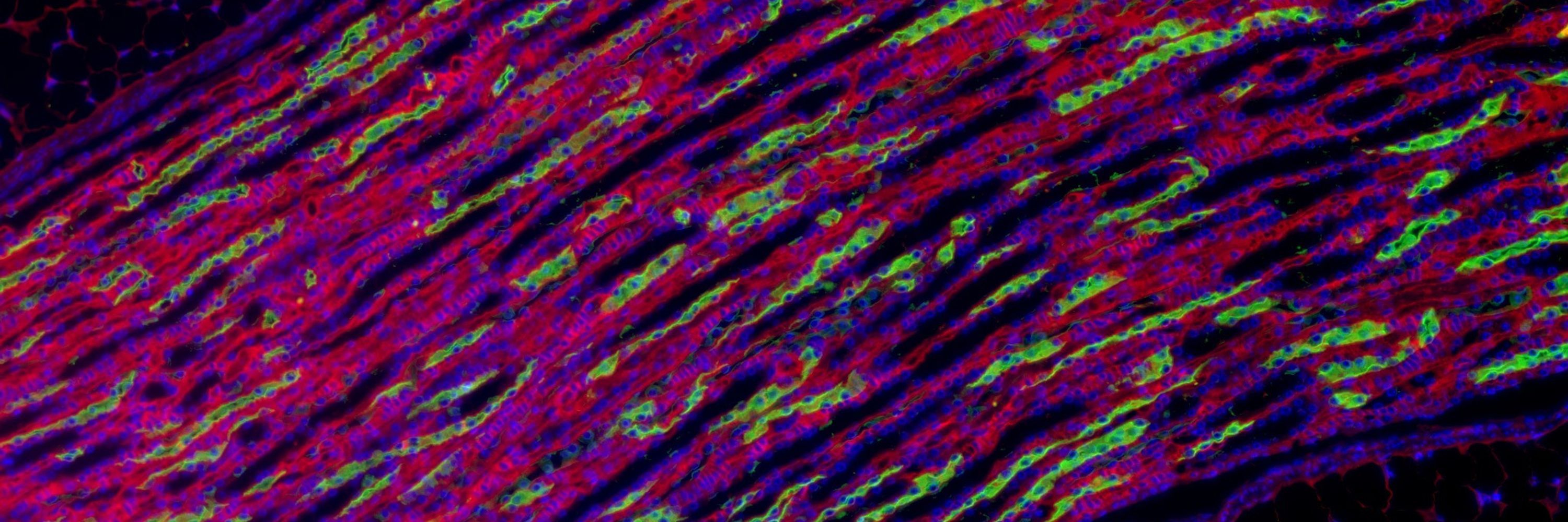
1/
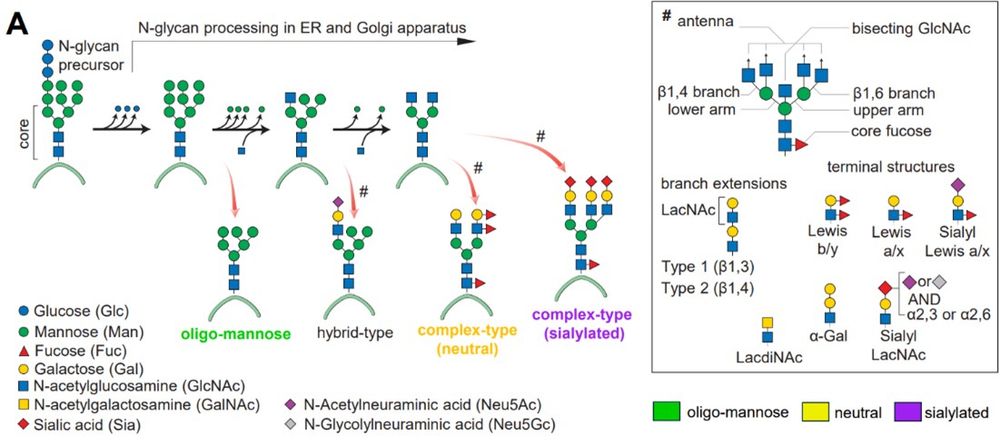
Hi 🦋! Did you know? Glycans—complex sugar structures—decorate almost every protein processed by the ER/Golgi. These N-glycans coat our cells, playing critical roles in cell interactions and immune processes. 🍬 Curious about how they vary? Let’s dive in! #GlycoTime
Hi 🦋! Did you know? Glycans—complex sugar structures—decorate almost every protein processed by the ER/Golgi. These N-glycans coat our cells, playing critical roles in cell interactions and immune processes. 🍬 Curious about how they vary? Let’s dive in! #GlycoTime
November 19, 2024 at 9:50 AM
1/
Hi 🦋! Did you know? Glycans—complex sugar structures—decorate almost every protein processed by the ER/Golgi. These N-glycans coat our cells, playing critical roles in cell interactions and immune processes. 🍬 Curious about how they vary? Let’s dive in! #GlycoTime
Hi 🦋! Did you know? Glycans—complex sugar structures—decorate almost every protein processed by the ER/Golgi. These N-glycans coat our cells, playing critical roles in cell interactions and immune processes. 🍬 Curious about how they vary? Let’s dive in! #GlycoTime