


Philip Hubbard
@philiphubbard.bsky.social
When #HHMIJanelia released the #Drosophila hemibrain in 2020, rendering all cell types at once with full shading/shadows was too hard, so the video showed them region by region. Technology has since advanced, and I went back and rendered them all together. I like how it shows the internal structure.
October 27, 2025 at 8:16 AM
When #HHMIJanelia released the #Drosophila hemibrain in 2020, rendering all cell types at once with full shading/shadows was too hard, so the video showed them region by region. Technology has since advanced, and I went back and rendered them all together. I like how it shows the internal structure.
On this day 10 years ago, #Tableau released Vizable for the iPad. It gave a way to explore data with gestures: pinch/unpinch to aggregate/disaggregate, swipe to filter out, etc. The development effort, led by Robin Stewart and Dave Story, was great fun. #DataViz #DataVis
vimeo.com/142648727
vimeo.com/142648727
October 20, 2025 at 8:46 AM
On this day 10 years ago, #Tableau released Vizable for the iPad. It gave a way to explore data with gestures: pinch/unpinch to aggregate/disaggregate, swipe to filter out, etc. The development effort, led by Robin Stewart and Dave Story, was great fun. #DataViz #DataVis
vimeo.com/142648727
vimeo.com/142648727
Some fly neurons are “dimorphic”, existing in both males and females but connecting to different neighboring neurons. Neighbors may be “isomorphic”, the same in both male and female; or sex specific; or dimorphic themselves. Here's an example, the type AOTU012, present in left and right instances.
October 8, 2025 at 9:11 AM
Some fly neurons are “dimorphic”, existing in both males and females but connecting to different neighboring neurons. Neighbors may be “isomorphic”, the same in both male and female; or sex specific; or dimorphic themselves. Here's an example, the type AOTU012, present in left and right instances.
A connectome spanning the brain and nerve cord reveals interesting pathways. Visual-motor pathways connect visual neurons, which help the fly detect objects, to motor neurons, which help the fly move in response. Here's one such pathway, from R1-R6 visual neurons to the DNg13 motor neuron.
October 7, 2025 at 8:27 AM
A connectome spanning the brain and nerve cord reveals interesting pathways. Visual-motor pathways connect visual neurons, which help the fly detect objects, to motor neurons, which help the fly move in response. Here's one such pathway, from R1-R6 visual neurons to the DNg13 motor neuron.
Come to #HHMIJanelia to work on the frontiers of AI and biology. Apply now for an AI Engineer position working on protein biosensors:
hhmi.wd1.myworkdayjobs.com/en-US/Extern...
More openings coming soon. Interesting AI is widespread at Janelia, like this diffusion model from Larissa Heinrich.
hhmi.wd1.myworkdayjobs.com/en-US/Extern...
More openings coming soon. Interesting AI is widespread at Janelia, like this diffusion model from Larissa Heinrich.
September 16, 2025 at 4:55 PM
Come to #HHMIJanelia to work on the frontiers of AI and biology. Apply now for an AI Engineer position working on protein biosensors:
hhmi.wd1.myworkdayjobs.com/en-US/Extern...
More openings coming soon. Interesting AI is widespread at Janelia, like this diffusion model from Larissa Heinrich.
hhmi.wd1.myworkdayjobs.com/en-US/Extern...
More openings coming soon. Interesting AI is widespread at Janelia, like this diffusion model from Larissa Heinrich.
The #HHMIJanelia FuncEWorm project is building the first cellular/molecular blueprint of an entire organism, the C. elegans nematode. Here's a video showing some of what's involved: light sheet fluorescence microscopy, electron microscopy, spatial transcriptomics.
www.janelia.org/project-team...
www.janelia.org/project-team...
August 14, 2025 at 5:08 PM
The #HHMIJanelia FuncEWorm project is building the first cellular/molecular blueprint of an entire organism, the C. elegans nematode. Here's a video showing some of what's involved: light sheet fluorescence microscopy, electron microscopy, spatial transcriptomics.
www.janelia.org/project-team...
www.janelia.org/project-team...
What does a neural network look like? Maybe like this view of Larissa Heinrich's U-net for organelle segmentation, with edges between layer inputs and outputs.
Want to help build networks like this? Join #HHMIJanelia's new AI initiative as a data engineer:
hhmi.wd1.myworkdayjobs.com/en-US/Extern...
Want to help build networks like this? Join #HHMIJanelia's new AI initiative as a data engineer:
hhmi.wd1.myworkdayjobs.com/en-US/Extern...
July 23, 2025 at 9:22 AM
What does a neural network look like? Maybe like this view of Larissa Heinrich's U-net for organelle segmentation, with edges between layer inputs and outputs.
Want to help build networks like this? Join #HHMIJanelia's new AI initiative as a data engineer:
hhmi.wd1.myworkdayjobs.com/en-US/Extern...
Want to help build networks like this? Join #HHMIJanelia's new AI initiative as a data engineer:
hhmi.wd1.myworkdayjobs.com/en-US/Extern...
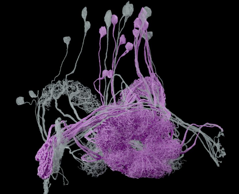
A final found image, with blue LPsP neurons that input to the grey PFL1s.
June 18, 2025 at 10:48 PM
A final found image, with blue LPsP neurons that input to the grey PFL1s.
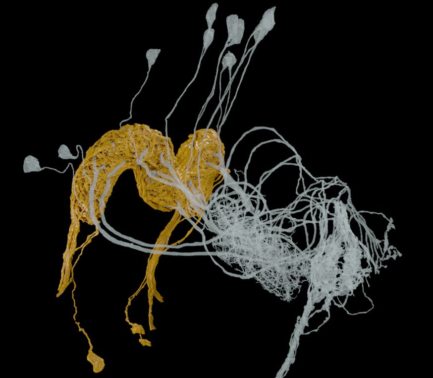
Here's another image I found, showing Delta7 neurons in gold providing more inputs to PFL1 neurons in grey. I like that the transparency makes the PFL1s look almost like glass.
June 18, 2025 at 10:48 PM
Here's another image I found, showing Delta7 neurons in gold providing more inputs to PFL1 neurons in grey. I like that the transparency makes the PFL1s look almost like glass.
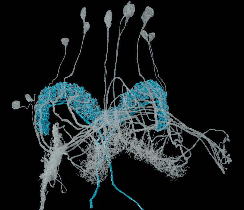
Cleaning up disk space, I found this image I made for someone not long after the release of the #HHMIJanelia #Drosophila hemibrain #connectome in 2020. It shows EPG neurons in pink providing inputs to PFL1 neurons in transparent grey. I'm not sure if the image was ever used.
June 18, 2025 at 10:46 PM
Cleaning up disk space, I found this image I made for someone not long after the release of the #HHMIJanelia #Drosophila hemibrain #connectome in 2020. It shows EPG neurons in pink providing inputs to PFL1 neurons in transparent grey. I'm not sure if the image was ever used.
When beauty and wonder seem elusive, try HHMI's Beautiful Biology site, for a reminder of what the scientific study of nature has to offer. Noah Green and the team update the collection every week and write accessible descriptions for everything.
www.hhmi.org/beautifulbiology
www.hhmi.org/beautifulbiology
March 28, 2025 at 9:02 AM
When beauty and wonder seem elusive, try HHMI's Beautiful Biology site, for a reminder of what the scientific study of nature has to offer. Noah Green and the team update the collection every week and write accessible descriptions for everything.
www.hhmi.org/beautifulbiology
www.hhmi.org/beautifulbiology
We made the experimental system with a toolkit of software packages built on the #Unity3D game engine. Here's an example of a "meadow" world: on top is the fly's view warped for a panorama of four display screens, below is some simulated fly motion.
github.com/JaneliaSciCo...
github.com/JaneliaSciCo...
March 10, 2025 at 9:25 AM
We made the experimental system with a toolkit of software packages built on the #Unity3D game engine. Here's an example of a "meadow" world: on top is the fly's view warped for a panorama of four display screens, below is some simulated fly motion.
github.com/JaneliaSciCo...
github.com/JaneliaSciCo...
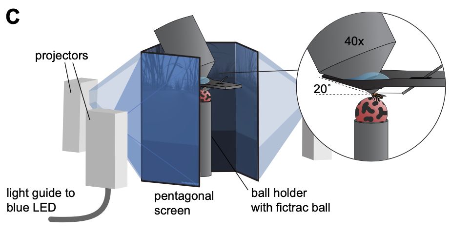
It's fascinating to see #Drosophila #Neuroscience experiments run in virtual reality (VR), with a fruit fly walking on a tiny trackball looking at tiny video displays. Here's a schematic of @hannah-haberkern.bsky.social 's rig, from our 2022 preprint:
doi.org/10.1101/2022...
doi.org/10.1101/2022...
March 10, 2025 at 9:22 AM
It's fascinating to see #Drosophila #Neuroscience experiments run in virtual reality (VR), with a fruit fly walking on a tiny trackball looking at tiny video displays. Here's a schematic of @hannah-haberkern.bsky.social 's rig, from our 2022 preprint:
doi.org/10.1101/2022...
doi.org/10.1101/2022...
I wanted a distraction from the news so I made this video, of some #Drosophila neurons with interesting morphologies: the mushroom body output neurons (MBONs). I used reconstructions from the FlyWire data set, and rendered with #Blender3D driven by neuVid.
February 23, 2025 at 7:39 PM
I wanted a distraction from the news so I made this video, of some #Drosophila neurons with interesting morphologies: the mushroom body output neurons (MBONs). I used reconstructions from the FlyWire data set, and rendered with #Blender3D driven by neuVid.
Color is enriching. Here is a meditation on coloring a ring of #Drosophila EPG neurons by heading angle. These colormaps---"cividis", "plasma", "viridis", "turbo"---aim to be "equiluminant" and/or "perceptually uniform", with the goal of faithfully depicting the smooth increase of the angle.

February 11, 2025 at 10:15 AM
Color is enriching. Here is a meditation on coloring a ring of #Drosophila EPG neurons by heading angle. These colormaps---"cividis", "plasma", "viridis", "turbo"---aim to be "equiluminant" and/or "perceptually uniform", with the goal of faithfully depicting the smooth increase of the angle.
Here's a #MicroscopyMonday segmentation of some neurons from the optic lobe, or visual processing area of the #Drosophila fruit fly's brain. The #connectome for the full optic lobe was released in 2024 by #HHMIJanelia, Google, and the University of Cambridge, with support from the Wellcome Trust.

February 3, 2025 at 9:41 AM
Here's a #MicroscopyMonday segmentation of some neurons from the optic lobe, or visual processing area of the #Drosophila fruit fly's brain. The #connectome for the full optic lobe was released in 2024 by #HHMIJanelia, Google, and the University of Cambridge, with support from the Wellcome Trust.
This segmentation of #MicroscopyMonday data reconstructs neurons of the "MANC", or male adult nerve cord, from the #Drosophila fruit fly. The nerve cord is like a spinal cord and connects the brain and muscles. #HHMIJanelia, Google, and the University of Cambridge released this work in 2023.

January 27, 2025 at 10:22 AM
This segmentation of #MicroscopyMonday data reconstructs neurons of the "MANC", or male adult nerve cord, from the #Drosophila fruit fly. The nerve cord is like a spinal cord and connects the brain and muscles. #HHMIJanelia, Google, and the University of Cambridge released this work in 2023.
On this day 5 years ago #HHMIJanelia and Google released the "hemibrain", a map of neural connections in much of the #Drosophila fly brain. At the time it was the largest such #connectome ever created. Here's a video from the release (polished a bit), showing the interesting shapes of the neurons.
January 22, 2025 at 9:35 AM
On this day 5 years ago #HHMIJanelia and Google released the "hemibrain", a map of neural connections in much of the #Drosophila fly brain. At the time it was the largest such #connectome ever created. Here's a video from the release (polished a bit), showing the interesting shapes of the neurons.
...then proofreaders from #HHMIJanelia made fixes. Here's a tool proofreaders used, to cleave false merges by designating seeds on different neurons, which Stuart Berg's graph algorithm then separated. The video's from 5 years ago so it looks a bit old fashioned, but the tool still works!
January 13, 2025 at 9:57 AM
...then proofreaders from #HHMIJanelia made fixes. Here's a tool proofreaders used, to cleave false merges by designating seeds on different neurons, which Stuart Berg's graph algorithm then separated. The video's from 5 years ago so it looks a bit old fashioned, but the tool still works!
A use of #MicroscopyMonday data is making segmentations. Here's one, of #Drosophila protocerebral bridge and nodulus neurons, from the 2020 Hemibrain by #HHMIJanelia and Google. Michał Januszewski and @stardazed0.bsky.social 's team made an initial segmentation with #AI flood-fill algorithms...

January 13, 2025 at 9:56 AM
A use of #MicroscopyMonday data is making segmentations. Here's one, of #Drosophila protocerebral bridge and nodulus neurons, from the 2020 Hemibrain by #HHMIJanelia and Google. Michał Januszewski and @stardazed0.bsky.social 's team made an initial segmentation with #AI flood-fill algorithms...
On this day 5 years ago, I released version 1.0 of neuVid. It takes high-level descriptions of #Neuroscience or #CellBiology videos and renders them with #Blender or VVD Viewer. Here's a montage of how I've used it. Thanks to #HHMIJanelia for support, especially Steve Plaza and Stuart Berg of FlyEM.
January 8, 2025 at 9:47 AM
On this day 5 years ago, I released version 1.0 of neuVid. It takes high-level descriptions of #Neuroscience or #CellBiology videos and renders them with #Blender or VVD Viewer. Here's a montage of how I've used it. Thanks to #HHMIJanelia for support, especially Steve Plaza and Stuart Berg of FlyEM.
This #FluorescenceFriday video shows 89 volumes from the #HHMIJanelia FlyLight Split-GAL4 collection: premotor and related neurons from the #Drosophila fruit fly's equivalent of a spinal cord. I gave a high-level description of the video to neuVid, which drove rendering by VVD Viewer.
January 3, 2025 at 9:59 AM
This #FluorescenceFriday video shows 89 volumes from the #HHMIJanelia FlyLight Split-GAL4 collection: premotor and related neurons from the #Drosophila fruit fly's equivalent of a spinal cord. I gave a high-level description of the video to neuVid, which drove rendering by VVD Viewer.
I used VVD Viewer for this #FluorescenceFriday video: 141 volumes from the #HHMIJanelia FlyLight Split-GAL4 #Drosophila driver collection. The animation comes from a high-level description processed by neuVid. Such a rapid tour of the data has questionable scientific value, but it's fun to watch!
December 20, 2024 at 9:57 AM
I used VVD Viewer for this #FluorescenceFriday video: 141 volumes from the #HHMIJanelia FlyLight Split-GAL4 #Drosophila driver collection. The animation comes from a high-level description processed by neuVid. Such a rapid tour of the data has questionable scientific value, but it's fun to watch!
My favorite volume renderer for fluorescence #microscopy is VVD Viewer, by Takashi Kawase and Hideo Otsuna at #HHMIJanelia. Takashi wrote fast GPU code. Hideo wrote a transfer function with 3D sampling and a final 2D power function, which together show just enough low data values for visual context.
December 19, 2024 at 9:56 AM
My favorite volume renderer for fluorescence #microscopy is VVD Viewer, by Takashi Kawase and Hideo Otsuna at #HHMIJanelia. Takashi wrote fast GPU code. Hideo wrote a transfer function with 3D sampling and a final 2D power function, which together show just enough low data values for visual context.
For fun, here's one more video of #CellBiology data from the OpenOrganelle collection. This one shows nuclei and mitochondria from a sample of mouse liver tissue, made with neuVid driving #Blender.
December 12, 2024 at 10:11 AM
For fun, here's one more video of #CellBiology data from the OpenOrganelle collection. This one shows nuclei and mitochondria from a sample of mouse liver tissue, made with neuVid driving #Blender.