


Julian Gargiulo
@nano-juli.bsky.social
Physicist and dad. Developing photonics tools for plasmonic chemistry and nanothermometry at UNSAM.
Drosophila brain imaging by #SnoutClub.
Photons are my own.
Drosophila brain imaging by #SnoutClub.
Photons are my own.
We are seeking assistance in locating a missing researcher in Germany. If anyone is based in Germany or has contacts in the area who might be able to help, please get in touch as soon as possible.
Any information or support you can provide would be greatly appreciated.
Any information or support you can provide would be greatly appreciated.

October 16, 2025 at 12:10 AM
We are seeking assistance in locating a missing researcher in Germany. If anyone is based in Germany or has contacts in the area who might be able to help, please get in touch as soon as possible.
Any information or support you can provide would be greatly appreciated.
Any information or support you can provide would be greatly appreciated.
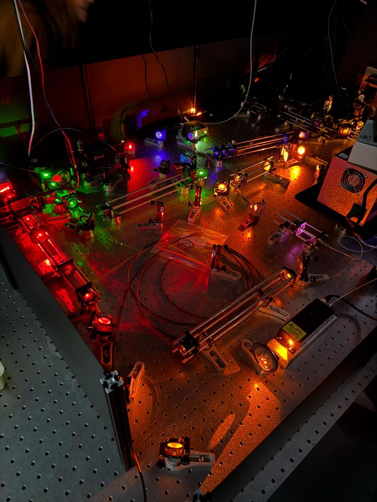
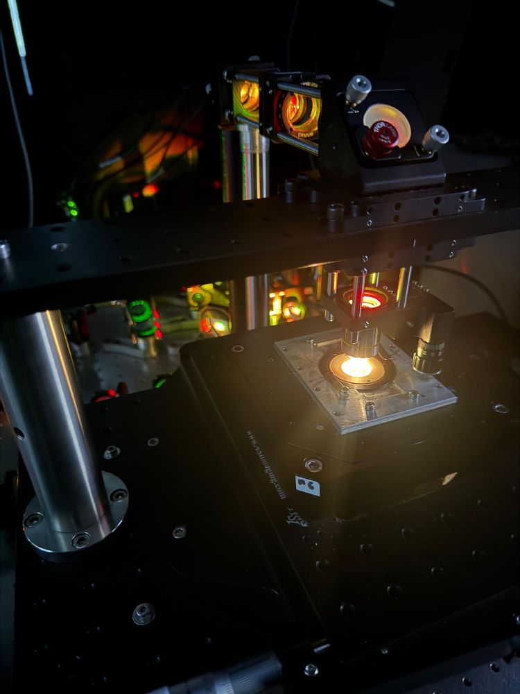
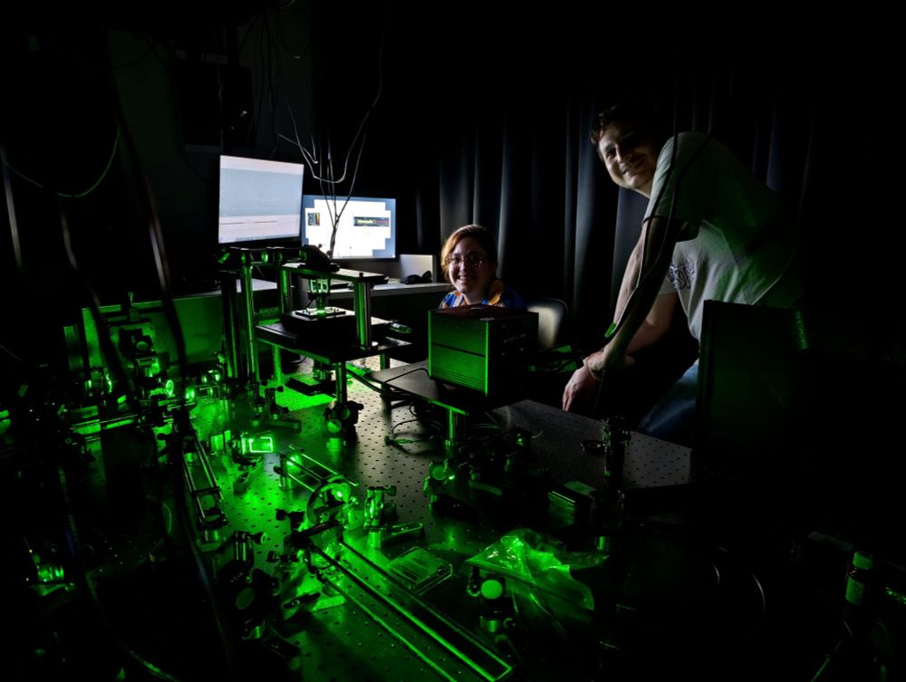
Lasers on at UNSAM.
June 11, 2025 at 9:43 PM
Lasers on at UNSAM.
May 7, 2025 at 10:55 PM
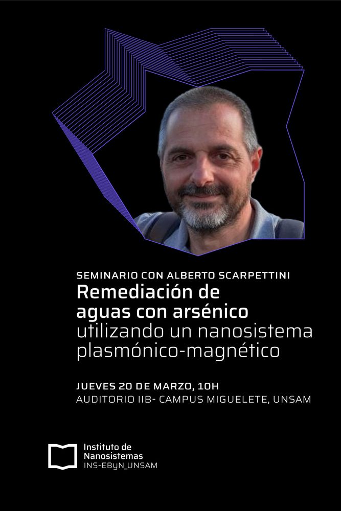
📣SEMINARIO INS
El investigador Alberto Scarpettini charlará sobre nanoestructuras híbridas núcleo-satélite formadas por satélites plasmónicos de oro soportados sobre núcleos magnéticos de óxidos de hierro para la remediación asistida por luz solar de agua que contiene arsénico
El investigador Alberto Scarpettini charlará sobre nanoestructuras híbridas núcleo-satélite formadas por satélites plasmónicos de oro soportados sobre núcleos magnéticos de óxidos de hierro para la remediación asistida por luz solar de agua que contiene arsénico
March 19, 2025 at 6:10 PM
📣SEMINARIO INS
El investigador Alberto Scarpettini charlará sobre nanoestructuras híbridas núcleo-satélite formadas por satélites plasmónicos de oro soportados sobre núcleos magnéticos de óxidos de hierro para la remediación asistida por luz solar de agua que contiene arsénico
El investigador Alberto Scarpettini charlará sobre nanoestructuras híbridas núcleo-satélite formadas por satélites plasmónicos de oro soportados sobre núcleos magnéticos de óxidos de hierro para la remediación asistida por luz solar de agua que contiene arsénico
It's been a long journey, only possible thanks to my lovely co-authors who are not (yet) in Bluesky.
Big thanks also to @amsikking.bsky.social , @andrewgyork.bsky.social and
@lumasullo.bsky.social, they have always been there to help.
Stay tuned for new imaging on living Drosophila!
6/6
Big thanks also to @amsikking.bsky.social , @andrewgyork.bsky.social and
@lumasullo.bsky.social, they have always been there to help.
Stay tuned for new imaging on living Drosophila!
6/6
February 26, 2025 at 2:48 AM
It's been a long journey, only possible thanks to my lovely co-authors who are not (yet) in Bluesky.
Big thanks also to @amsikking.bsky.social , @andrewgyork.bsky.social and
@lumasullo.bsky.social, they have always been there to help.
Stay tuned for new imaging on living Drosophila!
6/6
Big thanks also to @amsikking.bsky.social , @andrewgyork.bsky.social and
@lumasullo.bsky.social, they have always been there to help.
Stay tuned for new imaging on living Drosophila!
6/6
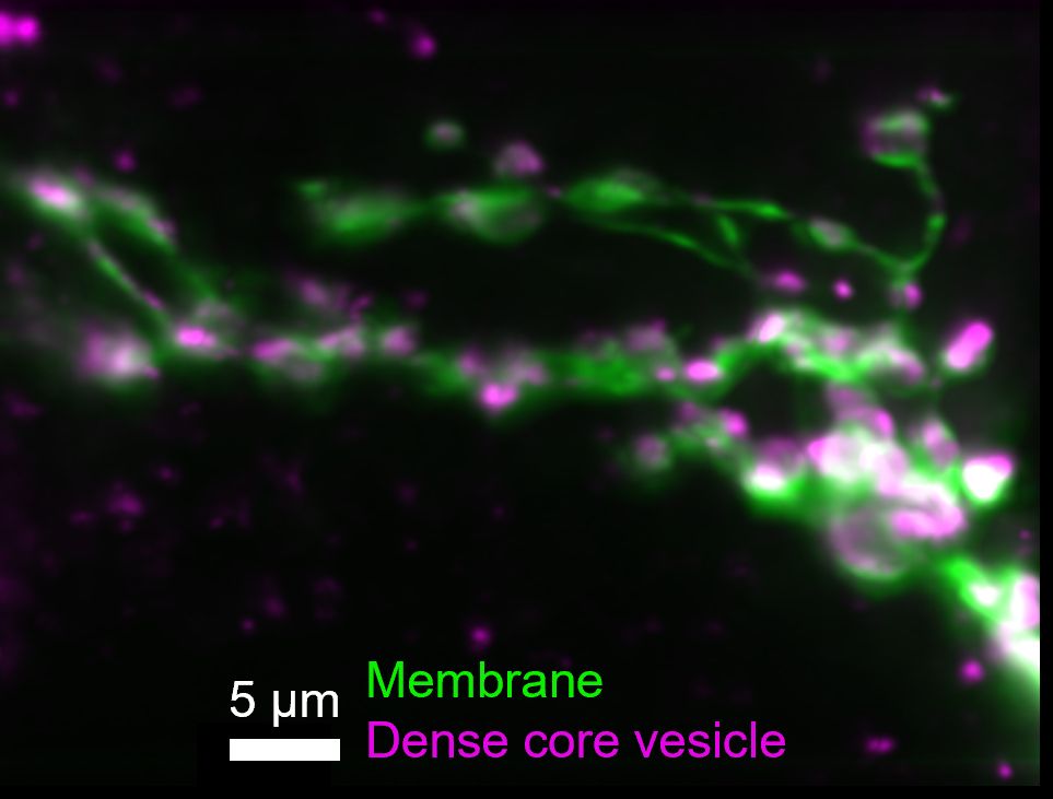
In addition, we can do 2 colours imaging...
Here the membrane is labelled in green (GFP), and dense core vesicles are labeled in magenta (mCherry).
We also show mitochondria imaging in the manuscript.
5/6
Here the membrane is labelled in green (GFP), and dense core vesicles are labeled in magenta (mCherry).
We also show mitochondria imaging in the manuscript.
5/6
February 26, 2025 at 2:48 AM
In addition, we can do 2 colours imaging...
Here the membrane is labelled in green (GFP), and dense core vesicles are labeled in magenta (mCherry).
We also show mitochondria imaging in the manuscript.
5/6
Here the membrane is labelled in green (GFP), and dense core vesicles are labeled in magenta (mCherry).
We also show mitochondria imaging in the manuscript.
5/6
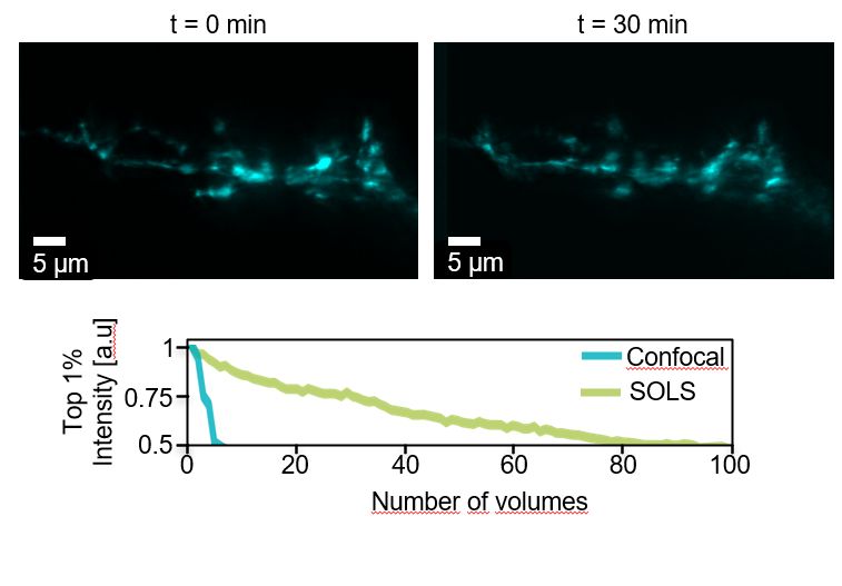
And what about the photobleaching?
We can image the fly for 30 minutes without bleaching it, and can get 10 times more images than with confocal microscopy!
Also, we don't rely on phototoxic pulsed infrared lasers like in 2-photon microscopy.
4/6
We can image the fly for 30 minutes without bleaching it, and can get 10 times more images than with confocal microscopy!
Also, we don't rely on phototoxic pulsed infrared lasers like in 2-photon microscopy.
4/6
February 26, 2025 at 2:48 AM
And what about the photobleaching?
We can image the fly for 30 minutes without bleaching it, and can get 10 times more images than with confocal microscopy!
Also, we don't rely on phototoxic pulsed infrared lasers like in 2-photon microscopy.
4/6
We can image the fly for 30 minutes without bleaching it, and can get 10 times more images than with confocal microscopy!
Also, we don't rely on phototoxic pulsed infrared lasers like in 2-photon microscopy.
4/6
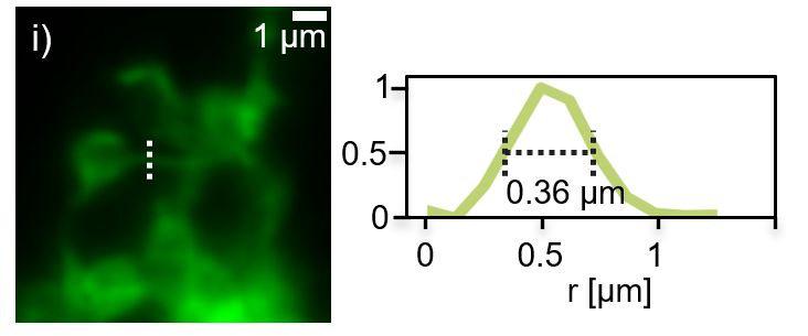
We imaged the axonal projections of small lateral ventral neurons (known as s-LNvs) in living adult flies.
The NA=1.1 allowed high spatial resolution.
Here, a line intensity profile showing features as small as 360 nm.
3/6
The NA=1.1 allowed high spatial resolution.
Here, a line intensity profile showing features as small as 360 nm.
3/6
February 26, 2025 at 2:48 AM
We imaged the axonal projections of small lateral ventral neurons (known as s-LNvs) in living adult flies.
The NA=1.1 allowed high spatial resolution.
Here, a line intensity profile showing features as small as 360 nm.
3/6
The NA=1.1 allowed high spatial resolution.
Here, a line intensity profile showing features as small as 360 nm.
3/6
We used a Single Objective Light-Sheet (#Snouty, for the friends).
It is less invasive than a lattice light sheet because it requires a smaller dissection area on the head´s cuticula, as it is only necessary to accommodate the light cone of a single objective. And it is simpler. 2/6
It is less invasive than a lattice light sheet because it requires a smaller dissection area on the head´s cuticula, as it is only necessary to accommodate the light cone of a single objective. And it is simpler. 2/6

February 26, 2025 at 2:48 AM
We used a Single Objective Light-Sheet (#Snouty, for the friends).
It is less invasive than a lattice light sheet because it requires a smaller dissection area on the head´s cuticula, as it is only necessary to accommodate the light cone of a single objective. And it is simpler. 2/6
It is less invasive than a lattice light sheet because it requires a smaller dissection area on the head´s cuticula, as it is only necessary to accommodate the light cone of a single objective. And it is simpler. 2/6
HIGH-RESOLUTION IN VIVO BRAIN IMAGING OF DROSOPHILA 🪰
New Preprint by Tassara et al.!
www.biorxiv.org/content/10.1...
#FluorescenceMonday #SnoutClub 1/6
New Preprint by Tassara et al.!
www.biorxiv.org/content/10.1...
#FluorescenceMonday #SnoutClub 1/6
February 26, 2025 at 2:48 AM
HIGH-RESOLUTION IN VIVO BRAIN IMAGING OF DROSOPHILA 🪰
New Preprint by Tassara et al.!
www.biorxiv.org/content/10.1...
#FluorescenceMonday #SnoutClub 1/6
New Preprint by Tassara et al.!
www.biorxiv.org/content/10.1...
#FluorescenceMonday #SnoutClub 1/6
Se viene el XX TOPFOT - XV EEOF, el evento del año de óptica en Argentina, en Buenos Aires del 13-16 Mayo.
Miren que lindo logo!
Hay tiempo hasta el 21 de Febrero para pedir ayudas económicas!
Acá pueden leer la segunda circular con toda la información:
drive.google.com/file/d/13_JS...
Miren que lindo logo!
Hay tiempo hasta el 21 de Febrero para pedir ayudas económicas!
Acá pueden leer la segunda circular con toda la información:
drive.google.com/file/d/13_JS...

February 17, 2025 at 2:11 PM
Se viene el XX TOPFOT - XV EEOF, el evento del año de óptica en Argentina, en Buenos Aires del 13-16 Mayo.
Miren que lindo logo!
Hay tiempo hasta el 21 de Febrero para pedir ayudas económicas!
Acá pueden leer la segunda circular con toda la información:
drive.google.com/file/d/13_JS...
Miren que lindo logo!
Hay tiempo hasta el 21 de Febrero para pedir ayudas económicas!
Acá pueden leer la segunda circular con toda la información:
drive.google.com/file/d/13_JS...
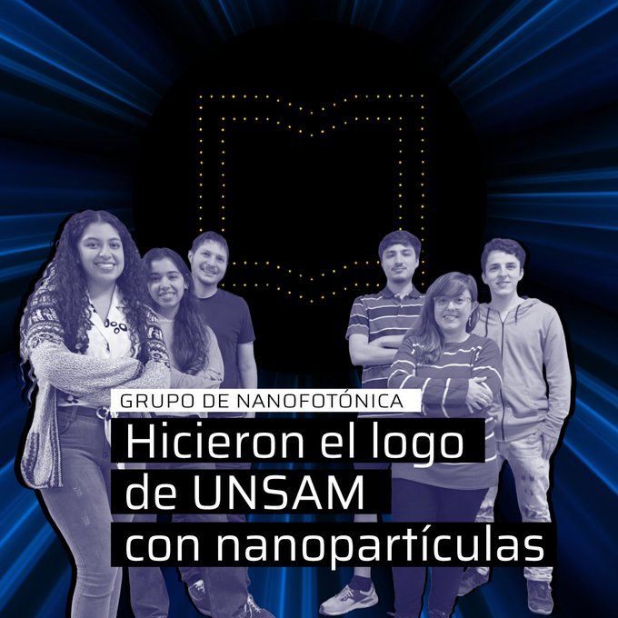
El grupo de Nanofotónica está liderado por Ianina Violi y Julián Gargiulo, quienes dirigen a la tesista doctoral María Cristina Mina y supervisan el trabajo de tres estudiantes de la licenciatura en Física de la UBA: Abril Pereyra, Valentín Salari e Iñaki González Vandam.
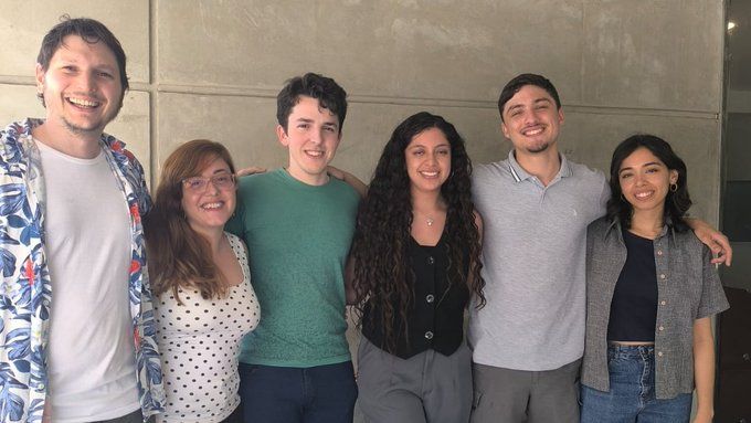
December 12, 2024 at 2:24 AM
El grupo de Nanofotónica está liderado por Ianina Violi y Julián Gargiulo, quienes dirigen a la tesista doctoral María Cristina Mina y supervisan el trabajo de tres estudiantes de la licenciatura en Física de la UBA: Abril Pereyra, Valentín Salari e Iñaki González Vandam.
El grupo de Nanofotónica trabaja en el laboratorio de láseres del INS. Fabrica dispositivos que permiten manipular la luz en la nanoescala, y tienen muchísimas aplicaciones como biosensores ultrasensibles, en celdas solares para la producción de combustibles o microfluídica.
December 12, 2024 at 2:24 AM
El grupo de Nanofotónica trabaja en el laboratorio de láseres del INS. Fabrica dispositivos que permiten manipular la luz en la nanoescala, y tienen muchísimas aplicaciones como biosensores ultrasensibles, en celdas solares para la producción de combustibles o microfluídica.
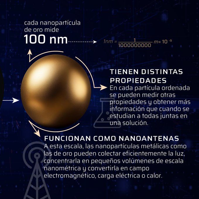
🧐Poder hacer este tipo de arreglos es muy útil para el estudio de nanomateriales, ya que una vez que están ordenados de a uno se les pueden medir propiedades a nivel de partícula única, mucho más ricos en información que cuando se estudian todas las partículas juntas en solución
December 12, 2024 at 2:24 AM
🧐Poder hacer este tipo de arreglos es muy útil para el estudio de nanomateriales, ya que una vez que están ordenados de a uno se les pueden medir propiedades a nivel de partícula única, mucho más ricos en información que cuando se estudian todas las partículas juntas en solución
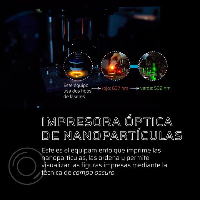
🔬Usamos una impresora óptica de nanopartículas, que permite usar la técnica de pinzas ópticas en la que un láser toma las nanopartículas una por una y las posiciona sobre un vidrio hasta crear un arreglo, en este caso el famoso librito de nuestra universidad.

December 12, 2024 at 2:24 AM
🔬Usamos una impresora óptica de nanopartículas, que permite usar la técnica de pinzas ópticas en la que un láser toma las nanopartículas una por una y las posiciona sobre un vidrio hasta crear un arreglo, en este caso el famoso librito de nuestra universidad.
💡En el grupo de Nanofotónica del Instituto de Nanosistemas sabemos cómo mover objetos con la luz.
¿Cómo hicimos este logo de
@unsamoficial
con nanopartículas de oro de apenas 100 nanómetros?
⬇️⬇️
Sale 🧵
¿Cómo hicimos este logo de
@unsamoficial
con nanopartículas de oro de apenas 100 nanómetros?
⬇️⬇️
Sale 🧵
December 12, 2024 at 2:24 AM
💡En el grupo de Nanofotónica del Instituto de Nanosistemas sabemos cómo mover objetos con la luz.
¿Cómo hicimos este logo de
@unsamoficial
con nanopartículas de oro de apenas 100 nanómetros?
⬇️⬇️
Sale 🧵
¿Cómo hicimos este logo de
@unsamoficial
con nanopartículas de oro de apenas 100 nanómetros?
⬇️⬇️
Sale 🧵