


MDIBL Light Microscopy Facility
@mdibl-lmf.bsky.social
Light Microscopy Facility @mdibiolab.bsky.social
https://lmf.mdibl.org/
https://lmf.mdibl.org/
Reposted by MDIBL Light Microscopy Facility
Big congratulations to Hannah Somers, Digital Image Analyst at MDI Bio Lab, whose striking visualization of a zebrafish’s brain was named an Image of Distinction in the Nikon Small World competition — and featured by #NationalGeographic!
#researchmatters #microscopymonday #lightsheet 🧪 🤝 🐟🎸 👩🔬
#researchmatters #microscopymonday #lightsheet 🧪 🤝 🐟🎸 👩🔬

MDI Bio Lab’s Hannah Somers’ Image Wins International Recognition
mdibl.org
November 3, 2025 at 3:01 PM
Big congratulations to Hannah Somers, Digital Image Analyst at MDI Bio Lab, whose striking visualization of a zebrafish’s brain was named an Image of Distinction in the Nikon Small World competition — and featured by #NationalGeographic!
#researchmatters #microscopymonday #lightsheet 🧪 🤝 🐟🎸 👩🔬
#researchmatters #microscopymonday #lightsheet 🧪 🤝 🐟🎸 👩🔬
Reposted by MDIBL Light Microscopy Facility
Congratulations to Frederic Bonnet and the @mdibl-lmf.bsky.social on these amazing achievements!! 🎉
#HappyFluorescenceFriday!
#microscopycommunity- 🎉 Winners of the BINA Image Contest!
1st Place: Frederic Bonnet – Hexagonal Vision: Compound Eye and Sensory Crown
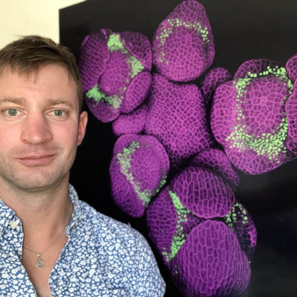
2nd Place: Nat Prunet – Allergy Season
3rd Place: Arlen Ramírez Corona – Blue, Red and Fish
People’s Choice: Mahgol Darvishmolla
#microscopycommunity- 🎉 Winners of the BINA Image Contest!
1st Place: Frederic Bonnet – Hexagonal Vision: Compound Eye and Sensory Crown
2nd Place: Nat Prunet – Allergy Season
3rd Place: Arlen Ramírez Corona – Blue, Red and Fish
People’s Choice: Mahgol Darvishmolla

October 17, 2025 at 7:17 PM
Congratulations to Frederic Bonnet and the @mdibl-lmf.bsky.social on these amazing achievements!! 🎉
Reposted by MDIBL Light Microscopy Facility
I love Frederic Bonnet's compound eye image that got first place 😍

October 14, 2025 at 4:03 PM
I love Frederic Bonnet's compound eye image that got first place 😍
Reposted by MDIBL Light Microscopy Facility
Grateful to see one my #superresolution #pollen images won 2nd place at the 2025 @bioimagingna.bsky.social image contest!
www.bioimagingnorthamerica.org/image-contes...
#microscopy #confocal #botany #plantscience #allergies #bioimaging
www.bioimagingnorthamerica.org/image-contes...
#microscopy #confocal #botany #plantscience #allergies #bioimaging

October 14, 2025 at 4:00 PM
Grateful to see one my #superresolution #pollen images won 2nd place at the 2025 @bioimagingna.bsky.social image contest!
www.bioimagingnorthamerica.org/image-contes...
#microscopy #confocal #botany #plantscience #allergies #bioimaging
www.bioimagingnorthamerica.org/image-contes...
#microscopy #confocal #botany #plantscience #allergies #bioimaging
Reposted by MDIBL Light Microscopy Facility
This #microscopymonday image of a planarian regenerating its head 7 days after decapitation is from MDI Bio Lab's Disney lab. The translucent area at the top is the blastema—a mass of stem cells that gives rise to new tissues during regeneration. MDIBL Light Microscopy Facility
🧪 🤝 🌎 #microscopy
🧪 🤝 🌎 #microscopy

October 13, 2025 at 2:00 PM
This #microscopymonday image of a planarian regenerating its head 7 days after decapitation is from MDI Bio Lab's Disney lab. The translucent area at the top is the blastema—a mass of stem cells that gives rise to new tissues during regeneration. MDIBL Light Microscopy Facility
🧪 🤝 🌎 #microscopy
🧪 🤝 🌎 #microscopy
Reposted by MDIBL Light Microscopy Facility

🎉 🔬 We’re thrilled to announce the winners of this year’s #NikonSmallWorld in Motion video #Microscopy competition: bit.ly/4mtjUk7
🥇 1st place goes to Jay McClellan of Saranac, Michigan (USA) for his video capturing self-pollination in a thymeleaf speedwell flower.
#Microscope #SciArt #SciComm
🥇 1st place goes to Jay McClellan of Saranac, Michigan (USA) for his video capturing self-pollination in a thymeleaf speedwell flower.
#Microscope #SciArt #SciComm
September 24, 2025 at 6:34 PM
🎉 🔬 We’re thrilled to announce the winners of this year’s #NikonSmallWorld in Motion video #Microscopy competition: bit.ly/4mtjUk7
🥇 1st place goes to Jay McClellan of Saranac, Michigan (USA) for his video capturing self-pollination in a thymeleaf speedwell flower.
#Microscope #SciArt #SciComm
🥇 1st place goes to Jay McClellan of Saranac, Michigan (USA) for his video capturing self-pollination in a thymeleaf speedwell flower.
#Microscope #SciArt #SciComm
Reposted by MDIBL Light Microscopy Facility
Thanks go out to MDI Bio Lab Digital Image Analyst @hsomers.bsky.social for this #microscopymonday view of an adult #zebrafish (brain vasculature) from the MesoSPIM.
#standwithscience #researchmatters 🧪 🤝 🐟🎸 #microscopy #fluorescentproteins #lightsheet
#standwithscience #researchmatters 🧪 🤝 🐟🎸 #microscopy #fluorescentproteins #lightsheet
September 22, 2025 at 12:20 PM
Thanks go out to MDI Bio Lab Digital Image Analyst @hsomers.bsky.social for this #microscopymonday view of an adult #zebrafish (brain vasculature) from the MesoSPIM.
#standwithscience #researchmatters 🧪 🤝 🐟🎸 #microscopy #fluorescentproteins #lightsheet
#standwithscience #researchmatters 🧪 🤝 🐟🎸 #microscopy #fluorescentproteins #lightsheet
Speaking with BioImaging North America about the Maine Microscopy Course! @bioimagingna.bsky.social @mdibiolab.bsky.social
www.youtube.com/watch?v=t90L...
#science #education #microscopy #EoE
www.youtube.com/watch?v=t90L...
#science #education #microscopy #EoE

Exchange of Experience Virtual Group | May 28 2025 | Frederic Bonnet
YouTube video by BioImaging North America
www.youtube.com
September 4, 2025 at 6:53 PM
Speaking with BioImaging North America about the Maine Microscopy Course! @bioimagingna.bsky.social @mdibiolab.bsky.social
www.youtube.com/watch?v=t90L...
#science #education #microscopy #EoE
www.youtube.com/watch?v=t90L...
#science #education #microscopy #EoE
Reposted by MDIBL Light Microscopy Facility
One of the people I'm indebted the most for creating stunningly beautiful #mesoSPIM samples was Martina Schaettin - who tragically passed away in March this year. To honor her memory, Elkhan Yusifov created an award - submit your best #microscopy images & videos! www.linkedin.com/company/mart...
August 22, 2025 at 5:32 PM
One of the people I'm indebted the most for creating stunningly beautiful #mesoSPIM samples was Martina Schaettin - who tragically passed away in March this year. To honor her memory, Elkhan Yusifov created an award - submit your best #microscopy images & videos! www.linkedin.com/company/mart...
Reposted by MDIBL Light Microscopy Facility
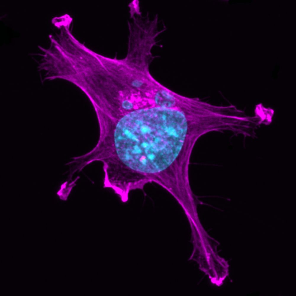
This mouse fibroblast cell captured by @mdibl-lmf.bsky.social in 2022 is labeled with a #fluorescence to visualize the actin cytoskeleton—a network of actin filaments & proteins that maintains cell shape, enables movement & supports key processes.
#researchmatters #microscopymonday #lightsheet 🧪 🤝
#researchmatters #microscopymonday #lightsheet 🧪 🤝

August 18, 2025 at 12:20 PM
This mouse fibroblast cell captured by @mdibl-lmf.bsky.social in 2022 is labeled with a #fluorescence to visualize the actin cytoskeleton—a network of actin filaments & proteins that maintains cell shape, enables movement & supports key processes.
#researchmatters #microscopymonday #lightsheet 🧪 🤝
#researchmatters #microscopymonday #lightsheet 🧪 🤝
Do you see chromatic aberration in your images? www.youtube.com/shorts/9lyix...
#science #microscopy #bioimaging #microscopymonday #chromaticaberration #bluesci #mdibl
#science #microscopy #bioimaging #microscopymonday #chromaticaberration #bluesci #mdibl

What is optical aberration ?
YouTube video by LMF@MDIBL
www.youtube.com
August 4, 2025 at 12:42 PM
Do you see chromatic aberration in your images? www.youtube.com/shorts/9lyix...
#science #microscopy #bioimaging #microscopymonday #chromaticaberration #bluesci #mdibl
#science #microscopy #bioimaging #microscopymonday #chromaticaberration #bluesci #mdibl
What is Köhler Illumination?
youtube.com/shorts/-1ct0...
#microscopy #microscopymonday #Köhler #Köhlerillumination #brightfield #science
youtube.com/shorts/-1ct0...
#microscopy #microscopymonday #Köhler #Köhlerillumination #brightfield #science

What is Köhler illumination ? #science #microscope #biology #optics
YouTube video by LMF@MDIBL
youtube.com
July 28, 2025 at 1:02 PM
What is Köhler Illumination?
youtube.com/shorts/-1ct0...
#microscopy #microscopymonday #Köhler #Köhlerillumination #brightfield #science
youtube.com/shorts/-1ct0...
#microscopy #microscopymonday #Köhler #Köhlerillumination #brightfield #science
Reposted by MDIBL Light Microscopy Facility
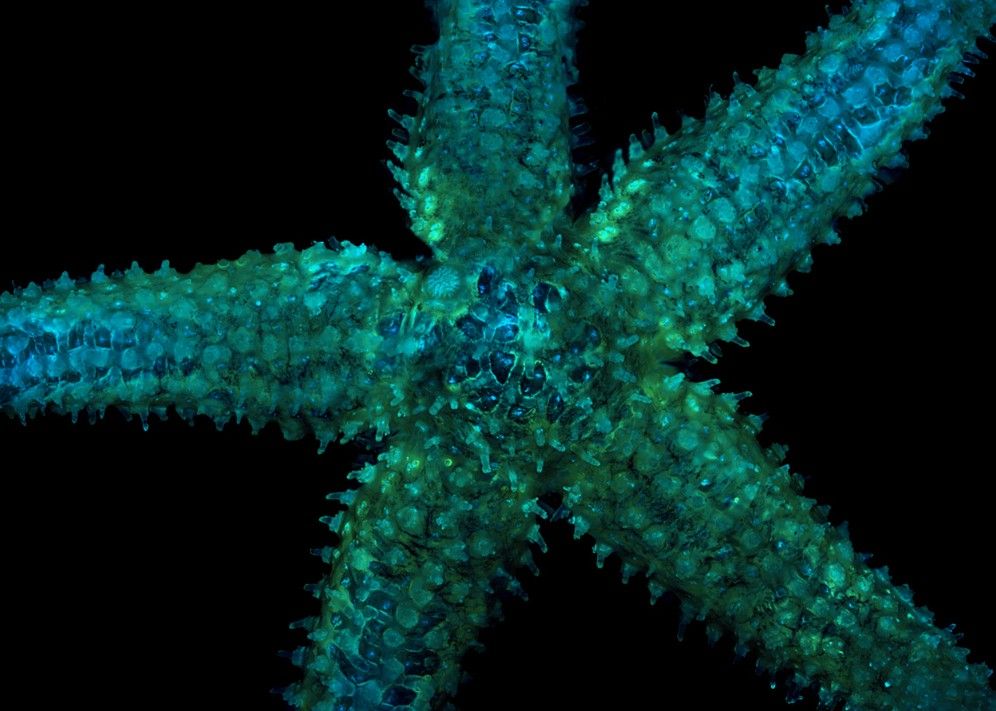
A beautiful #microscopymonday sea star captured by @hsomers.bsky.social in 2022.
Sample: Asterias amurensis (Northern Sea Star)
Label: Nuclear staining with DAPI in Cyan and auto-fluorescence in green
#standwithscience #researchmatters #microscopy #fluorescentproteins #lightsheet 🧪 🤝 #wildlife
Sample: Asterias amurensis (Northern Sea Star)
Label: Nuclear staining with DAPI in Cyan and auto-fluorescence in green
#standwithscience #researchmatters #microscopy #fluorescentproteins #lightsheet 🧪 🤝 #wildlife
July 28, 2025 at 12:13 PM
A beautiful #microscopymonday sea star captured by @hsomers.bsky.social in 2022.
Sample: Asterias amurensis (Northern Sea Star)
Label: Nuclear staining with DAPI in Cyan and auto-fluorescence in green
#standwithscience #researchmatters #microscopy #fluorescentproteins #lightsheet 🧪 🤝 #wildlife
Sample: Asterias amurensis (Northern Sea Star)
Label: Nuclear staining with DAPI in Cyan and auto-fluorescence in green
#standwithscience #researchmatters #microscopy #fluorescentproteins #lightsheet 🧪 🤝 #wildlife
Reposted by MDIBL Light Microscopy Facility
Reminder! Application Deadline: August 8, 2025
From mastering iPSC culture to directed differentiation and organoid creation, participants will gain skills they can immediately apply to their own research workflows.
#standwithscience #researchmatters 🧪 🤝 🐟🎸 🍎 👩🔬 🖥️🧬
From mastering iPSC culture to directed differentiation and organoid creation, participants will gain skills they can immediately apply to their own research workflows.
#standwithscience #researchmatters 🧪 🤝 🐟🎸 🍎 👩🔬 🖥️🧬
Applications of Human iPSCs and Organoids 2025
mdibl.org
July 24, 2025 at 11:15 AM
Reminder! Application Deadline: August 8, 2025
From mastering iPSC culture to directed differentiation and organoid creation, participants will gain skills they can immediately apply to their own research workflows.
#standwithscience #researchmatters 🧪 🤝 🐟🎸 🍎 👩🔬 🖥️🧬
From mastering iPSC culture to directed differentiation and organoid creation, participants will gain skills they can immediately apply to their own research workflows.
#standwithscience #researchmatters 🧪 🤝 🐟🎸 🍎 👩🔬 🖥️🧬
Reposted by MDIBL Light Microscopy Facility
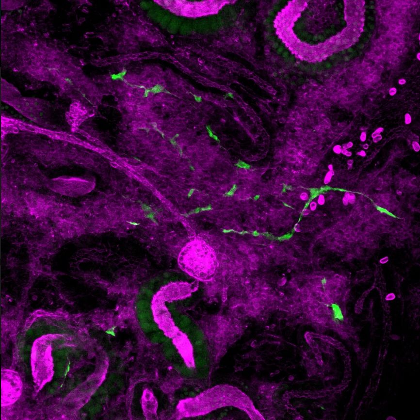
This #microscopymonday image shows GFP+ stem cells moving through the complex environment of a #zebrafish kidney.
It was captured in 2023 by Senior Research Scientist Caramai Kamei.
#standwithscience #researchmatters #microscopy #fluorescentproteins #lightsheet 🧪 🤝 🐟🎸
It was captured in 2023 by Senior Research Scientist Caramai Kamei.
#standwithscience #researchmatters #microscopy #fluorescentproteins #lightsheet 🧪 🤝 🐟🎸
July 21, 2025 at 12:24 PM
This #microscopymonday image shows GFP+ stem cells moving through the complex environment of a #zebrafish kidney.
It was captured in 2023 by Senior Research Scientist Caramai Kamei.
#standwithscience #researchmatters #microscopy #fluorescentproteins #lightsheet 🧪 🤝 🐟🎸
It was captured in 2023 by Senior Research Scientist Caramai Kamei.
#standwithscience #researchmatters #microscopy #fluorescentproteins #lightsheet 🧪 🤝 🐟🎸
Reposted by MDIBL Light Microscopy Facility
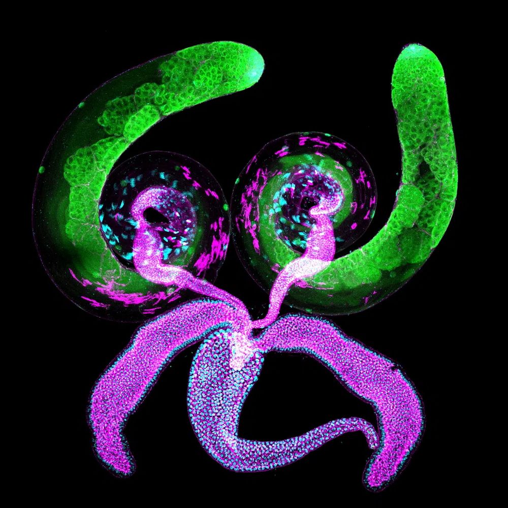
This #microscopymonday image is of a #Drosophila male reproductive system: paired coiled testes, seminal vesicles & accessory glands, and a single ejaculatory duct. Each coiled testis is about 1.5mm. Image captured by MDI Bio Lab Sr. Scientist Travis Carney.
#standwithscience #researchmatters 🧪 🤝
#standwithscience #researchmatters 🧪 🤝
June 9, 2025 at 1:36 PM
This #microscopymonday image is of a #Drosophila male reproductive system: paired coiled testes, seminal vesicles & accessory glands, and a single ejaculatory duct. Each coiled testis is about 1.5mm. Image captured by MDI Bio Lab Sr. Scientist Travis Carney.
#standwithscience #researchmatters 🧪 🤝
#standwithscience #researchmatters 🧪 🤝
What is Numerical Aperture?
#microscopy #na #numericalaperture #microscopymonday #science #bioimaging
www.youtube.com/shorts/x1XUs...
#microscopy #na #numericalaperture #microscopymonday #science #bioimaging
www.youtube.com/shorts/x1XUs...

What is Numerical Aperture (NA) ?
YouTube video by LMF@MDIBL
www.youtube.com
July 21, 2025 at 7:09 PM
What is Numerical Aperture?
#microscopy #na #numericalaperture #microscopymonday #science #bioimaging
www.youtube.com/shorts/x1XUs...
#microscopy #na #numericalaperture #microscopymonday #science #bioimaging
www.youtube.com/shorts/x1XUs...
Reposted by MDIBL Light Microscopy Facility
This image from 2024 is by MDI Bio Lab Senior Scientist Travis Carney. It shows the neurons in the brain of a #Drosophila fruit fly larva. The images are color-coded projections, so different focal planes are in different colors.
#standwithscience #researchmatters #microscopymonday #lightsheet 🧪 🤝
#standwithscience #researchmatters #microscopymonday #lightsheet 🧪 🤝

June 30, 2025 at 12:14 PM
This image from 2024 is by MDI Bio Lab Senior Scientist Travis Carney. It shows the neurons in the brain of a #Drosophila fruit fly larva. The images are color-coded projections, so different focal planes are in different colors.
#standwithscience #researchmatters #microscopymonday #lightsheet 🧪 🤝
#standwithscience #researchmatters #microscopymonday #lightsheet 🧪 🤝
Do you know the PSF of your microscope?
#PSF #Pointspreadfunction #microscopy #microscopymonday #introduction #science #biology
youtube.com/shorts/K7Xr1...
#PSF #Pointspreadfunction #microscopy #microscopymonday #introduction #science #biology
youtube.com/shorts/K7Xr1...

What is the Point Spread Function (PSF) of a microscope
YouTube video by LMF@MDIBL
youtube.com
July 14, 2025 at 12:50 PM
Do you know the PSF of your microscope?
#PSF #Pointspreadfunction #microscopy #microscopymonday #introduction #science #biology
youtube.com/shorts/K7Xr1...
#PSF #Pointspreadfunction #microscopy #microscopymonday #introduction #science #biology
youtube.com/shorts/K7Xr1...
Reposted by MDIBL Light Microscopy Facility
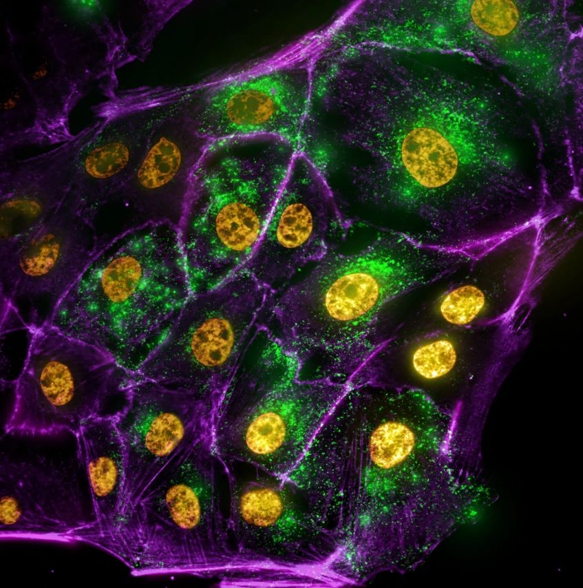
This colorized #microscopymonday image from 2023 by Frederic Bonnet shows kidney cells of a pig: the nuclei in yellow, the cytoplasmic vesicle in green and the membrane in magenta.
#standwithscience #researchmatters @mdibl-lmf.bsky.social
#microscopy #fluorescentproteins #lightsheet 🧪 🤝 🐟🎸 🍎 👩🔬
#standwithscience #researchmatters @mdibl-lmf.bsky.social
#microscopy #fluorescentproteins #lightsheet 🧪 🤝 🐟🎸 🍎 👩🔬
July 14, 2025 at 12:26 PM
This colorized #microscopymonday image from 2023 by Frederic Bonnet shows kidney cells of a pig: the nuclei in yellow, the cytoplasmic vesicle in green and the membrane in magenta.
#standwithscience #researchmatters @mdibl-lmf.bsky.social
#microscopy #fluorescentproteins #lightsheet 🧪 🤝 🐟🎸 🍎 👩🔬
#standwithscience #researchmatters @mdibl-lmf.bsky.social
#microscopy #fluorescentproteins #lightsheet 🧪 🤝 🐟🎸 🍎 👩🔬
Do you follow best practices for visualizing microscopy images?
#microscopy #microscopymonday #imaging #visualization #science
youtube.com/shorts/_HlXa...
#microscopy #microscopymonday #imaging #visualization #science
youtube.com/shorts/_HlXa...

What are best practices for visualizing microscopy images ?
YouTube video by LMF@MDIBL
youtube.com
July 7, 2025 at 5:26 PM
Do you follow best practices for visualizing microscopy images?
#microscopy #microscopymonday #imaging #visualization #science
youtube.com/shorts/_HlXa...
#microscopy #microscopymonday #imaging #visualization #science
youtube.com/shorts/_HlXa...
Reposted by MDIBL Light Microscopy Facility

SGLT2 Inhibition Ameliorates Age-Dependent Renovascular Rarefaction https://www.biorxiv.org/content/10.1101/2025.06.27.654312v1
June 29, 2025 at 4:06 AM
SGLT2 Inhibition Ameliorates Age-Dependent Renovascular Rarefaction https://www.biorxiv.org/content/10.1101/2025.06.27.654312v1
Reposted by MDIBL Light Microscopy Facility
Deadline Approaching! July 7, 2025
Maine #Microscopy: Foundations and Fundamentals 2025, August 5-13 at MDI Bio Lab!
Optimize your experiment planning, imaging and analysis skills to strengthen your research and accelerate discoveries in your field.
#standwithscience #researchmatters 🧪 🤝 🍎 👩🔬
Maine #Microscopy: Foundations and Fundamentals 2025, August 5-13 at MDI Bio Lab!
Optimize your experiment planning, imaging and analysis skills to strengthen your research and accelerate discoveries in your field.
#standwithscience #researchmatters 🧪 🤝 🍎 👩🔬

Maine Microscopy: Foundations and Fundamentals 2025
mdibl.org
June 25, 2025 at 1:08 PM
Deadline Approaching! July 7, 2025
Maine #Microscopy: Foundations and Fundamentals 2025, August 5-13 at MDI Bio Lab!
Optimize your experiment planning, imaging and analysis skills to strengthen your research and accelerate discoveries in your field.
#standwithscience #researchmatters 🧪 🤝 🍎 👩🔬
Maine #Microscopy: Foundations and Fundamentals 2025, August 5-13 at MDI Bio Lab!
Optimize your experiment planning, imaging and analysis skills to strengthen your research and accelerate discoveries in your field.
#standwithscience #researchmatters 🧪 🤝 🍎 👩🔬
Reposted by MDIBL Light Microscopy Facility
Maine #Microscopy: Foundations & Fundamentals 2025, August 5-13
For Maine grad students, postdoc trainees, research assistants & junior faculty. Understand current technology and how technical decisions impact final image products.
Application Deadline: July 7. Register today!
🧪 🤝 🍎 👩🔬 🖥️ 🧬
For Maine grad students, postdoc trainees, research assistants & junior faculty. Understand current technology and how technical decisions impact final image products.
Application Deadline: July 7. Register today!
🧪 🤝 🍎 👩🔬 🖥️ 🧬
Maine Microscopy: Foundations and Fundamentals 2025
mdibl.org
June 13, 2025 at 12:11 PM
Maine #Microscopy: Foundations & Fundamentals 2025, August 5-13
For Maine grad students, postdoc trainees, research assistants & junior faculty. Understand current technology and how technical decisions impact final image products.
Application Deadline: July 7. Register today!
🧪 🤝 🍎 👩🔬 🖥️ 🧬
For Maine grad students, postdoc trainees, research assistants & junior faculty. Understand current technology and how technical decisions impact final image products.
Application Deadline: July 7. Register today!
🧪 🤝 🍎 👩🔬 🖥️ 🧬
Reposted by MDIBL Light Microscopy Facility

Happy #MicroscopyMonday!
#microscopycommunity - join @bioimagingna.bsky.social May 28, to hear Fredric Bonnet of @MDIBL_LMF speak on “Maine Microscopy: Democratizing Microscopy Education for Local Scientists” as part of the BINA EoE Virtual Group
Learn more and register here: buff.ly/F3rZzx7
#microscopycommunity - join @bioimagingna.bsky.social May 28, to hear Fredric Bonnet of @MDIBL_LMF speak on “Maine Microscopy: Democratizing Microscopy Education for Local Scientists” as part of the BINA EoE Virtual Group
Learn more and register here: buff.ly/F3rZzx7

May 26, 2025 at 4:06 PM
Happy #MicroscopyMonday!
#microscopycommunity - join @bioimagingna.bsky.social May 28, to hear Fredric Bonnet of @MDIBL_LMF speak on “Maine Microscopy: Democratizing Microscopy Education for Local Scientists” as part of the BINA EoE Virtual Group
Learn more and register here: buff.ly/F3rZzx7
#microscopycommunity - join @bioimagingna.bsky.social May 28, to hear Fredric Bonnet of @MDIBL_LMF speak on “Maine Microscopy: Democratizing Microscopy Education for Local Scientists” as part of the BINA EoE Virtual Group
Learn more and register here: buff.ly/F3rZzx7