


Luuk Loeff
@lloeff.bsky.social
Assistant Prof. at Leiden University Medical Center
Host-Defence | Structural biology | Single-molecule Biophysics
http://www.loefflab.com
Host-Defence | Structural biology | Single-molecule Biophysics
http://www.loefflab.com
Had a great day at @ebi.embl.org Grenoble for the Next-Gen in Infection Biology micro symposium! Big thanks to @kowaeva.bsky.social for the invite!


June 6, 2025 at 8:24 PM
Had a great day at @ebi.embl.org Grenoble for the Next-Gen in Infection Biology micro symposium! Big thanks to @kowaeva.bsky.social for the invite!
In sum, we combined single-particle cryo-EM with biochemical experiments to reveal the mechanistic basis for the protective function of PYROXD1. Our results further underscore the notion of co-evolution of PYROXD1 with RTCB as a mechanism to enable the tRNA-LC to function under aerobic conditions.

March 11, 2025 at 10:14 AM
In sum, we combined single-particle cryo-EM with biochemical experiments to reveal the mechanistic basis for the protective function of PYROXD1. Our results further underscore the notion of co-evolution of PYROXD1 with RTCB as a mechanism to enable the tRNA-LC to function under aerobic conditions.
The initial step of RTCB-catalyzed RNA ligation involves Archease mediated guanylylation of the RTCB catalytic center. We performed mass spectrometry and show that PYROXD1 selectively interacts with the apo-enzyme form of RTCB, while guanylylated RTCB remains active under oxidative stress.

March 11, 2025 at 10:14 AM
The initial step of RTCB-catalyzed RNA ligation involves Archease mediated guanylylation of the RTCB catalytic center. We performed mass spectrometry and show that PYROXD1 selectively interacts with the apo-enzyme form of RTCB, while guanylylated RTCB remains active under oxidative stress.
Superposition of the RTCB–PYROXD1 complex structure with the crystal structure of NAD(P)H-free PYROXD1 reveals structural rearrangements in PYROXD1 induced by NADH binding and RTCB recruitment.

March 11, 2025 at 10:14 AM
Superposition of the RTCB–PYROXD1 complex structure with the crystal structure of NAD(P)H-free PYROXD1 reveals structural rearrangements in PYROXD1 induced by NADH binding and RTCB recruitment.
The structure of the RTCB-PYROXD1 complex
reveals that the PYROXD1 CTD engages with the
catalytic center cleft of RTCB, thereby blocking the
substrate binding sites and rendering the catalytic
center inaccessible. Mutation of the interacting residues resulted in a loss of protection.
reveals that the PYROXD1 CTD engages with the
catalytic center cleft of RTCB, thereby blocking the
substrate binding sites and rendering the catalytic
center inaccessible. Mutation of the interacting residues resulted in a loss of protection.

March 11, 2025 at 10:14 AM
The structure of the RTCB-PYROXD1 complex
reveals that the PYROXD1 CTD engages with the
catalytic center cleft of RTCB, thereby blocking the
substrate binding sites and rendering the catalytic
center inaccessible. Mutation of the interacting residues resulted in a loss of protection.
reveals that the PYROXD1 CTD engages with the
catalytic center cleft of RTCB, thereby blocking the
substrate binding sites and rendering the catalytic
center inaccessible. Mutation of the interacting residues resulted in a loss of protection.
The structure shows clear density of both the nicotinamide and flavin dinucleotides within the catalytic center of PYROXD1 that are required for complex formation.

March 11, 2025 at 10:14 AM
The structure shows clear density of both the nicotinamide and flavin dinucleotides within the catalytic center of PYROXD1 that are required for complex formation.
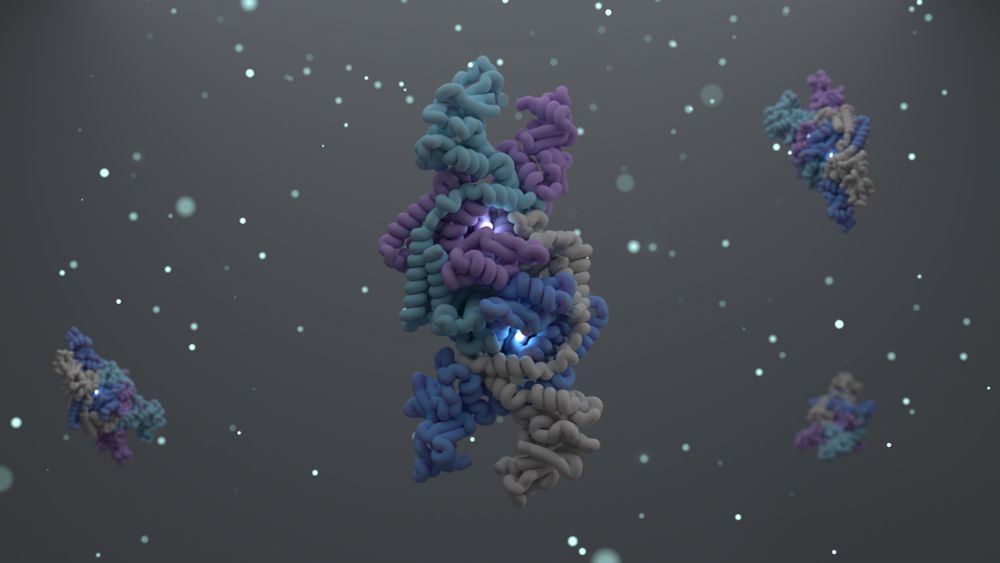
To uncover the mechanism of oxidative stress protection by PYROXD1 we used cryo-EM to obtain a structure of the RTCB-PYROXD1 complex.

March 11, 2025 at 10:14 AM
To uncover the mechanism of oxidative stress protection by PYROXD1 we used cryo-EM to obtain a structure of the RTCB-PYROXD1 complex.
The human tRNA ligase RTCB contains an active site that contains a highly reactive cystine group that is sensitive to oxidative inactivation.

March 11, 2025 at 10:14 AM
The human tRNA ligase RTCB contains an active site that contains a highly reactive cystine group that is sensitive to oxidative inactivation.
Our data suggests that Shedu specifically targets free DNA ends, providing self vs non-self-recognition. Our findings underline that recognition of pathogen-specific nucleic acid structures is a conserved feature of innate immunity across all domains of life.

December 31, 2024 at 3:38 PM
Our data suggests that Shedu specifically targets free DNA ends, providing self vs non-self-recognition. Our findings underline that recognition of pathogen-specific nucleic acid structures is a conserved feature of innate immunity across all domains of life.
Next, we generated gene edited phages to carrying the most frequent mutations (shoutout to @benadler.bsky.social from @doudna-lab.bsky.social). These experiments show that loss UvsW is responsible for the escape phenotype.

December 31, 2024 at 3:38 PM
Next, we generated gene edited phages to carrying the most frequent mutations (shoutout to @benadler.bsky.social from @doudna-lab.bsky.social). These experiments show that loss UvsW is responsible for the escape phenotype.
Lastly, we isolated phages that escape the Shedu immune system. All the escape mutants carry mutations in UvsW a helicase that is involved in the recombination-dependent DNA replication pathway of T-even phages, a process that relies on the generation of free DNA ends.

December 31, 2024 at 3:38 PM
Lastly, we isolated phages that escape the Shedu immune system. All the escape mutants carry mutations in UvsW a helicase that is involved in the recombination-dependent DNA replication pathway of T-even phages, a process that relies on the generation of free DNA ends.
To validate DNA-end targeting in vivo, we transformed cells with linear plasmid DNA fragments carrying short homology arms. Cells expressing SduA were 10x less efficient in assembling the fragments into a functional plasmid compared to the control.

December 31, 2024 at 3:38 PM
To validate DNA-end targeting in vivo, we transformed cells with linear plasmid DNA fragments carrying short homology arms. Cells expressing SduA were 10x less efficient in assembling the fragments into a functional plasmid compared to the control.
The end-bound DNA is projected over the active site of protomer SduA.2 and protrudes towards the active site of SduA.4. This positions the phosphate backbone of nucleotide 14 towards the active site, and results in cleavage at a fixed distance from the 5’-end.

December 31, 2024 at 3:38 PM
The end-bound DNA is projected over the active site of protomer SduA.2 and protrudes towards the active site of SduA.4. This positions the phosphate backbone of nucleotide 14 towards the active site, and results in cleavage at a fixed distance from the 5’-end.
The structure shows that Shedu binds dsDNA ends, using its N-terminal clamp. To be able to fit the DNA in the clamp in the unwound, while base pairing is retained.

December 31, 2024 at 3:38 PM
The structure shows that Shedu binds dsDNA ends, using its N-terminal clamp. To be able to fit the DNA in the clamp in the unwound, while base pairing is retained.
But how does Shedu provide phage defense and how does it discriminate self from non-self? 🤔 After exhausting many options, the answer came from a cryo-EM structure of Shedu bound to a dsDNA substrate.

December 31, 2024 at 3:38 PM
But how does Shedu provide phage defense and how does it discriminate self from non-self? 🤔 After exhausting many options, the answer came from a cryo-EM structure of Shedu bound to a dsDNA substrate.
DUF4263 adopts a PD(D/E)XK like fold consistent with its predicted nuclease function. The conserved residues that coordinate a magnesium ion point towards its active site. Mutational analysis shows that these residues are essential for both phage defense and DNA cleavage.

December 31, 2024 at 3:38 PM
DUF4263 adopts a PD(D/E)XK like fold consistent with its predicted nuclease function. The conserved residues that coordinate a magnesium ion point towards its active site. Mutational analysis shows that these residues are essential for both phage defense and DNA cleavage.
Given the putative nuclease function of Shedu, we performed cleavage assays on oligos and observed that SduA is a dsDNA nuclease.

December 31, 2024 at 3:38 PM
Given the putative nuclease function of Shedu, we performed cleavage assays on oligos and observed that SduA is a dsDNA nuclease.
Each protomer is composed of two domains connected by an alpha-helical linker. In absence of the N-term domain, DUF4263 elicits toxicity in cells. This indicates that the N-term domain regulates the activity of DUF4263.

December 31, 2024 at 3:38 PM
Each protomer is composed of two domains connected by an alpha-helical linker. In absence of the N-term domain, DUF4263 elicits toxicity in cells. This indicates that the N-term domain regulates the activity of DUF4263.
To understand the defense mechanism of Shedu we purified SduA from E.coli and determined its structure using cryo-EM. The structure shows SduA forms a tetrameric complex through its DUF4263 domain, while the N-terminal domains form clamp like structures on each end.

December 31, 2024 at 3:38 PM
To understand the defense mechanism of Shedu we purified SduA from E.coli and determined its structure using cryo-EM. The structure shows SduA forms a tetrameric complex through its DUF4263 domain, while the N-terminal domains form clamp like structures on each end.
We tested several orthologs of SduA for phage defense and could show robust phage defense in E. coli. Infection assays in liquid culture show that Shedu uses a mechanism that is distinct from abortive infection, suggesting that Shedu can distinguish self from non-self.

December 31, 2024 at 3:38 PM
We tested several orthologs of SduA for phage defense and could show robust phage defense in E. coli. Infection assays in liquid culture show that Shedu uses a mechanism that is distinct from abortive infection, suggesting that Shedu can distinguish self from non-self.
Shedu systems are characterized by the presence of a DUF4263 domain. By performing phylogenetic analysis, we found that SduA can be grouped into two clusters (Type I & II) based on their size and position of the signature domain.

December 31, 2024 at 3:38 PM
Shedu systems are characterized by the presence of a DUF4263 domain. By performing phylogenetic analysis, we found that SduA can be grouped into two clusters (Type I & II) based on their size and position of the signature domain.
What a great way to end the year! ✨
Today in @cellpress.bsky.social we report the structure and function of the Shedu anti-phage defense system.
tinyurl.com/4crj6dnx
A long 🧵...
Today in @cellpress.bsky.social we report the structure and function of the Shedu anti-phage defense system.
tinyurl.com/4crj6dnx
A long 🧵...
December 31, 2024 at 3:38 PM
What a great way to end the year! ✨
Today in @cellpress.bsky.social we report the structure and function of the Shedu anti-phage defense system.
tinyurl.com/4crj6dnx
A long 🧵...
Today in @cellpress.bsky.social we report the structure and function of the Shedu anti-phage defense system.
tinyurl.com/4crj6dnx
A long 🧵...
To fill up my empty feed, my first post on #bluesky.
Earlier this year we showed how the DdmDE system from pandemic Vibrio cholerae defends against invading plasmids. A great collaboration between the labs of Martin Jinek (UZH) and @mblokesch.bsky.social (EPFL).
www.science.org/doi/10.1126/...
Earlier this year we showed how the DdmDE system from pandemic Vibrio cholerae defends against invading plasmids. A great collaboration between the labs of Martin Jinek (UZH) and @mblokesch.bsky.social (EPFL).
www.science.org/doi/10.1126/...

November 12, 2024 at 1:12 PM
To fill up my empty feed, my first post on #bluesky.
Earlier this year we showed how the DdmDE system from pandemic Vibrio cholerae defends against invading plasmids. A great collaboration between the labs of Martin Jinek (UZH) and @mblokesch.bsky.social (EPFL).
www.science.org/doi/10.1126/...
Earlier this year we showed how the DdmDE system from pandemic Vibrio cholerae defends against invading plasmids. A great collaboration between the labs of Martin Jinek (UZH) and @mblokesch.bsky.social (EPFL).
www.science.org/doi/10.1126/...