


Linda Sinclair
@lindavsinclair.bsky.social
Immunologist, working with Cantrell Lab. Interested in LOTS - focused on nutrient and metabolic control of the immune system. (opinions are my own)
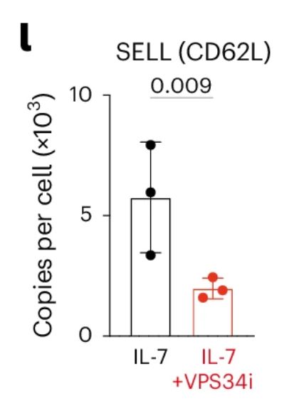
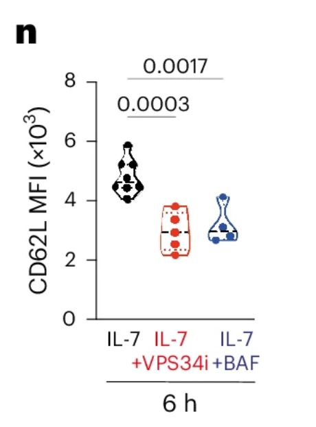
12/n The proteins fuelled by autophagy in naïve CD8 Tcells included essential proteins for naïve cell homeostasis : eg KLF2 and CD62L
March 1, 2025 at 11:18 AM
12/n The proteins fuelled by autophagy in naïve CD8 Tcells included essential proteins for naïve cell homeostasis : eg KLF2 and CD62L
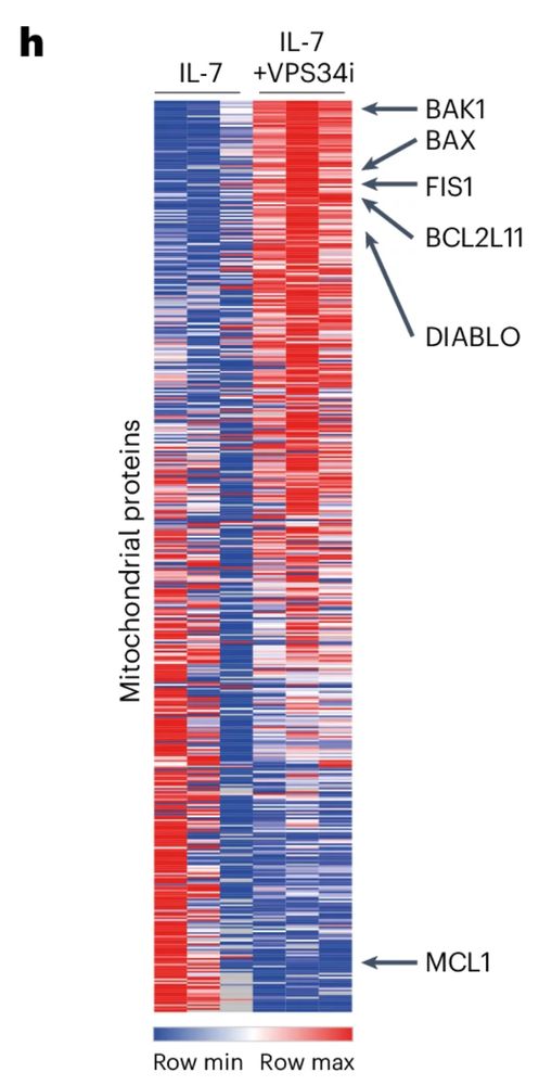
11/n Proteomics on naïve CD8 Tcells +/- VPS34 inhibition showed the predominant substrates are mitochondrial proteins.
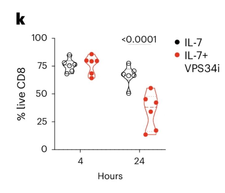
In naïve CD8 T cells mitochondria are constantly pruned to remain healthy. Blocking autophagy results in accumulation of pro-apoptotic proteins eg BAK1 and BIM, and cell death.
In naïve CD8 T cells mitochondria are constantly pruned to remain healthy. Blocking autophagy results in accumulation of pro-apoptotic proteins eg BAK1 and BIM, and cell death.
March 1, 2025 at 11:18 AM
11/n Proteomics on naïve CD8 Tcells +/- VPS34 inhibition showed the predominant substrates are mitochondrial proteins.
In naïve CD8 T cells mitochondria are constantly pruned to remain healthy. Blocking autophagy results in accumulation of pro-apoptotic proteins eg BAK1 and BIM, and cell death.
In naïve CD8 T cells mitochondria are constantly pruned to remain healthy. Blocking autophagy results in accumulation of pro-apoptotic proteins eg BAK1 and BIM, and cell death.
8/n What are the substrates of autophagy in CD8 Tcells?
They are different in naïve cells and effector cells!
- Proteomics on CTLs showed AA deprivation drives a loss in 55% of proteins ~ ½ of this loss was protected by VPS34 inhibition=autophagy. ½ was not (ie degraded by other mechanisms).
They are different in naïve cells and effector cells!
- Proteomics on CTLs showed AA deprivation drives a loss in 55% of proteins ~ ½ of this loss was protected by VPS34 inhibition=autophagy. ½ was not (ie degraded by other mechanisms).

March 1, 2025 at 11:18 AM
8/n What are the substrates of autophagy in CD8 Tcells?
They are different in naïve cells and effector cells!
- Proteomics on CTLs showed AA deprivation drives a loss in 55% of proteins ~ ½ of this loss was protected by VPS34 inhibition=autophagy. ½ was not (ie degraded by other mechanisms).
They are different in naïve cells and effector cells!
- Proteomics on CTLs showed AA deprivation drives a loss in 55% of proteins ~ ½ of this loss was protected by VPS34 inhibition=autophagy. ½ was not (ie degraded by other mechanisms).
4/n So, we used Ian Ganley's autophagy flux reporter (mCherry-GFP:LC3b) mouse: Flux can be measured by comparing the level of GFP (quenched in autolysosome) to mCherry (stable) fluorescence.
*Naïve CD8 Tcells have very autophagy flux, which is REDUCED in response to TCR activation*
*Naïve CD8 Tcells have very autophagy flux, which is REDUCED in response to TCR activation*


March 1, 2025 at 11:18 AM
4/n So, we used Ian Ganley's autophagy flux reporter (mCherry-GFP:LC3b) mouse: Flux can be measured by comparing the level of GFP (quenched in autolysosome) to mCherry (stable) fluorescence.
*Naïve CD8 Tcells have very autophagy flux, which is REDUCED in response to TCR activation*
*Naïve CD8 Tcells have very autophagy flux, which is REDUCED in response to TCR activation*
2/n The basis and framework for this behemoth study was all grown from Tom's PhD work. Tom noted protein and RNA expression of key autophagy machinery/cargo adaptors showed a disconnect during activation and differentiation of CD8 T cells. Protein INCREASED while mRNA DECREASED.
What was going on?
What was going on?


March 1, 2025 at 11:18 AM
2/n The basis and framework for this behemoth study was all grown from Tom's PhD work. Tom noted protein and RNA expression of key autophagy machinery/cargo adaptors showed a disconnect during activation and differentiation of CD8 T cells. Protein INCREASED while mRNA DECREASED.
What was going on?
What was going on?
