


Josquin Courte, PhD
@josquincourte.bsky.social
Scientist.
Microfluidics, synthetic biology, neurodegeneration, tissue engineering.
I want to develop better in vitro disease models.
Currently looking for positions in the biotech sector in Switzerland / France / Belgium.
Also DM, TTRPG enthusiast.
Microfluidics, synthetic biology, neurodegeneration, tissue engineering.
I want to develop better in vitro disease models.
Currently looking for positions in the biotech sector in Switzerland / France / Belgium.
Also DM, TTRPG enthusiast.
For the 1st goal, we have an idea of how to proceed:
Have the OFF transceivers express P-cad, and the ON transceiver N-cad.
This just requires a NOT gate downstream of synNotch.
...We have the plasmids already cloned, now we just need a brave student or postdoc to use them!
Have the OFF transceivers express P-cad, and the ON transceiver N-cad.
This just requires a NOT gate downstream of synNotch.
...We have the plasmids already cloned, now we just need a brave student or postdoc to use them!

December 17, 2024 at 2:03 PM
For the 1st goal, we have an idea of how to proceed:
Have the OFF transceivers express P-cad, and the ON transceiver N-cad.
This just requires a NOT gate downstream of synNotch.
...We have the plasmids already cloned, now we just need a brave student or postdoc to use them!
Have the OFF transceivers express P-cad, and the ON transceiver N-cad.
This just requires a NOT gate downstream of synNotch.
...We have the plasmids already cloned, now we just need a brave student or postdoc to use them!
We then simulated elongation circuits in this “realistically bound” parameter space.
To better understand how our in vitro results compared to in silico ones, we used a visualization approach with a “morphospace” framework, inspired by @ricardsole.bsky.social
To better understand how our in vitro results compared to in silico ones, we used a visualization approach with a “morphospace” framework, inspired by @ricardsole.bsky.social

December 17, 2024 at 2:03 PM
We then simulated elongation circuits in this “realistically bound” parameter space.
To better understand how our in vitro results compared to in silico ones, we used a visualization approach with a “morphospace” framework, inspired by @ricardsole.bsky.social
To better understand how our in vitro results compared to in silico ones, we used a visualization approach with a “morphospace” framework, inspired by @ricardsole.bsky.social
In the second class, we induced N-cad (+/- p21) in activated transceivers, and this too led to tissue elongation.

December 17, 2024 at 2:03 PM
In the second class, we induced N-cad (+/- p21) in activated transceivers, and this too led to tissue elongation.
At this point, we implemented 2/3 target design principles.
So, what about enforcing the segregation of the “growing tip” region?
=> We found that inducing N-cad upon transceivers activation (so, in the “structural support” region) would lead to better segregation! (We are not sure why)
So, what about enforcing the segregation of the “growing tip” region?
=> We found that inducing N-cad upon transceivers activation (so, in the “structural support” region) would lead to better segregation! (We are not sure why)

December 17, 2024 at 2:03 PM
At this point, we implemented 2/3 target design principles.
So, what about enforcing the segregation of the “growing tip” region?
=> We found that inducing N-cad upon transceivers activation (so, in the “structural support” region) would lead to better segregation! (We are not sure why)
So, what about enforcing the segregation of the “growing tip” region?
=> We found that inducing N-cad upon transceivers activation (so, in the “structural support” region) would lead to better segregation! (We are not sure why)
Coming back to our design: we then screened candidate effectors for controlling tissue fluidity.
Here, we believe we share the first screen of this type, as we had to look for inspiration in many publications!
Our two best hits targeted actomyosin contractility: constitutively active RHOA & MLCK.
Here, we believe we share the first screen of this type, as we had to look for inspiration in many publications!
Our two best hits targeted actomyosin contractility: constitutively active RHOA & MLCK.

December 17, 2024 at 2:03 PM
Coming back to our design: we then screened candidate effectors for controlling tissue fluidity.
Here, we believe we share the first screen of this type, as we had to look for inspiration in many publications!
Our two best hits targeted actomyosin contractility: constitutively active RHOA & MLCK.
Here, we believe we share the first screen of this type, as we had to look for inspiration in many publications!
Our two best hits targeted actomyosin contractility: constitutively active RHOA & MLCK.
… The most efficient growth inhibitors also increased tissue rounding speed!
(We do not yet know why.)
p21 was the only effector which both decreased tissue growth and fluidity, so we decided to build upon its induction.
(We do not yet know why.)
p21 was the only effector which both decreased tissue growth and fluidity, so we decided to build upon its induction.

December 17, 2024 at 2:03 PM
… The most efficient growth inhibitors also increased tissue rounding speed!
(We do not yet know why.)
p21 was the only effector which both decreased tissue growth and fluidity, so we decided to build upon its induction.
(We do not yet know why.)
p21 was the only effector which both decreased tissue growth and fluidity, so we decided to build upon its induction.
With this method, we first screened effectors to inhibit tissue growth.
We found some promising ones, like p53.
… but there was a hidden issue with those…
We found some promising ones, like p53.
… but there was a hidden issue with those…

December 17, 2024 at 2:03 PM
With this method, we first screened effectors to inhibit tissue growth.
We found some promising ones, like p53.
… but there was a hidden issue with those…
We found some promising ones, like p53.
… but there was a hidden issue with those…
We first identified a transceiver clone with a fate propagation speed in the right range. (the "Fast" clone here)

December 17, 2024 at 2:03 PM
We first identified a transceiver clone with a fate propagation speed in the right range. (the "Fast" clone here)
To verify if this would work, we first implemented the circuit in our own CompuCell3D framework: pubs.acs.org/doi/abs/10.1021/acssynbio.0c00369
… and this worked great! (...before physics were completely parametrized!)
(color code mistake here, the growing tip is blue instead of gray!)
… and this worked great! (...before physics were completely parametrized!)
(color code mistake here, the growing tip is blue instead of gray!)
December 17, 2024 at 2:03 PM
To verify if this would work, we first implemented the circuit in our own CompuCell3D framework: pubs.acs.org/doi/abs/10.1021/acssynbio.0c00369
… and this worked great! (...before physics were completely parametrized!)
(color code mistake here, the growing tip is blue instead of gray!)
… and this worked great! (...before physics were completely parametrized!)
(color code mistake here, the growing tip is blue instead of gray!)
So, how to exploit this circuit design?
We got the idea that if transceivers changed their properties based on their activation status, this could result in tissue elongation.
For that, the cells must only proliferate when OFF (grey), and become collectively very rigid when ON (red).
We got the idea that if transceivers changed their properties based on their activation status, this could result in tissue elongation.
For that, the cells must only proliferate when OFF (grey), and become collectively very rigid when ON (red).

December 17, 2024 at 2:03 PM
So, how to exploit this circuit design?
We got the idea that if transceivers changed their properties based on their activation status, this could result in tissue elongation.
For that, the cells must only proliferate when OFF (grey), and become collectively very rigid when ON (red).
We got the idea that if transceivers changed their properties based on their activation status, this could result in tissue elongation.
For that, the cells must only proliferate when OFF (grey), and become collectively very rigid when ON (red).
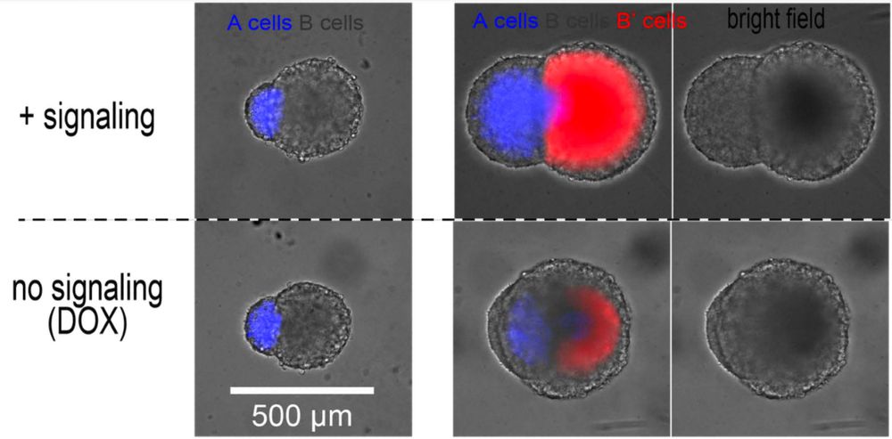
This circuit can be used to propagate a change in cell fate in 2D in a wave-like pattern… And also in 3D!
In this animation, a spheroid of “sender” cells (red+green=yellow) is fused with a spheroid of “transceiver” cells. Activated transceivers become green.
In this animation, a spheroid of “sender” cells (red+green=yellow) is fused with a spheroid of “transceiver” cells. Activated transceivers become green.
December 17, 2024 at 2:03 PM
This circuit can be used to propagate a change in cell fate in 2D in a wave-like pattern… And also in 3D!
In this animation, a spheroid of “sender” cells (red+green=yellow) is fused with a spheroid of “transceiver” cells. Activated transceivers become green.
In this animation, a spheroid of “sender” cells (red+green=yellow) is fused with a spheroid of “transceiver” cells. Activated transceivers become green.
But, how to translate this concept to a synthetic gene circuit?
We were very lucky… We could just take advantage of another circuit we developed in the lab, the “transceiver” circuit!
(here: www.nature.com/articles/s41467-024-53078-8)
We were very lucky… We could just take advantage of another circuit we developed in the lab, the “transceiver” circuit!
(here: www.nature.com/articles/s41467-024-53078-8)

December 17, 2024 at 2:03 PM
But, how to translate this concept to a synthetic gene circuit?
We were very lucky… We could just take advantage of another circuit we developed in the lab, the “transceiver” circuit!
(here: www.nature.com/articles/s41467-024-53078-8)
We were very lucky… We could just take advantage of another circuit we developed in the lab, the “transceiver” circuit!
(here: www.nature.com/articles/s41467-024-53078-8)
Looking at other (very different!) examples, one can notice a common design principle:
A combination of local material addition (to the tip) with mechanical resistance to rounding forces (of the bulk).
A combination of local material addition (to the tip) with mechanical resistance to rounding forces (of the bulk).

December 17, 2024 at 2:03 PM
Looking at other (very different!) examples, one can notice a common design principle:
A combination of local material addition (to the tip) with mechanical resistance to rounding forces (of the bulk).
A combination of local material addition (to the tip) with mechanical resistance to rounding forces (of the bulk).
So, how did we do that?
First, of course, we got our initial inspiration from the literature on axial elongation.
Notably this wonderful 2018 publication from @campaslab.bsky.social where the team demonstrated how tissue fluidity supports axial elongation in the zebrafish embryo!
First, of course, we got our initial inspiration from the literature on axial elongation.
Notably this wonderful 2018 publication from @campaslab.bsky.social where the team demonstrated how tissue fluidity supports axial elongation in the zebrafish embryo!

December 17, 2024 at 2:03 PM
So, how did we do that?
First, of course, we got our initial inspiration from the literature on axial elongation.
Notably this wonderful 2018 publication from @campaslab.bsky.social where the team demonstrated how tissue fluidity supports axial elongation in the zebrafish embryo!
First, of course, we got our initial inspiration from the literature on axial elongation.
Notably this wonderful 2018 publication from @campaslab.bsky.social where the team demonstrated how tissue fluidity supports axial elongation in the zebrafish embryo!
While we were aiming to generate baguettes, we obtained something which is more in the range of olives / cocktail sausages, but, well, you have to start somewhere!
Here is a 20-ish posts summary, check my repost of Leonardo’s recap for a shorter version 😉
Here is a 20-ish posts summary, check my repost of Leonardo’s recap for a shorter version 😉

December 17, 2024 at 2:03 PM
While we were aiming to generate baguettes, we obtained something which is more in the range of olives / cocktail sausages, but, well, you have to start somewhere!
Here is a 20-ish posts summary, check my repost of Leonardo’s recap for a shorter version 😉
Here is a 20-ish posts summary, check my repost of Leonardo’s recap for a shorter version 😉
Delighted to share our last preprint!!! 🎉🎉🎉
biorxiv.org/content/10.1101/2024.12.11.627621v1
We share the first synthetic gene circuit guiding the self-organization of mammalian tissues in elongating structures by dynamically tuning growth, viscosity and adhesion.
biorxiv.org/content/10.1101/2024.12.11.627621v1
We share the first synthetic gene circuit guiding the self-organization of mammalian tissues in elongating structures by dynamically tuning growth, viscosity and adhesion.
December 17, 2024 at 2:03 PM
Delighted to share our last preprint!!! 🎉🎉🎉
biorxiv.org/content/10.1101/2024.12.11.627621v1
We share the first synthetic gene circuit guiding the self-organization of mammalian tissues in elongating structures by dynamically tuning growth, viscosity and adhesion.
biorxiv.org/content/10.1101/2024.12.11.627621v1
We share the first synthetic gene circuit guiding the self-organization of mammalian tissues in elongating structures by dynamically tuning growth, viscosity and adhesion.
An exciting result about controling signaling with density (doi.org/10.1038/s41467-024-53078-8)
=> As distal parts of rod-shaped aggregates are less dense., we get "free" spatial patterning in 3D!
=> Some interesting potential for feedback loops, if this circuit controls tissue physics...
=> As distal parts of rod-shaped aggregates are less dense., we get "free" spatial patterning in 3D!
=> Some interesting potential for feedback loops, if this circuit controls tissue physics...

December 6, 2024 at 2:53 PM
An exciting result about controling signaling with density (doi.org/10.1038/s41467-024-53078-8)
=> As distal parts of rod-shaped aggregates are less dense., we get "free" spatial patterning in 3D!
=> Some interesting potential for feedback loops, if this circuit controls tissue physics...
=> As distal parts of rod-shaped aggregates are less dense., we get "free" spatial patterning in 3D!
=> Some interesting potential for feedback loops, if this circuit controls tissue physics...
We then demonstrated that this principle could be harnessed to guide tissue patterning guided by a novel lab-made synthetic circuit we named "transceiver". Those outcomes were well predicted by a computational model taking cellular density into account.


November 25, 2024 at 1:34 PM
We then demonstrated that this principle could be harnessed to guide tissue patterning guided by a novel lab-made synthetic circuit we named "transceiver". Those outcomes were well predicted by a computational model taking cellular density into account.
Cell crowding inhibits signaling by slectively shutting down the expression of membrane proteins, which might be linked to their shorter half-life.

November 25, 2024 at 1:34 PM
Cell crowding inhibits signaling by slectively shutting down the expression of membrane proteins, which might be linked to their shorter half-life.
Here, we describe a new paradigm for synthetic gene circuits to "read" local physics: synthetic cell-cell communication can be tuned by local cellular crowding!
This is true in 2D and 3D, in mammalian cell fibroblasts and mouse stem cells.
This is true in 2D and 3D, in mammalian cell fibroblasts and mouse stem cells.


November 25, 2024 at 1:34 PM
Here, we describe a new paradigm for synthetic gene circuits to "read" local physics: synthetic cell-cell communication can be tuned by local cellular crowding!
This is true in 2D and 3D, in mammalian cell fibroblasts and mouse stem cells.
This is true in 2D and 3D, in mammalian cell fibroblasts and mouse stem cells.