


Jaroslav Icha
@ichajaroslav.bsky.social
Light microscopy at Bruker Fluorescence Microscopy division.
#lightsheet #multiphoton #superresolution #screening
Cell biologist in my previous life. Alumnus @mpicbg.bsky.social
@turkubioscience.bsky.social
#lightsheet #multiphoton #superresolution #screening
Cell biologist in my previous life. Alumnus @mpicbg.bsky.social
@turkubioscience.bsky.social
Are you interested in long time lapse imaging of organoids?
In this example, retinal organoids were imaged in high throughput for 3 days. The aim was to collect sufficient data to be able to use machine learning for predicting organoid differentiation just from brightfield images.
In this example, retinal organoids were imaged in high throughput for 3 days. The aim was to collect sufficient data to be able to use machine learning for predicting organoid differentiation just from brightfield images.

October 15, 2025 at 1:14 PM
Are you interested in long time lapse imaging of organoids?
In this example, retinal organoids were imaged in high throughput for 3 days. The aim was to collect sufficient data to be able to use machine learning for predicting organoid differentiation just from brightfield images.
In this example, retinal organoids were imaged in high throughput for 3 days. The aim was to collect sufficient data to be able to use machine learning for predicting organoid differentiation just from brightfield images.
We have just installed a third HIVE server in Czechia. After Prague and Brno, we are adding Olomouc!
www.bruker.com/de/products-...
Researchers often get HIVE to handle data from Luxendo as well as other companies' light-sheet scopes but it is a rather universal solution for big data management.
www.bruker.com/de/products-...
Researchers often get HIVE to handle data from Luxendo as well as other companies' light-sheet scopes but it is a rather universal solution for big data management.


September 11, 2025 at 9:54 AM
We have just installed a third HIVE server in Czechia. After Prague and Brno, we are adding Olomouc!
www.bruker.com/de/products-...
Researchers often get HIVE to handle data from Luxendo as well as other companies' light-sheet scopes but it is a rather universal solution for big data management.
www.bruker.com/de/products-...
Researchers often get HIVE to handle data from Luxendo as well as other companies' light-sheet scopes but it is a rather universal solution for big data management.
Happy to be at the #V4SDB 2025 developmental biology conference. Amazing quality of the talks and a beautiful venue in the High Tatras in Slovakia.
@v4sdb.bsky.social
v4sdb2025.img.cas.cz/programme/
@v4sdb.bsky.social
v4sdb2025.img.cas.cz/programme/


September 6, 2025 at 7:56 AM
Happy to be at the #V4SDB 2025 developmental biology conference. Amazing quality of the talks and a beautiful venue in the High Tatras in Slovakia.
@v4sdb.bsky.social
v4sdb2025.img.cas.cz/programme/
@v4sdb.bsky.social
v4sdb2025.img.cas.cz/programme/
We have just installed the high throughput microscope Acquifer IM from Bruker in the lab of Prof. Buchtova in Brno, Inst. of Animal Physiology and Genetics. This microscope is a versatile widefield fluorescence microscope, which is suited for imaging larger samples like organoids or fish embryos.


August 16, 2025 at 11:47 AM
We have just installed the high throughput microscope Acquifer IM from Bruker in the lab of Prof. Buchtova in Brno, Inst. of Animal Physiology and Genetics. This microscope is a versatile widefield fluorescence microscope, which is suited for imaging larger samples like organoids or fish embryos.
Post no. 2 on lightsheet microscopy in neuroscience.
Imaging whole cleared brains
This is probably the most obvious use of lightsheet but I want to highlight something specific, read on.
First, why is lightsheet uniquely suited for imaging whole cleared brains?
Imaging whole cleared brains
This is probably the most obvious use of lightsheet but I want to highlight something specific, read on.
First, why is lightsheet uniquely suited for imaging whole cleared brains?
August 8, 2025 at 7:19 AM
Post no. 2 on lightsheet microscopy in neuroscience.
Imaging whole cleared brains
This is probably the most obvious use of lightsheet but I want to highlight something specific, read on.
First, why is lightsheet uniquely suited for imaging whole cleared brains?
Imaging whole cleared brains
This is probably the most obvious use of lightsheet but I want to highlight something specific, read on.
First, why is lightsheet uniquely suited for imaging whole cleared brains?
If you want to run a (drug) screen on something bigger than just 2D cells - like zebrafish embryos or organoids, we have a perfect instrument for you, the Acquifer IM.
My colleagues summarized this into a new app note. Take a look:
www.bruker.com/en/products-...
My colleagues summarized this into a new app note. Take a look:
www.bruker.com/en/products-...

August 1, 2025 at 12:28 PM
If you want to run a (drug) screen on something bigger than just 2D cells - like zebrafish embryos or organoids, we have a perfect instrument for you, the Acquifer IM.
My colleagues summarized this into a new app note. Take a look:
www.bruker.com/en/products-...
My colleagues summarized this into a new app note. Take a look:
www.bruker.com/en/products-...
I’ve decided to do a few posts throughout the summer about using lightsheet microscopy in neuroscience, as part of my own learning.
Today: calcium imaging
Lightsheet with its widefield detection can be really fast and gentle to the sample, enabling 100s of frames per second imaging.
Today: calcium imaging
Lightsheet with its widefield detection can be really fast and gentle to the sample, enabling 100s of frames per second imaging.
July 30, 2025 at 7:04 AM
I’ve decided to do a few posts throughout the summer about using lightsheet microscopy in neuroscience, as part of my own learning.
Today: calcium imaging
Lightsheet with its widefield detection can be really fast and gentle to the sample, enabling 100s of frames per second imaging.
Today: calcium imaging
Lightsheet with its widefield detection can be really fast and gentle to the sample, enabling 100s of frames per second imaging.
Photomanipulation.
Something available on most Bruker Fluorescence Microscopy microscopes. Multiphotons, Lightsheets, high throughput systems. Over the years we specialized in this and accumulated a lot of practical know-how.
Something available on most Bruker Fluorescence Microscopy microscopes. Multiphotons, Lightsheets, high throughput systems. Over the years we specialized in this and accumulated a lot of practical know-how.
July 23, 2025 at 7:49 AM
Photomanipulation.
Something available on most Bruker Fluorescence Microscopy microscopes. Multiphotons, Lightsheets, high throughput systems. Over the years we specialized in this and accumulated a lot of practical know-how.
Something available on most Bruker Fluorescence Microscopy microscopes. Multiphotons, Lightsheets, high throughput systems. Over the years we specialized in this and accumulated a lot of practical know-how.

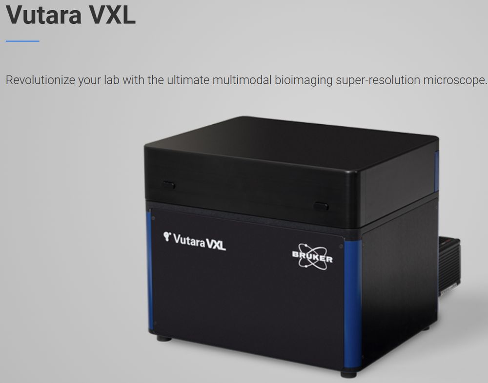
Corentin Rousset now introducing Bruker's Vutara VXL microscope to the EMBO course in superresolution microscopy in Prague.
Practicals to follow later today with the generous help of Dr. Christoph Spahn. We will try z-stack acquisitions, STORM PAINT and PALM on bacteria. Looking forward!
Practicals to follow later today with the generous help of Dr. Christoph Spahn. We will try z-stack acquisitions, STORM PAINT and PALM on bacteria. Looking forward!

June 20, 2025 at 12:04 PM
Corentin Rousset now introducing Bruker's Vutara VXL microscope to the EMBO course in superresolution microscopy in Prague.
Practicals to follow later today with the generous help of Dr. Christoph Spahn. We will try z-stack acquisitions, STORM PAINT and PALM on bacteria. Looking forward!
Practicals to follow later today with the generous help of Dr. Christoph Spahn. We will try z-stack acquisitions, STORM PAINT and PALM on bacteria. Looking forward!
An opportunity to test @brukercorporation.bsky.social SMLM microscope Vutara VXL in Prague on 18th and 19th June before our practical at the @embo.org superres microscopy course @imgprague.bsky.social
meetings.embo.org/event/25-sup...
Let me know if interested.
meetings.embo.org/event/25-sup...
Let me know if interested.


May 27, 2025 at 11:47 AM
An opportunity to test @brukercorporation.bsky.social SMLM microscope Vutara VXL in Prague on 18th and 19th June before our practical at the @embo.org superres microscopy course @imgprague.bsky.social
meetings.embo.org/event/25-sup...
Let me know if interested.
meetings.embo.org/event/25-sup...
Let me know if interested.
We (@brukercorporation.bsky.social) are going to participate im #EMBO Super-resolution in light microscopy course in Prague in June @imgprague.bsky.social.
We are bringing Vutara VXL microscope for hands-on sessions.
bruker.com/en/products-...
Demo of VXL outside course possible, let me know.
We are bringing Vutara VXL microscope for hands-on sessions.
bruker.com/en/products-...
Demo of VXL outside course possible, let me know.

April 1, 2025 at 3:26 PM
We (@brukercorporation.bsky.social) are going to participate im #EMBO Super-resolution in light microscopy course in Prague in June @imgprague.bsky.social.
We are bringing Vutara VXL microscope for hands-on sessions.
bruker.com/en/products-...
Demo of VXL outside course possible, let me know.
We are bringing Vutara VXL microscope for hands-on sessions.
bruker.com/en/products-...
Demo of VXL outside course possible, let me know.
My colleagues put together an ebook on tissue clearing (for #lightsheet microscopy).
If you are like me and have only experience with live imaging and are a bit confused with the different acronyms flying around, I highly recommend you take a look!
I counted 37 methods!
Pdf below or message me.
If you are like me and have only experience with live imaging and are a bit confused with the different acronyms flying around, I highly recommend you take a look!
I counted 37 methods!
Pdf below or message me.


March 5, 2025 at 12:02 PM
My colleagues put together an ebook on tissue clearing (for #lightsheet microscopy).
If you are like me and have only experience with live imaging and are a bit confused with the different acronyms flying around, I highly recommend you take a look!
I counted 37 methods!
Pdf below or message me.
If you are like me and have only experience with live imaging and are a bit confused with the different acronyms flying around, I highly recommend you take a look!
I counted 37 methods!
Pdf below or message me.