


Seth Donoughe
@donoughe.bsky.social
AI+Bio researcher, working to make sure new tech makes the world better.
Also interested in predictive biology, qbio, morphogenesis, evolution, embryos, wings, eggs, dataviz, generative art, illustration
www.sethdonoughe.com
Also interested in predictive biology, qbio, morphogenesis, evolution, embryos, wings, eggs, dataviz, generative art, illustration
www.sethdonoughe.com
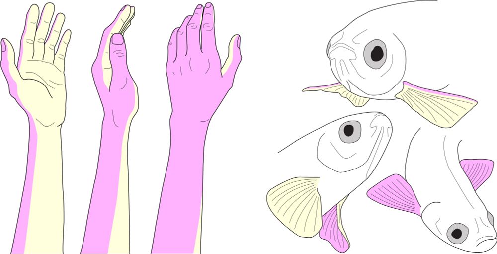
Great thread on the "palm" of a fish's fin
(and those illustrations...!)
(and those illustrations...!)

We take for granted that our hands have two sides and can articulate in an endless number of ways. But what about a fish’s fin? Can a fish know something “like the back of its fin,” or have its future told with a fin palm reading? Check out this bluetorial to find out 👇🧪🧬🐟 doi.org/10.1101/2025...
July 8, 2025 at 12:15 AM
Great thread on the "palm" of a fish's fin
(and those illustrations...!)
(and those illustrations...!)
Reposted by Seth Donoughe

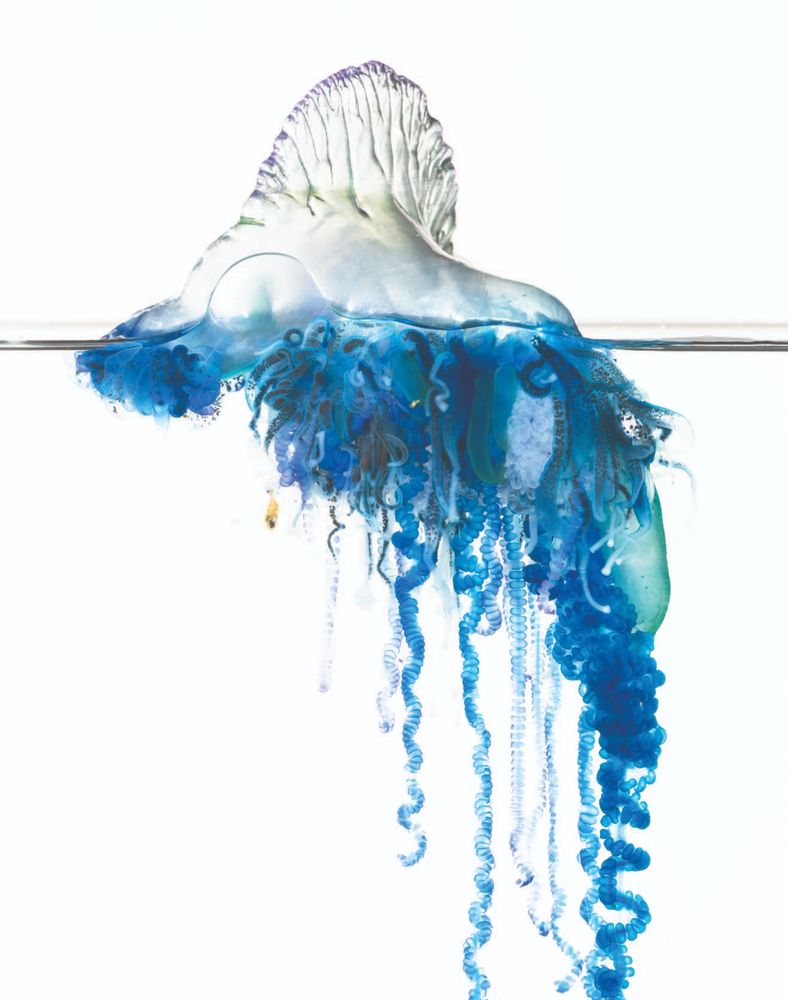
Excited to share our study on sailing siphonophores, AKA bluebottles or man-o'-war! 🌊 we received hundreds of samples from scientists around the world, part of a huge effort to sequence genomes and test for multiple species 🧬 out today in @currentbiology.bsky.social doi.org/10.1016/j.cu... 🦑🧪📌
June 19, 2025 at 7:51 PM
Excited to share our study on sailing siphonophores, AKA bluebottles or man-o'-war! 🌊 we received hundreds of samples from scientists around the world, part of a huge effort to sequence genomes and test for multiple species 🧬 out today in @currentbiology.bsky.social doi.org/10.1016/j.cu... 🦑🧪📌
"given the very likely course of AI improvements over the next few years, how EXACTLY should I change my work in my field right now" is an extremely practical question.
I liked this exercise by @jefftk.com to think through some specific answers for his research group's work
I liked this exercise by @jefftk.com to think through some specific answers for his research group's work

Thinking about the Nucleic Acid Observatory, AI will likely speed up some aspects of the creation of a detection system more than others. To the extent that we expect rapid advances in AI we should prioritize the work that we expect to bottleneck our future AI-accelerated work.
AI Advances and Detection Strategy
data.securebio.org
April 25, 2025 at 10:35 AM
"given the very likely course of AI improvements over the next few years, how EXACTLY should I change my work in my field right now" is an extremely practical question.
I liked this exercise by @jefftk.com to think through some specific answers for his research group's work
I liked this exercise by @jefftk.com to think through some specific answers for his research group's work
Career update: About a year ago, I became the AIxBio group leader at SecureBio, a research non-profit.
We study how rapidly accelerating AI is changing biology research, with a particular focus on making sure we mostly get the upsides.
Check out this thread for our new paper:
We study how rapidly accelerating AI is changing biology research, with a particular focus on making sure we mostly get the upsides.
Check out this thread for our new paper:

Can AIs provide expert-level troubleshooting assistance for work with viruses?
We built a new benchmark to answer that question.
To our surprise, we found that leading models outperform the vast majority of practicing virologists we sampled. 🧵 1/13
We built a new benchmark to answer that question.
To our surprise, we found that leading models outperform the vast majority of practicing virologists we sampled. 🧵 1/13

April 23, 2025 at 10:01 PM
Career update: About a year ago, I became the AIxBio group leader at SecureBio, a research non-profit.
We study how rapidly accelerating AI is changing biology research, with a particular focus on making sure we mostly get the upsides.
Check out this thread for our new paper:
We study how rapidly accelerating AI is changing biology research, with a particular focus on making sure we mostly get the upsides.
Check out this thread for our new paper:
A scanning electron micrograph zoom-in of a single wing scale from a pantry moth (Plodia interpunctella)—you know, the ones in your baking supplies.
Can't wait until somebody discovers how the underlying cell takes on such a wild shape & how the ECM is laid down.
Width of the image is ~8 microns.
Can't wait until somebody discovers how the underlying cell takes on such a wild shape & how the ECM is laid down.
Width of the image is ~8 microns.

December 1, 2024 at 8:55 PM
A scanning electron micrograph zoom-in of a single wing scale from a pantry moth (Plodia interpunctella)—you know, the ones in your baking supplies.
Can't wait until somebody discovers how the underlying cell takes on such a wild shape & how the ECM is laid down.
Width of the image is ~8 microns.
Can't wait until somebody discovers how the underlying cell takes on such a wild shape & how the ECM is laid down.
Width of the image is ~8 microns.
As insect wings develop, they form "crumpled up" in a very particular way. Unlike origami, there are no crisp folds.
Then, at adulthood, the wings emerge and open up.
Here is a cricket hindwing that I prepped from a juvenile, compared to an adult wing (from Turchyn & Popadić, FEVO 2024) 🧪
Then, at adulthood, the wings emerge and open up.
Here is a cricket hindwing that I prepped from a juvenile, compared to an adult wing (from Turchyn & Popadić, FEVO 2024) 🧪


November 20, 2024 at 3:37 PM
As insect wings develop, they form "crumpled up" in a very particular way. Unlike origami, there are no crisp folds.
Then, at adulthood, the wings emerge and open up.
Here is a cricket hindwing that I prepped from a juvenile, compared to an adult wing (from Turchyn & Popadić, FEVO 2024) 🧪
Then, at adulthood, the wings emerge and open up.
Here is a cricket hindwing that I prepped from a juvenile, compared to an adult wing (from Turchyn & Popadić, FEVO 2024) 🧪
Geometric order at multiple scales.
This is an optical slice into the eggshell of a fruit fly.
Every dot is the cross-section of a tiny pillar holding up the "roof" that is the outer surface of the egg. The black polygons are cavities left where cellular interfaces used to be. 🧪
This is an optical slice into the eggshell of a fruit fly.
Every dot is the cross-section of a tiny pillar holding up the "roof" that is the outer surface of the egg. The black polygons are cavities left where cellular interfaces used to be. 🧪

November 20, 2024 at 3:18 AM
Geometric order at multiple scales.
This is an optical slice into the eggshell of a fruit fly.
Every dot is the cross-section of a tiny pillar holding up the "roof" that is the outer surface of the egg. The black polygons are cavities left where cellular interfaces used to be. 🧪
This is an optical slice into the eggshell of a fruit fly.
Every dot is the cross-section of a tiny pillar holding up the "roof" that is the outer surface of the egg. The black polygons are cavities left where cellular interfaces used to be. 🧪
You know those "veins" on the wings of insects? In some species, they join together with surprisingly complex mechanical linkages.
This is an electron micrograph we captured of a vein joint from a damselfly. Note how the cuticle spikes allow more flexibility in one direction than the other. 🧪
This is an electron micrograph we captured of a vein joint from a damselfly. Note how the cuticle spikes allow more flexibility in one direction than the other. 🧪

August 4, 2023 at 2:31 PM
You know those "veins" on the wings of insects? In some species, they join together with surprisingly complex mechanical linkages.
This is an electron micrograph we captured of a vein joint from a damselfly. Note how the cuticle spikes allow more flexibility in one direction than the other. 🧪
This is an electron micrograph we captured of a vein joint from a damselfly. Note how the cuticle spikes allow more flexibility in one direction than the other. 🧪
A larva of the "lesser house fly" Fannia canicularis, illustrated in the Manual of Nearctic Diptera.
This book has astonishingly detailed and loving drawings of larvae. 🧪
This book has astonishingly detailed and loving drawings of larvae. 🧪

August 2, 2023 at 7:07 PM
A larva of the "lesser house fly" Fannia canicularis, illustrated in the Manual of Nearctic Diptera.
This book has astonishingly detailed and loving drawings of larvae. 🧪
This book has astonishingly detailed and loving drawings of larvae. 🧪
~Embryo gymnastics~
In some insect species, the developing embryo does half of a backwards somersault (a movement called "katatrepsis").
Kozo Miyakawa captured this process in a dragonfly in a 1987 paper w/ tons of pen & ink illustrations. 🧪
(freely available: http://aesj.co-site.jp/raiejp.html)
In some insect species, the developing embryo does half of a backwards somersault (a movement called "katatrepsis").
Kozo Miyakawa captured this process in a dragonfly in a 1987 paper w/ tons of pen & ink illustrations. 🧪
(freely available: http://aesj.co-site.jp/raiejp.html)

July 12, 2023 at 11:02 PM
~Embryo gymnastics~
In some insect species, the developing embryo does half of a backwards somersault (a movement called "katatrepsis").
Kozo Miyakawa captured this process in a dragonfly in a 1987 paper w/ tons of pen & ink illustrations. 🧪
(freely available: http://aesj.co-site.jp/raiejp.html)
In some insect species, the developing embryo does half of a backwards somersault (a movement called "katatrepsis").
Kozo Miyakawa captured this process in a dragonfly in a 1987 paper w/ tons of pen & ink illustrations. 🧪
(freely available: http://aesj.co-site.jp/raiejp.html)
The old descriptive embryology papers had some gems of hand-drawn illustration.
These are drawings of the developing embryo of a cabbage white butterfly (Pieris rapae), a species that is often an unwelcome muncher of your kale and bok choy plants.
by L.E.S. Eastham (Phil. Trans. Roy. Soc., 1930) 🧪
These are drawings of the developing embryo of a cabbage white butterfly (Pieris rapae), a species that is often an unwelcome muncher of your kale and bok choy plants.
by L.E.S. Eastham (Phil. Trans. Roy. Soc., 1930) 🧪

July 11, 2023 at 11:12 AM
The old descriptive embryology papers had some gems of hand-drawn illustration.
These are drawings of the developing embryo of a cabbage white butterfly (Pieris rapae), a species that is often an unwelcome muncher of your kale and bok choy plants.
by L.E.S. Eastham (Phil. Trans. Roy. Soc., 1930) 🧪
These are drawings of the developing embryo of a cabbage white butterfly (Pieris rapae), a species that is often an unwelcome muncher of your kale and bok choy plants.
by L.E.S. Eastham (Phil. Trans. Roy. Soc., 1930) 🧪
Tardigrade eggs! 🧪
Some bizarre surfaces on these things. I'm trying (and failing) to imagine the cellular processes that give rise to the branched surface structures. Any ideas? Let me know.
From Vecchi et al 2021: https://jyx.jyu.fi/handle/123456789/75978
Some bizarre surfaces on these things. I'm trying (and failing) to imagine the cellular processes that give rise to the branched surface structures. Any ideas? Let me know.
From Vecchi et al 2021: https://jyx.jyu.fi/handle/123456789/75978

July 10, 2023 at 10:49 PM
Tardigrade eggs! 🧪
Some bizarre surfaces on these things. I'm trying (and failing) to imagine the cellular processes that give rise to the branched surface structures. Any ideas? Let me know.
From Vecchi et al 2021: https://jyx.jyu.fi/handle/123456789/75978
Some bizarre surfaces on these things. I'm trying (and failing) to imagine the cellular processes that give rise to the branched surface structures. Any ideas? Let me know.
From Vecchi et al 2021: https://jyx.jyu.fi/handle/123456789/75978
Portrait of a two-spotted field cricket (Gryllus bimaculatus), taken with my collaborator Taro Nakamura 🧪
No need for chopsticks or fork if you have eating utensils built-in on your face.
No need for chopsticks or fork if you have eating utensils built-in on your face.

July 10, 2023 at 12:02 PM
Portrait of a two-spotted field cricket (Gryllus bimaculatus), taken with my collaborator Taro Nakamura 🧪
No need for chopsticks or fork if you have eating utensils built-in on your face.
No need for chopsticks or fork if you have eating utensils built-in on your face.
Wings from a speckle-winged rangeland grasshopper (Arphia conspersa).
This specimen was collected by Wayne Schennum in CO in 1973, & then I recently imaged it at the Chicago Field Museum. Wayne passed away in 2021, leaving a wealth of notes & specimens that are still giving rise to new discoveries.
This specimen was collected by Wayne Schennum in CO in 1973, & then I recently imaged it at the Chicago Field Museum. Wayne passed away in 2021, leaving a wealth of notes & specimens that are still giving rise to new discoveries.

July 6, 2023 at 4:17 PM
Wings from a speckle-winged rangeland grasshopper (Arphia conspersa).
This specimen was collected by Wayne Schennum in CO in 1973, & then I recently imaged it at the Chicago Field Museum. Wayne passed away in 2021, leaving a wealth of notes & specimens that are still giving rise to new discoveries.
This specimen was collected by Wayne Schennum in CO in 1973, & then I recently imaged it at the Chicago Field Museum. Wayne passed away in 2021, leaving a wealth of notes & specimens that are still giving rise to new discoveries.