


Stefano Di Talia
@ditalialab.bsky.social
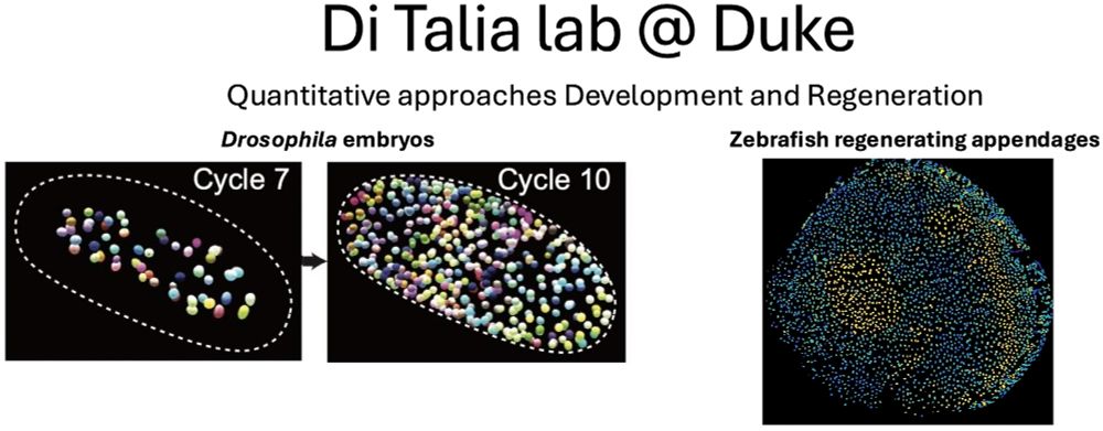
Quantitative Developmental Biology @Duke
Always a treat to see Victoria and get updated about her science. Of course, she is always up to interesting and new stuff :)

September 28, 2025 at 1:58 PM
Always a treat to see Victoria and get updated about her science. Of course, she is always up to interesting and new stuff :)
Just got back from a great visit to Vienna, spending time both at @impvienna.bsky.social and @istaresearch.bsky.social. Followed by a great visit to @embl.org. Amazing science, great and supportive colleagues and most notably incredible young trainees. It was so much fun to meet everyone.

September 28, 2025 at 1:58 PM
Just got back from a great visit to Vienna, spending time both at @impvienna.bsky.social and @istaresearch.bsky.social. Followed by a great visit to @embl.org. Amazing science, great and supportive colleagues and most notably incredible young trainees. It was so much fun to meet everyone.
Great dinner with current and former members of the lab, as well as current and former Duke members and friends of the lab. Great to catch up with @ceriweber.bsky.social ,Ben, @woonyunghur.bsky.social and share stories. Great @socdevbio.bsky.social meeting!

June 22, 2025 at 2:53 AM
Great dinner with current and former members of the lab, as well as current and former Duke members and friends of the lab. Great to catch up with @ceriweber.bsky.social ,Ben, @woonyunghur.bsky.social and share stories. Great @socdevbio.bsky.social meeting!
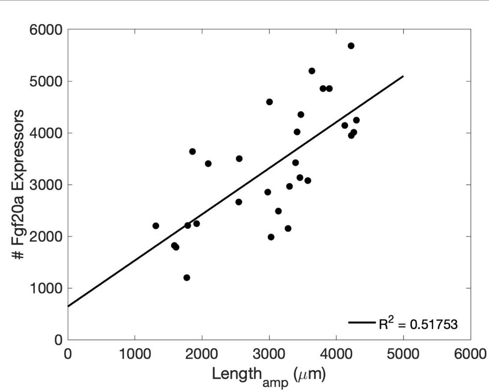
Next, we turned our attention to the ligand producing source and specifically to the cells of the basal epidermis that produce Fgf20a, which is upstream in the regeneration cascade and absolutely required. We found that the number of Fgf20a producing cells scale with the amount of tissue removed.
January 27, 2025 at 3:23 PM
Next, we turned our attention to the ligand producing source and specifically to the cells of the basal epidermis that produce Fgf20a, which is upstream in the regeneration cascade and absolutely required. We found that the number of Fgf20a producing cells scale with the amount of tissue removed.
Based on several estimates, we concluded that diffusion was unlikely to play a major role in the establishment of the gradients. Thus we focused on the role of advection (transport) of ligands by tissue growth and found that such a model could perfectly explain our data
January 27, 2025 at 3:23 PM
Based on several estimates, we concluded that diffusion was unlikely to play a major role in the establishment of the gradients. Thus we focused on the role of advection (transport) of ligands by tissue growth and found that such a model could perfectly explain our data
To be more clear, this is the behavior of the gradients across space and time
January 27, 2025 at 3:23 PM
To be more clear, this is the behavior of the gradients across space and time
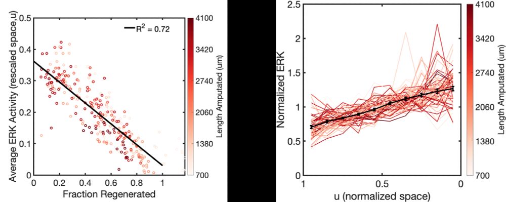
This led us to ask how Erk activity is controlled in space and time. We realized that Erk activity can be described mathematically throughout the entire regeneration process as the product of two functions, one describing time-dependency (left) and one spatial-dependency (right)
January 27, 2025 at 3:23 PM
This led us to ask how Erk activity is controlled in space and time. We realized that Erk activity can be described mathematically throughout the entire regeneration process as the product of two functions, one describing time-dependency (left) and one spatial-dependency (right)
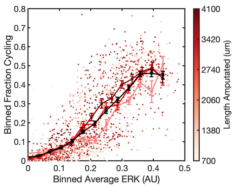
By quantifying Erk activity and proliferation in all osteoblasts of the regenerate, we found that Erk activity is predictive of the probability of cells to proliferate independently of the amount of tissue removed
January 27, 2025 at 3:23 PM
By quantifying Erk activity and proliferation in all osteoblasts of the regenerate, we found that Erk activity is predictive of the probability of cells to proliferate independently of the amount of tissue removed
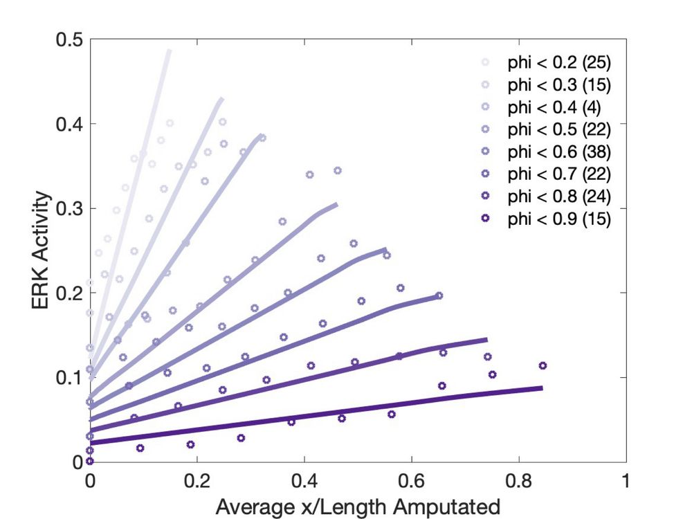
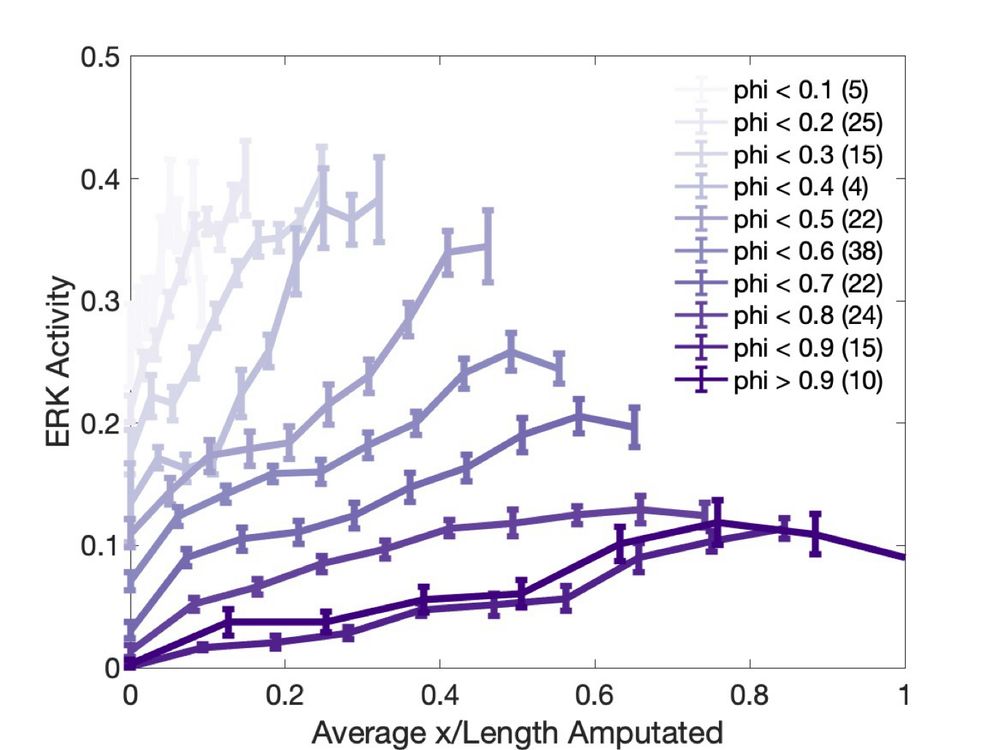
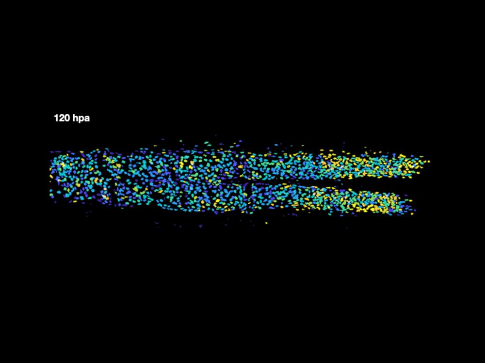
To understand how this scaling is encoded, we turned our attention to Fgf/Erk pathway as it has a well established role in controlling growth in many contexts, including fin regeneration. To characterize the spatiotemporal dynamics of the pathway, we use the Erk biosensor to measure activity.
January 27, 2025 at 3:23 PM
To understand how this scaling is encoded, we turned our attention to Fgf/Erk pathway as it has a well established role in controlling growth in many contexts, including fin regeneration. To characterize the spatiotemporal dynamics of the pathway, we use the Erk biosensor to measure activity.
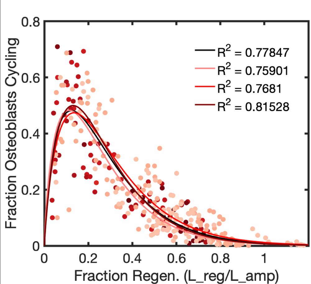
We then went on to demonstrate that proliferation perfectly scales when different amounts of tissue are removed by amputation, so that the probability of proliferation depends only on fraction of tissue that has regenerated.
January 27, 2025 at 3:23 PM
We then went on to demonstrate that proliferation perfectly scales when different amounts of tissue are removed by amputation, so that the probability of proliferation depends only on fraction of tissue that has regenerated.
Here we asked how the memory of bone size is encoded during zebrafish regeneration. This process nicely illustrates the concept of positional (geometric) memory, that is the ability of the regenerating fin to grow back to its pre-injury size and shape.
January 27, 2025 at 3:23 PM
Here we asked how the memory of bone size is encoded during zebrafish regeneration. This process nicely illustrates the concept of positional (geometric) memory, that is the ability of the regenerating fin to grow back to its pre-injury size and shape.
Please spread the word!
We are looking for postdoc(s) interested in quantitative problems in Development and Regeneration. Previous experience in quantitative biology is not required. We will support you and provide a great training environment if you are committed to learn new things.
We are looking for postdoc(s) interested in quantitative problems in Development and Regeneration. Previous experience in quantitative biology is not required. We will support you and provide a great training environment if you are committed to learn new things.
December 12, 2024 at 2:03 PM
Please spread the word!
We are looking for postdoc(s) interested in quantitative problems in Development and Regeneration. Previous experience in quantitative biology is not required. We will support you and provide a great training environment if you are committed to learn new things.
We are looking for postdoc(s) interested in quantitative problems in Development and Regeneration. Previous experience in quantitative biology is not required. We will support you and provide a great training environment if you are committed to learn new things.