


Reposted
RAC9229. M70s Sole of foot 5mm pigmented lesion. #Dermpath #HelpWithDiagnosis please.


September 15, 2025 at 3:40 PM
RAC9229. M70s Sole of foot 5mm pigmented lesion. #Dermpath #HelpWithDiagnosis please.
Reposted

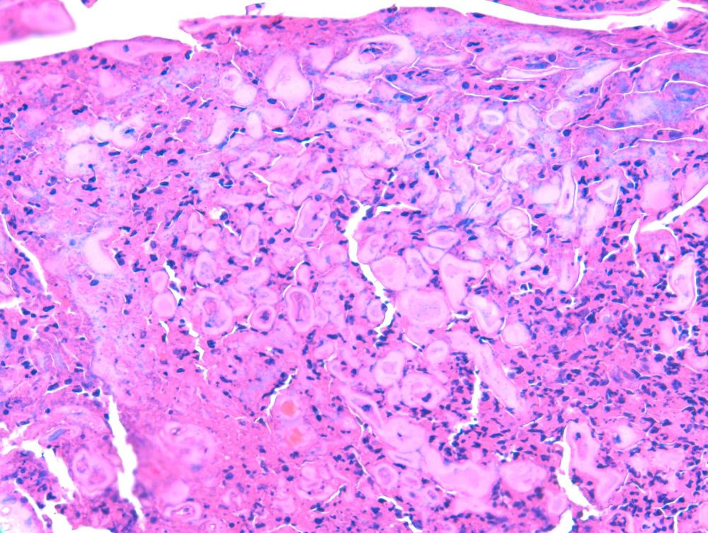
Image Quiz!
A patient presents with multiple pruritic, scaly patches on the back. A skin biopsy was performed, and a PAS special stain is shown. What's the best diagnosis?
A. Candidiasis
B. Tinea corporis
C. Tinea cruris
D. Tinea versicolor
See comments for answer.
#Pathology #PathologyOutlines
A patient presents with multiple pruritic, scaly patches on the back. A skin biopsy was performed, and a PAS special stain is shown. What's the best diagnosis?
A. Candidiasis
B. Tinea corporis
C. Tinea cruris
D. Tinea versicolor
See comments for answer.
#Pathology #PathologyOutlines

September 16, 2025 at 6:14 PM
Image Quiz!
A patient presents with multiple pruritic, scaly patches on the back. A skin biopsy was performed, and a PAS special stain is shown. What's the best diagnosis?
A. Candidiasis
B. Tinea corporis
C. Tinea cruris
D. Tinea versicolor
See comments for answer.
#Pathology #PathologyOutlines
A patient presents with multiple pruritic, scaly patches on the back. A skin biopsy was performed, and a PAS special stain is shown. What's the best diagnosis?
A. Candidiasis
B. Tinea corporis
C. Tinea cruris
D. Tinea versicolor
See comments for answer.
#Pathology #PathologyOutlines
Reposted
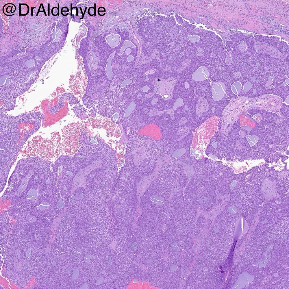
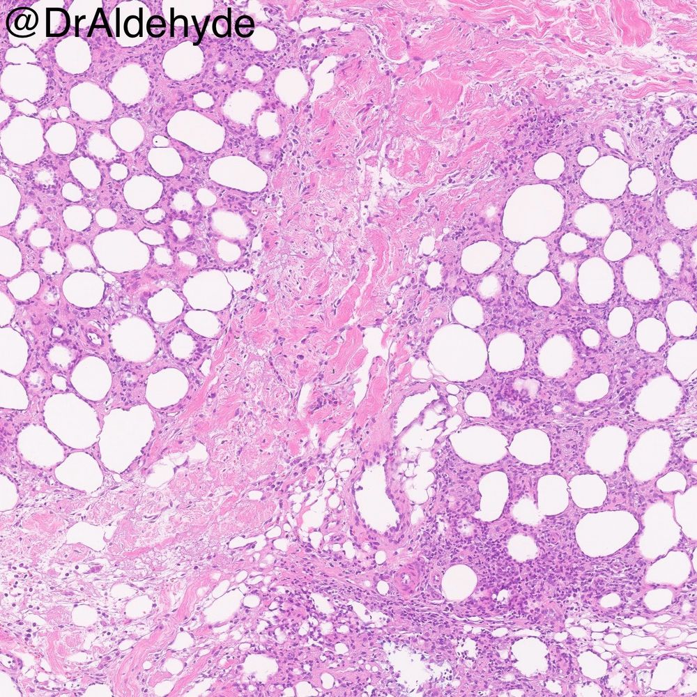
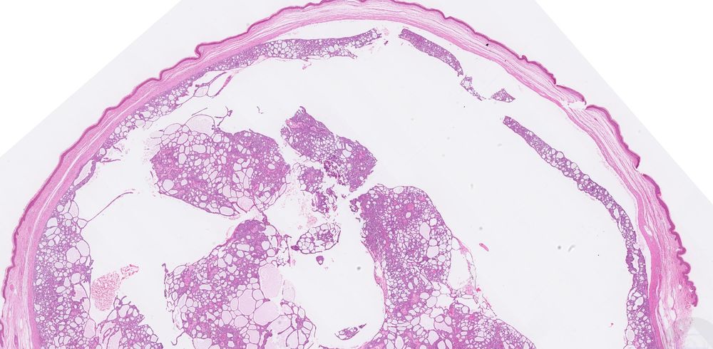
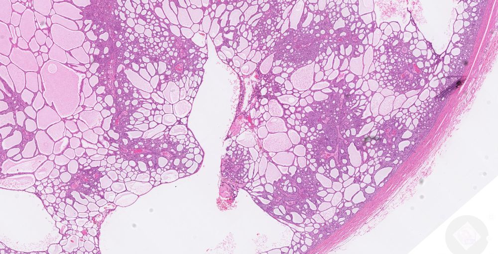
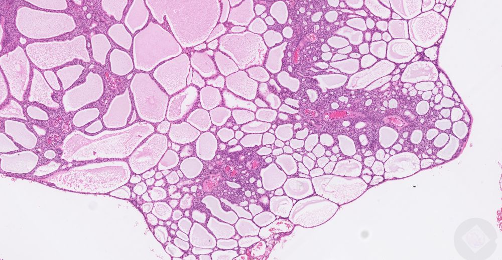
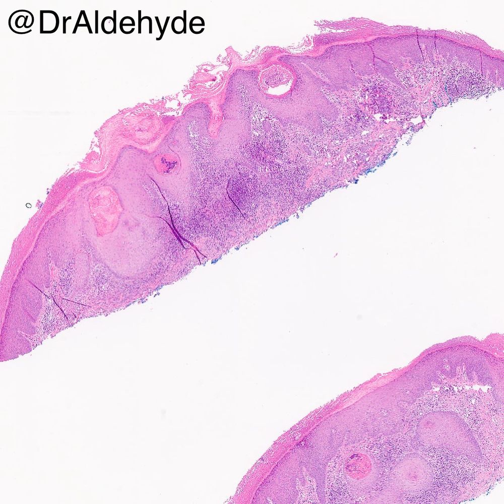
RAC9231 M30s Cyst in the foreskin. #ItsNeverACyst #Dermpath


September 16, 2025 at 5:15 PM
RAC9231 M30s Cyst in the foreskin. #ItsNeverACyst #Dermpath
Reposted
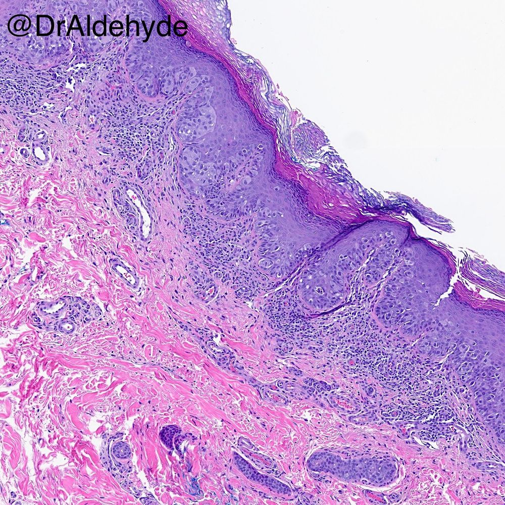
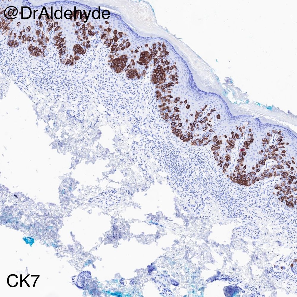
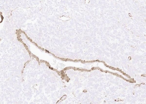
RAC9232 Lab gives you this p53 staining for signing off as control tissue. [M80s Right Cheek. Tumour 47mm diameter. Signed out by myself without IHC as FSCC-some KA-like features]. Comments to lab....??? #Dermpath #Immunohistochemistry


September 17, 2025 at 11:19 AM
RAC9232 Lab gives you this p53 staining for signing off as control tissue. [M80s Right Cheek. Tumour 47mm diameter. Signed out by myself without IHC as FSCC-some KA-like features]. Comments to lab....??? #Dermpath #Immunohistochemistry


Reposted
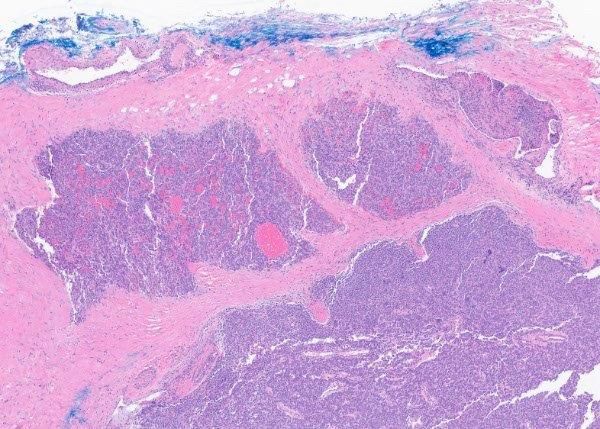
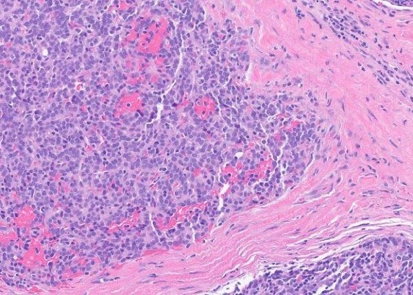
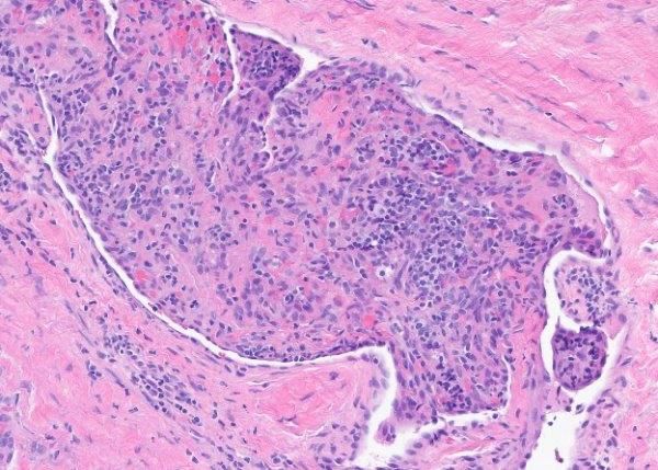
RAC9225: F50s. 20mm, red macular lesion with dilated vasculature on dermoscopy. No significant response to topical steroids. Doubled in size. Diagnostic uncertainty. Exclude BCC / dermal inflammatory reaction/other. PBx 4mm #Dermpath #SpotDiagnosis


September 6, 2025 at 8:24 AM
RAC9225: F50s. 20mm, red macular lesion with dilated vasculature on dermoscopy. No significant response to topical steroids. Doubled in size. Diagnostic uncertainty. Exclude BCC / dermal inflammatory reaction/other. PBx 4mm #Dermpath #SpotDiagnosis
Reposted

@racarr51.bsky.social Lesion clinically suggestive of keratoacanthoma


September 10, 2025 at 9:55 AM
@racarr51.bsky.social Lesion clinically suggestive of keratoacanthoma
Reposted

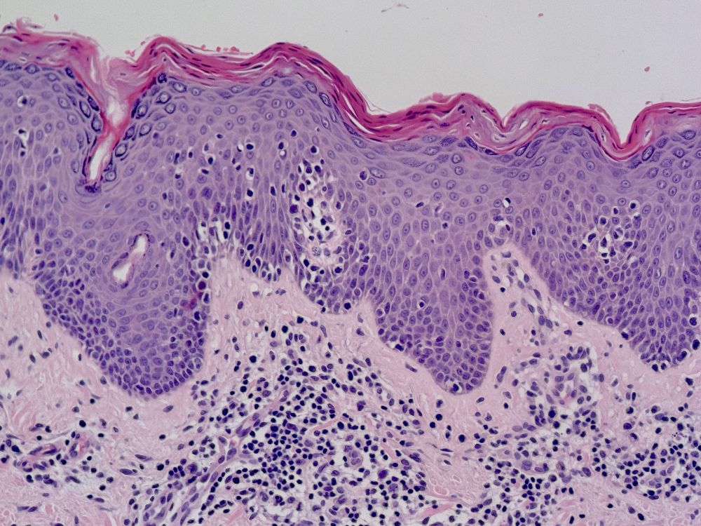
#MycosisFungoides #CTCL
Atypical intraepidermal lymphocytes tagging along the basal epidermis with halos. No spongiosis. This case really doesn’t need immunohistochemisry to confirm #classic 😉
#pathology
#dermpath
#bluepath
Atypical intraepidermal lymphocytes tagging along the basal epidermis with halos. No spongiosis. This case really doesn’t need immunohistochemisry to confirm #classic 😉
#pathology
#dermpath
#bluepath
April 19, 2025 at 3:40 PM
#MycosisFungoides #CTCL
Atypical intraepidermal lymphocytes tagging along the basal epidermis with halos. No spongiosis. This case really doesn’t need immunohistochemisry to confirm #classic 😉
#pathology
#dermpath
#bluepath
Atypical intraepidermal lymphocytes tagging along the basal epidermis with halos. No spongiosis. This case really doesn’t need immunohistochemisry to confirm #classic 😉
#pathology
#dermpath
#bluepath
Reposted

"Granulation tissue" is called like that because it forms small granules due to new vessel formation during the wound healing repair. Under the microscope, it looks like what you see in these pictures. #Pathology #GranulationTissue


May 2, 2025 at 6:43 PM
"Granulation tissue" is called like that because it forms small granules due to new vessel formation during the wound healing repair. Under the microscope, it looks like what you see in these pictures. #Pathology #GranulationTissue


Reposted
🚨Spoiler alert: Primary cutaneous NUT adnexal carcinoma can metastasize—but long-term outcomes remain unclear. We're about to report an almost 20-year follow-up on this new entity. Stay tuned! 😆🔬🧬
#dermpath #pathology #molpath #IHCPath #MDACCPath
#dermpath #pathology #molpath #IHCPath #MDACCPath
May 31, 2025 at 11:53 PM
🚨Spoiler alert: Primary cutaneous NUT adnexal carcinoma can metastasize—but long-term outcomes remain unclear. We're about to report an almost 20-year follow-up on this new entity. Stay tuned! 😆🔬🧬
#dermpath #pathology #molpath #IHCPath #MDACCPath
#dermpath #pathology #molpath #IHCPath #MDACCPath
Reposted
🚨Spoiler alert: Here’s a key pitfall almost no #dermpath textbook covers—SPTCLs can often show marked gamma/delta T-cell populations, mimicking pcGDTCL. A diagnostic trap you’ll want to know about. Our paper is coming soon—stay tuned! 😆🔬
#dermpath #hemepath #MDACCPath
#dermpath #hemepath #MDACCPath
June 10, 2025 at 9:21 PM
🚨Spoiler alert: Here’s a key pitfall almost no #dermpath textbook covers—SPTCLs can often show marked gamma/delta T-cell populations, mimicking pcGDTCL. A diagnostic trap you’ll want to know about. Our paper is coming soon—stay tuned! 😆🔬
#dermpath #hemepath #MDACCPath
#dermpath #hemepath #MDACCPath


Reposted
It’s been a while since I shared a case.
Did you know this immunoprofile, especially the diffuse TRPS1 immunoreactivity, strongly favors primary EMPD over secondary EMPD? Give it a try if you haven’t used it in your practice. 😆🔬
#dermpath #IHCPath #MDACCPath
Did you know this immunoprofile, especially the diffuse TRPS1 immunoreactivity, strongly favors primary EMPD over secondary EMPD? Give it a try if you haven’t used it in your practice. 😆🔬
#dermpath #IHCPath #MDACCPath
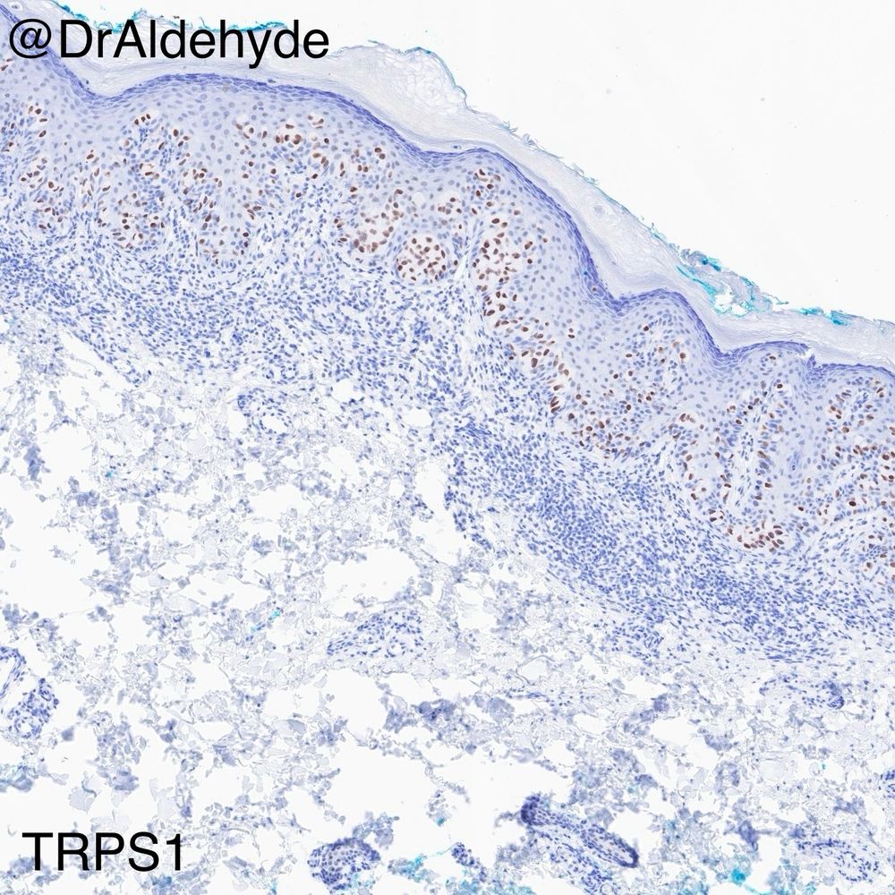
August 6, 2025 at 6:22 PM
It’s been a while since I shared a case.
Did you know this immunoprofile, especially the diffuse TRPS1 immunoreactivity, strongly favors primary EMPD over secondary EMPD? Give it a try if you haven’t used it in your practice. 😆🔬
#dermpath #IHCPath #MDACCPath
Did you know this immunoprofile, especially the diffuse TRPS1 immunoreactivity, strongly favors primary EMPD over secondary EMPD? Give it a try if you haven’t used it in your practice. 😆🔬
#dermpath #IHCPath #MDACCPath
Reposted
RAC9212: Spot diagnosis. Male 30s Eyelid


July 30, 2025 at 11:57 AM
RAC9212: Spot diagnosis. Male 30s Eyelid


Reposted

Dr. John R. Goodlad shared this case which he'll be presenting at our October Hematopathology course in Palm Springs: my.uscap.org/app/program/...
Male aged 12 years presents with hypopigmented patches on back
Histology: Epidermotropic infiltrate of CD8 positive T-cells
#PathSky
Male aged 12 years presents with hypopigmented patches on back
Histology: Epidermotropic infiltrate of CD8 positive T-cells
#PathSky

August 21, 2025 at 11:46 PM
Dr. John R. Goodlad shared this case which he'll be presenting at our October Hematopathology course in Palm Springs: my.uscap.org/app/program/...
Male aged 12 years presents with hypopigmented patches on back
Histology: Epidermotropic infiltrate of CD8 positive T-cells
#PathSky
Male aged 12 years presents with hypopigmented patches on back
Histology: Epidermotropic infiltrate of CD8 positive T-cells
#PathSky
Reposted

#PathSky 15F, thumb mass. There are no relevant positive immunostains. However, FISH for DDIT3 amplification (not rearrangement) is positive. This is an excellent example of a GLI1-amplified sarcoma. This is a great paper from the MSKCC group. pubmed.ncbi.nlm.nih.gov/38934567/
March 8, 2025 at 3:27 PM
Reposted
2025 Mth 3 RAC6518 on Kiko
kikoxp.com/posts/56258
kikoxp.com/posts/56258

KiKo XP | RAC6518
F60.
kikoxp.com
March 11, 2025 at 7:30 AM
2025 Mth 3 RAC6518 on Kiko
kikoxp.com/posts/56258
kikoxp.com/posts/56258
Reposted

Reposted
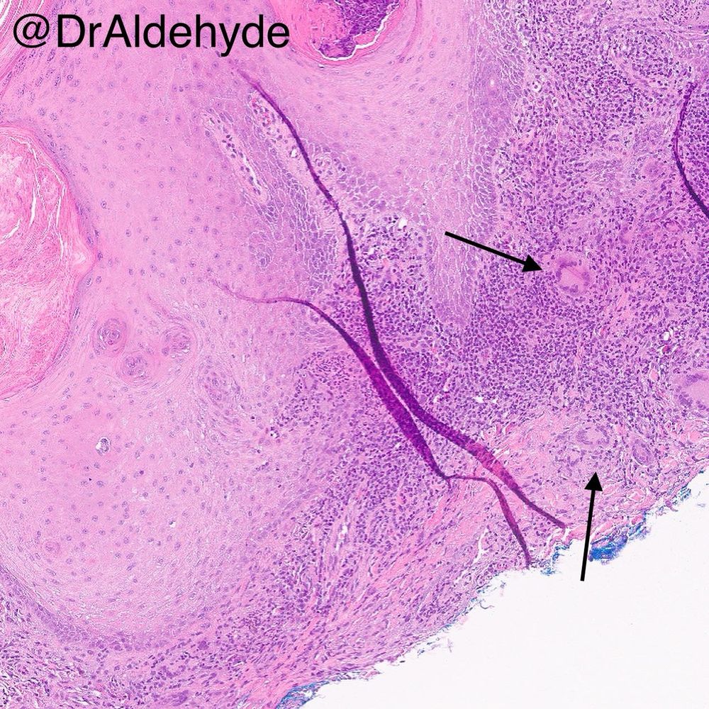
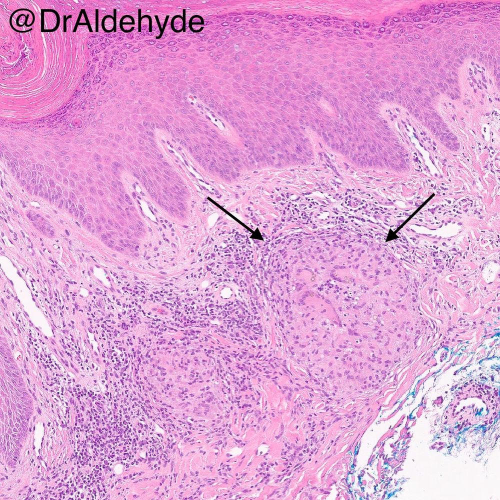
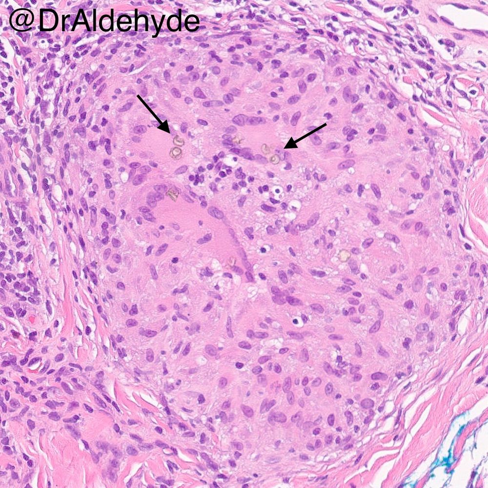
Do you see what I see? 😆🔬
Pseudoepitheliomatous hyperplasia and dermal granulomatous inflammation with multinucleated giant cells. That’s it? Nope, keep looking and you will find the cause of these histopathologic changes—Medlar bodies of chromoblastomycosis!
#dermpath #IDpath #MDACCpath
Pseudoepitheliomatous hyperplasia and dermal granulomatous inflammation with multinucleated giant cells. That’s it? Nope, keep looking and you will find the cause of these histopathologic changes—Medlar bodies of chromoblastomycosis!
#dermpath #IDpath #MDACCpath
March 19, 2025 at 1:44 AM
Do you see what I see? 😆🔬
Pseudoepitheliomatous hyperplasia and dermal granulomatous inflammation with multinucleated giant cells. That’s it? Nope, keep looking and you will find the cause of these histopathologic changes—Medlar bodies of chromoblastomycosis!
#dermpath #IDpath #MDACCpath
Pseudoepitheliomatous hyperplasia and dermal granulomatous inflammation with multinucleated giant cells. That’s it? Nope, keep looking and you will find the cause of these histopathologic changes—Medlar bodies of chromoblastomycosis!
#dermpath #IDpath #MDACCpath