


BIC_Bordeaux
@bic-bordeaux.bsky.social
Imaging facility located in Bordeaux. We offer service in light and electron microscopy, and in image treatment and analyse.
November #ImageOfTheMonth goes to M.Pujol from LPCO lab (UMR 5629). Acquired with Transmission Electron Microscopy, it shows a polystyrene (PS) latex obtained by post-dispersion of a yogurt cup made of PS in water using a surfactant. The resulting particles are highly heterogeneous and large.

November 3, 2025 at 11:02 AM
November #ImageOfTheMonth goes to M.Pujol from LPCO lab (UMR 5629). Acquired with Transmission Electron Microscopy, it shows a polystyrene (PS) latex obtained by post-dispersion of a yogurt cup made of PS in water using a surfactant. The resulting particles are highly heterogeneous and large.
October #ImageOfTheMonth is from O. Barba-Vila from @iins-bordeaux.bsky.social Slice of the gustatory cortex showing axons from the gustatory thalamus (G), basolateral amygdala (R) and a thick-tufted layer 5 neuron (W), recorded through patch clamp and imaged via biocytin-streptavidin labelling

October 7, 2025 at 7:48 AM
October #ImageOfTheMonth is from O. Barba-Vila from @iins-bordeaux.bsky.social Slice of the gustatory cortex showing axons from the gustatory thalamus (G), basolateral amygdala (R) and a thick-tufted layer 5 neuron (W), recorded through patch clamp and imaged via biocytin-streptavidin labelling
September #ImageOfTheMonth is from @rkinet.bsky.social from @imn-bordeaux.bsky.social
This is a maximum projection of a confocal image of a microglia cell (blue) and its nucleus (red), internalising multiple nanoparticles (yellow), one day after supranigral injection in C57B6-J mouse
This is a maximum projection of a confocal image of a microglia cell (blue) and its nucleus (red), internalising multiple nanoparticles (yellow), one day after supranigral injection in C57B6-J mouse

September 8, 2025 at 9:09 AM
September #ImageOfTheMonth is from @rkinet.bsky.social from @imn-bordeaux.bsky.social
This is a maximum projection of a confocal image of a microglia cell (blue) and its nucleus (red), internalising multiple nanoparticles (yellow), one day after supranigral injection in C57B6-J mouse
This is a maximum projection of a confocal image of a microglia cell (blue) and its nucleus (red), internalising multiple nanoparticles (yellow), one day after supranigral injection in C57B6-J mouse
August #ImageOfTheMonth is from L.Congiu from
@imn-bordeaux.bsky.social. This is a 3D reconstruction of a CD68-positive particles (white) within the cell body of microglia cells (Red) in the mouse cortex. The confocal Z-stack was processed with Imaris software.
@imn-bordeaux.bsky.social. This is a 3D reconstruction of a CD68-positive particles (white) within the cell body of microglia cells (Red) in the mouse cortex. The confocal Z-stack was processed with Imaris software.

August 7, 2025 at 6:56 PM
August #ImageOfTheMonth is from L.Congiu from
@imn-bordeaux.bsky.social. This is a 3D reconstruction of a CD68-positive particles (white) within the cell body of microglia cells (Red) in the mouse cortex. The confocal Z-stack was processed with Imaris software.
@imn-bordeaux.bsky.social. This is a 3D reconstruction of a CD68-positive particles (white) within the cell body of microglia cells (Red) in the mouse cortex. The confocal Z-stack was processed with Imaris software.
July #ImageOfTheMonth is from A.Drouet from
@iins-bordeaux.bsky.social. This image represents a neurosphere (150x150 µm) formed from dissociated hippocampal neurons cultured for 14 days under non-adhesive conditions. Yellow shows GFP expressing neurons and nuclei are showed in cyan.
@iins-bordeaux.bsky.social. This image represents a neurosphere (150x150 µm) formed from dissociated hippocampal neurons cultured for 14 days under non-adhesive conditions. Yellow shows GFP expressing neurons and nuclei are showed in cyan.

July 4, 2025 at 9:36 AM
July #ImageOfTheMonth is from A.Drouet from
@iins-bordeaux.bsky.social. This image represents a neurosphere (150x150 µm) formed from dissociated hippocampal neurons cultured for 14 days under non-adhesive conditions. Yellow shows GFP expressing neurons and nuclei are showed in cyan.
@iins-bordeaux.bsky.social. This image represents a neurosphere (150x150 µm) formed from dissociated hippocampal neurons cultured for 14 days under non-adhesive conditions. Yellow shows GFP expressing neurons and nuclei are showed in cyan.
June #ImageOfTheMonth is from L.Boutaleb from @inrae-bfp.bsky.social. They study the impact of heat stress on cell division and DNA replication on the tomato meristem. On this confocal image, EdU staining (magenta) detects replicating DNA and Hoechst labeling (cyan) shows all cell nuclei.

June 5, 2025 at 8:50 AM
June #ImageOfTheMonth is from L.Boutaleb from @inrae-bfp.bsky.social. They study the impact of heat stress on cell division and DNA replication on the tomato meristem. On this confocal image, EdU staining (magenta) detects replicating DNA and Hoechst labeling (cyan) shows all cell nuclei.
May #ImageOfTheMonth was from C.Mazocco & M.Darricau from @imn-bordeaux.bsky.social. Staining on spinal cord slices reveal TDP43 protein in the nucleus of neurons (Red). Motoneurons and synaptic terminations were stained targetting ChAT (Cyan).

June 5, 2025 at 8:44 AM
May #ImageOfTheMonth was from C.Mazocco & M.Darricau from @imn-bordeaux.bsky.social. Staining on spinal cord slices reveal TDP43 protein in the nucleus of neurons (Red). Motoneurons and synaptic terminations were stained targetting ChAT (Cyan).
April #ImageOfTheMonth is from H. Vaïtinadapoulé (BiiO EA 2521). Here is a guttae, a “drop-like deposits” of extracellular matrix components on the surface of the cornea. This image was acquired using scanning electron microscopy with the help of F.Decoeur from the EM unit of the BIC.

April 1, 2025 at 4:19 PM
April #ImageOfTheMonth is from H. Vaïtinadapoulé (BiiO EA 2521). Here is a guttae, a “drop-like deposits” of extracellular matrix components on the surface of the cornea. This image was acquired using scanning electron microscopy with the help of F.Decoeur from the EM unit of the BIC.
March #ImageOfTheMonth goes to M.Bonhivers from MFP lab (UMR 5234).
Trypanosoma brucei cell was extracted in detergent, expanded 4.2 fold using U-ExM, labelled for the microtubule cytoskeleton (magenta) and for the Hook Complex (yellow), and images usinf scanning confocal microscopy.
Trypanosoma brucei cell was extracted in detergent, expanded 4.2 fold using U-ExM, labelled for the microtubule cytoskeleton (magenta) and for the Hook Complex (yellow), and images usinf scanning confocal microscopy.

March 3, 2025 at 11:00 AM
March #ImageOfTheMonth goes to M.Bonhivers from MFP lab (UMR 5234).
Trypanosoma brucei cell was extracted in detergent, expanded 4.2 fold using U-ExM, labelled for the microtubule cytoskeleton (magenta) and for the Hook Complex (yellow), and images usinf scanning confocal microscopy.
Trypanosoma brucei cell was extracted in detergent, expanded 4.2 fold using U-ExM, labelled for the microtubule cytoskeleton (magenta) and for the Hook Complex (yellow), and images usinf scanning confocal microscopy.
February #ImageOfTheMonth is from
@tdhellemmes.bsky.social
. GFP expressing Relaxin3 neurons from the nucleus incertus and their projections are followed through the whole mouse brain using Adipoclear+ method and Light Sheet microscopy. Scale:1mm
@tdhellemmes.bsky.social
. GFP expressing Relaxin3 neurons from the nucleus incertus and their projections are followed through the whole mouse brain using Adipoclear+ method and Light Sheet microscopy. Scale:1mm

February 3, 2025 at 5:01 PM
February #ImageOfTheMonth is from
@tdhellemmes.bsky.social
. GFP expressing Relaxin3 neurons from the nucleus incertus and their projections are followed through the whole mouse brain using Adipoclear+ method and Light Sheet microscopy. Scale:1mm
@tdhellemmes.bsky.social
. GFP expressing Relaxin3 neurons from the nucleus incertus and their projections are followed through the whole mouse brain using Adipoclear+ method and Light Sheet microscopy. Scale:1mm
Happy new year! Let's start 2025 with an #ImageOfTheMonth from N. Bollier @inrae-bfp.bsky.social. Scanning confocal 3D reconstruction of a tomato shoot apical meristem. The fixed meristem was cleared using clearsee and the cell walls are stained by calcofluor white.

January 5, 2025 at 3:38 PM
Happy new year! Let's start 2025 with an #ImageOfTheMonth from N. Bollier @inrae-bfp.bsky.social. Scanning confocal 3D reconstruction of a tomato shoot apical meristem. The fixed meristem was cleared using clearsee and the cell walls are stained by calcofluor white.
Hello, the BIC is starting its Bluesky experience with its December #ImageOfTheMonth. Beautiful rat neurons imaged in STED 😍 by T.Cloâtre from @iins-bordeaux.bsky.social !

December 12, 2024 at 10:30 AM
Hello, the BIC is starting its Bluesky experience with its December #ImageOfTheMonth. Beautiful rat neurons imaged in STED 😍 by T.Cloâtre from @iins-bordeaux.bsky.social !
Le BIC vous souhaite de bonnes fêtes de fin d'année !

December 12, 2024 at 10:22 AM
Le BIC vous souhaite de bonnes fêtes de fin d'année !
Aujourd'hui le BIC reçoit le cristal collectif du CNRS. Tous les membres de l'équipe tiennent à remercier les utilisateurs grâce à qui nous évoluons au quotidien.
Félicitations aux autres Lauréats aquitains des #TalentsCNRS !
Félicitations aux autres Lauréats aquitains des #TalentsCNRS !


December 12, 2024 at 10:22 AM
Aujourd'hui le BIC reçoit le cristal collectif du CNRS. Tous les membres de l'équipe tiennent à remercier les utilisateurs grâce à qui nous évoluons au quotidien.
Félicitations aux autres Lauréats aquitains des #TalentsCNRS !
Félicitations aux autres Lauréats aquitains des #TalentsCNRS !
Today all BIC staff is out for some fun on and around the lake. On the process of considering coming back to the dark imaging rooms or not...

December 12, 2024 at 10:22 AM
Today all BIC staff is out for some fun on and around the lake. On the process of considering coming back to the dark imaging rooms or not...
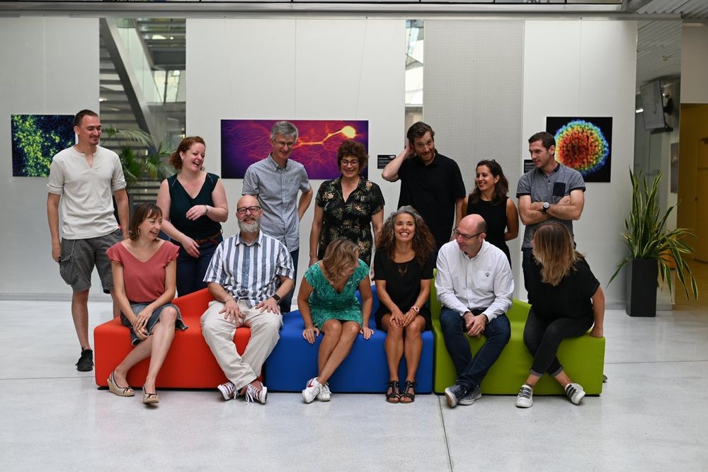
We started our official back to school week with the official picture to advertise our "Cristal collectif du CNRS 2022Like kids we were not really focused first and our photographer was very patient 😅😎thanks @avecunz
cnrs.fr/fr/personne/bo…
cnrs.fr/fr/personne/bo…
December 12, 2024 at 10:22 AM
We started our official back to school week with the official picture to advertise our "Cristal collectif du CNRS 2022Like kids we were not really focused first and our photographer was very patient 😅😎thanks @avecunz
cnrs.fr/fr/personne/bo…
cnrs.fr/fr/personne/bo…

Fin de la première semaine de manip. Malgré notre habitude de la paillasse on a transpiré pour assurer la logistique 🥵 Au taquet pour le deuxième protocole la semaine prochaine 💪#BlobCNRS

December 12, 2024 at 10:21 AM
Fin de la première semaine de manip. Malgré notre habitude de la paillasse on a transpiré pour assurer la logistique 🥵 Au taquet pour le deuxième protocole la semaine prochaine 💪#BlobCNRS
Breaking news! @BIC_Bordeaux has just been awarded the prestigious “Cristal collectif du CNRS 2022”. We are very proud that our long term team efforts are acknowledged with this award😊🔬
cnrs.fr/fr/personne/cr…
cnrs.fr/fr/personne/cr…

December 12, 2024 at 10:21 AM
Breaking news! @BIC_Bordeaux has just been awarded the prestigious “Cristal collectif du CNRS 2022”. We are very proud that our long term team efforts are acknowledged with this award😊🔬
cnrs.fr/fr/personne/cr…
cnrs.fr/fr/personne/cr…

Apparently our students are involved in some post-it contest with the neighboor lab. And of course they started with an obvious piece of art 🔬🤩

December 12, 2024 at 10:21 AM
Apparently our students are involved in some post-it contest with the neighboor lab. And of course they started with an obvious piece of art 🔬🤩
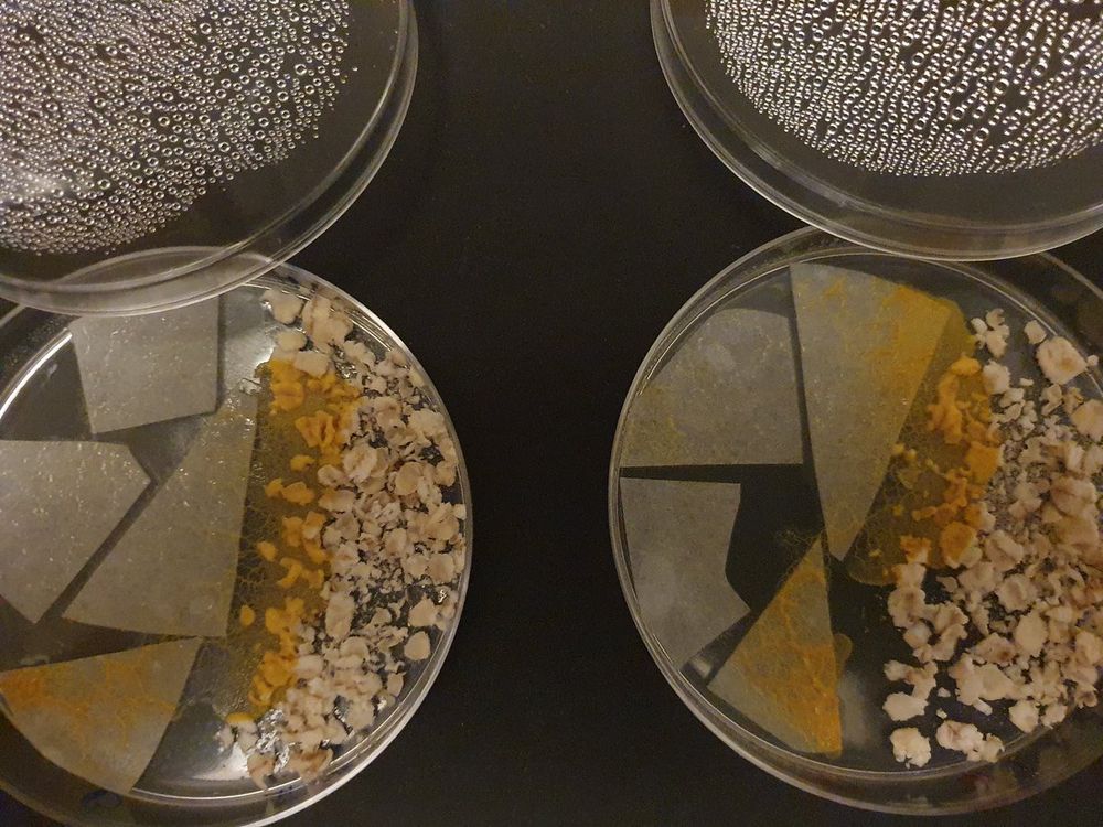
Dernière étape de préparation: les géloses pour la phase de réveil. Grâce @fab_cordelieres et à son imprimante 3D on parallélise et on normalise le volume d'agar dans chaque boîte 😅 #BlobCNRS


December 12, 2024 at 10:21 AM
Dernière étape de préparation: les géloses pour la phase de réveil. Grâce @fab_cordelieres et à son imprimante 3D on parallélise et on normalise le volume d'agar dans chaque boîte 😅 #BlobCNRS
Avant de se lancer dans le réveil et les premiers protocoles on continue de préparer notre espace de travail... Enfin on s'amuse avec notre espace de travail @fab_cordelieres @Seb_BIC @MonicaFM_BIC @ChristelPoujol #BlobCNRS

December 12, 2024 at 10:21 AM
Avant de se lancer dans le réveil et les premiers protocoles on continue de préparer notre espace de travail... Enfin on s'amuse avec notre espace de travail @fab_cordelieres @Seb_BIC @MonicaFM_BIC @ChristelPoujol #BlobCNRS
