

Patricia Ramos
@apramos.bsky.social
Dev Biologist, postdoc @BarrigaLab_@PolDresden. Postdoc
@NordenLab, MSCA fellow_optic cup morphogenesis, Phd @michalis_averof_parhyale eye evo-devo. #morphogenesis #evo_devo #imaging #image_analysis
@NordenLab, MSCA fellow_optic cup morphogenesis, Phd @michalis_averof_parhyale eye evo-devo. #morphogenesis #evo_devo #imaging #image_analysis
Fantastic interaction with @carldmodes.bsky.social
and @liormoneta.bsky.social , who did all the modelling. It was great to work in such an interchangeable setting between biology and physics, to lear about #ShapeProgramming and shed a little more light on how organs gain their 3D shape.
and @liormoneta.bsky.social , who did all the modelling. It was great to work in such an interchangeable setting between biology and physics, to lear about #ShapeProgramming and shed a little more light on how organs gain their 3D shape.
September 1, 2025 at 9:22 PM
Fantastic interaction with @carldmodes.bsky.social
and @liormoneta.bsky.social , who did all the modelling. It was great to work in such an interchangeable setting between biology and physics, to lear about #ShapeProgramming and shed a little more light on how organs gain their 3D shape.
and @liormoneta.bsky.social , who did all the modelling. It was great to work in such an interchangeable setting between biology and physics, to lear about #ShapeProgramming and shed a little more light on how organs gain their 3D shape.
This work also had the great contributions of A. Szalapak,
@louise-dagher.bsky.social and
@the-chaotician.bsky.social . Special thanks also to
@lcferme.bsky.social for the stardist model! And of course to Caren and all the @nordenlab.bsky.social
@louise-dagher.bsky.social and
@the-chaotician.bsky.social . Special thanks also to
@lcferme.bsky.social for the stardist model! And of course to Caren and all the @nordenlab.bsky.social
September 1, 2025 at 9:22 PM
This work also had the great contributions of A. Szalapak,
@louise-dagher.bsky.social and
@the-chaotician.bsky.social . Special thanks also to
@lcferme.bsky.social for the stardist model! And of course to Caren and all the @nordenlab.bsky.social
@louise-dagher.bsky.social and
@the-chaotician.bsky.social . Special thanks also to
@lcferme.bsky.social for the stardist model! And of course to Caren and all the @nordenlab.bsky.social
Finally, we disrupted apical patterned behaviors experimentally. The optic cup completely failed to start the invagination! (side note, the phenotype was so striking that I thought the microscope had crashed while imaging)
September 1, 2025 at 9:22 PM
Finally, we disrupted apical patterned behaviors experimentally. The optic cup completely failed to start the invagination! (side note, the phenotype was so striking that I thought the microscope had crashed while imaging)
Our model became even cooler: we converted tissue segmentations into networks. Like this we could test the strain patterns in the REAL geometry of the tissue

September 1, 2025 at 9:22 PM
Our model became even cooler: we converted tissue segmentations into networks. Like this we could test the strain patterns in the REAL geometry of the tissue
But can the shape transitions at the apical surface be sufficient to drive basal surface invagination? Updating our model with basal surfaces: apical strain patterns alone are enough to initiate invagination—even if basal surfaces are passive

September 1, 2025 at 9:22 PM
But can the shape transitions at the apical surface be sufficient to drive basal surface invagination? Updating our model with basal surfaces: apical strain patterns alone are enough to initiate invagination—even if basal surfaces are passive
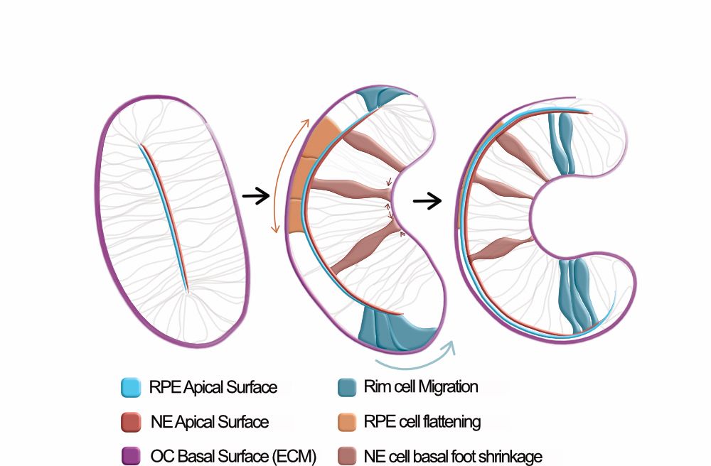
First, we needed to understand how are the apical surfaces curving. We went back and forth between experiments and modelling (inspired by shape programming). We found each apical surface displays different cell behaviors, generating its own in-plane strain patterns,

September 1, 2025 at 9:22 PM
First, we needed to understand how are the apical surfaces curving. We went back and forth between experiments and modelling (inspired by shape programming). We found each apical surface displays different cell behaviors, generating its own in-plane strain patterns,
But the surprise came from the apical surfaces. They undergo shape transitions before invagination starts. Could they be driving the process?
September 1, 2025 at 9:22 PM
But the surprise came from the apical surfaces. They undergo shape transitions before invagination starts. Could they be driving the process?
Separating the basal surface of the two epithelia (NE and RPE), we saw that they change their shape at the same time!
September 1, 2025 at 9:22 PM
Separating the basal surface of the two epithelia (NE and RPE), we saw that they change their shape at the same time!
We followed 3D shape changes of the forming optic cup, by tracking the basal surface
September 1, 2025 at 9:22 PM
We followed 3D shape changes of the forming optic cup, by tracking the basal surface
Many things happen at the same time: 2 epithelial layers differentiate, cells migrate, other cells flatten and others constrict their basal surface, and the lens grows in. We went through (a LOT of) negative results to try to understand which processes were triggering the onset of the invagination
September 1, 2025 at 9:22 PM
Many things happen at the same time: 2 epithelial layers differentiate, cells migrate, other cells flatten and others constrict their basal surface, and the lens grows in. We went through (a LOT of) negative results to try to understand which processes were triggering the onset of the invagination
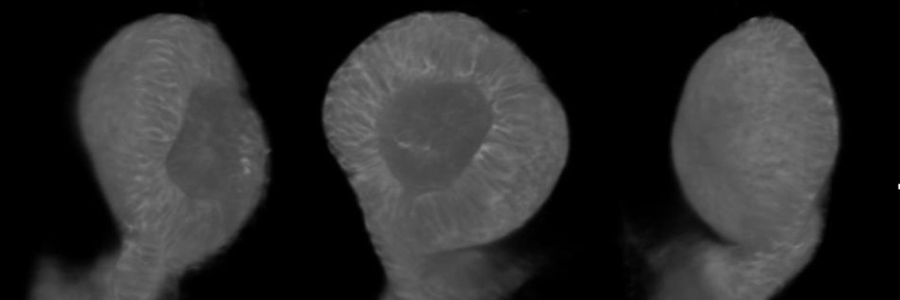
We used the Optic Cup formation as a system. The shape transition from a flat vesicle to an hemispheric eye precursor is just mesmerizing
September 1, 2025 at 9:22 PM
We used the Optic Cup formation as a system. The shape transition from a flat vesicle to an hemispheric eye precursor is just mesmerizing
We departed from a bigger question: how 3D shape emerges in complex tissues, from the interaction of multiple cell behaviors and mechanical inputs?
September 1, 2025 at 9:22 PM
We departed from a bigger question: how 3D shape emerges in complex tissues, from the interaction of multiple cell behaviors and mechanical inputs?
Briefly, we found that OC invagination onset relies on active, collective cell behaviors, that create patterned strains at the apical surfaces. In addition, OC morphogenesis involves the cooperative reinforcement of two independent active patterns. More details bellow 👇
September 1, 2025 at 9:22 PM
Briefly, we found that OC invagination onset relies on active, collective cell behaviors, that create patterned strains at the apical surfaces. In addition, OC morphogenesis involves the cooperative reinforcement of two independent active patterns. More details bellow 👇