


Aafke Gros
@aafkegros.bsky.social
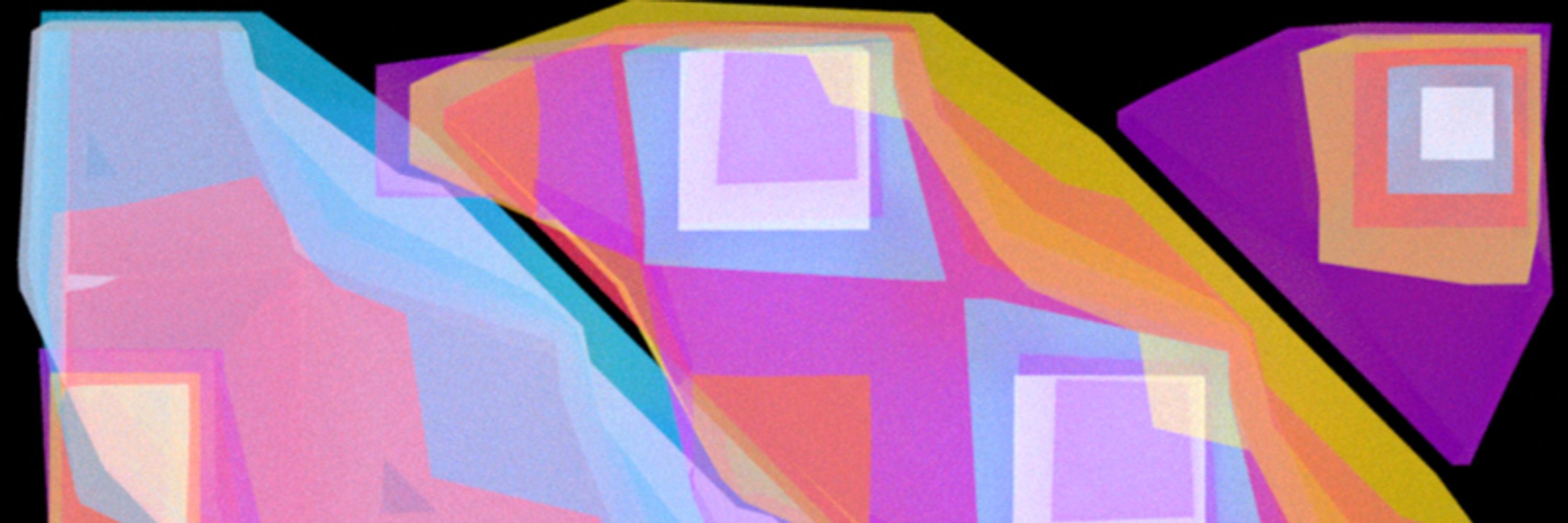
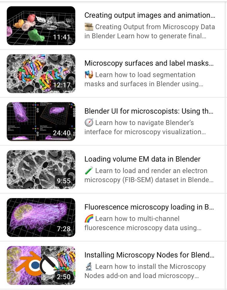
Check out Microscopy Nodes for handling microscopy data in Blender! Now loading .tif and OME-Zarr with big volume support :)
she/her | IPA: ˈafkə ɡrɔs
she/her | IPA: ˈafkə ɡrɔs
This is accompanied by a new set of tutorials showing exactly how you can start making beautiful microscopy visualizations in @blender.org!
Following along is also easy with the example OME-Zarr @openmicroscopy.org datasets 😄
Find the tutorials here: www.youtube.com/playlist?lis...
Following along is also easy with the example OME-Zarr @openmicroscopy.org datasets 😄
Find the tutorials here: www.youtube.com/playlist?lis...
May 23, 2025 at 1:13 PM
This is accompanied by a new set of tutorials showing exactly how you can start making beautiful microscopy visualizations in @blender.org!
Following along is also easy with the example OME-Zarr @openmicroscopy.org datasets 😄
Find the tutorials here: www.youtube.com/playlist?lis...
Following along is also easy with the example OME-Zarr @openmicroscopy.org datasets 😄
Find the tutorials here: www.youtube.com/playlist?lis...
In the context of our @reviewcommons.org revision process, I'm happy to announce Microscopy Nodes v2.2.0!
This packs lots of new fun features, including new color management 🌈, clearer transparency handling 🫥, custom default settings 🔧 and more!
Preprint at doi.org/10.1101/2025...
This packs lots of new fun features, including new color management 🌈, clearer transparency handling 🫥, custom default settings 🔧 and more!
Preprint at doi.org/10.1101/2025...
May 23, 2025 at 1:13 PM
In the context of our @reviewcommons.org revision process, I'm happy to announce Microscopy Nodes v2.2.0!
This packs lots of new fun features, including new color management 🌈, clearer transparency handling 🫥, custom default settings 🔧 and more!
Preprint at doi.org/10.1101/2025...
This packs lots of new fun features, including new color management 🌈, clearer transparency handling 🫥, custom default settings 🔧 and more!
Preprint at doi.org/10.1101/2025...
And it also works in 2D 😁 Here we extract a circular projection instead of a spherical, and apply a 1D Fourier transform.
Here we can see how the channel distributions all align in the projection after we optically activate Rac1 in one part of the cell. 💡
With @jpassmore.bsky.social , Lukas Kapitein
Here we can see how the channel distributions all align in the projection after we optically activate Rac1 in one part of the cell. 💡
With @jpassmore.bsky.social , Lukas Kapitein
January 30, 2025 at 10:11 AM
And it also works in 2D 😁 Here we extract a circular projection instead of a spherical, and apply a 1D Fourier transform.
Here we can see how the channel distributions all align in the projection after we optically activate Rac1 in one part of the cell. 💡
With @jpassmore.bsky.social , Lukas Kapitein
Here we can see how the channel distributions all align in the projection after we optically activate Rac1 in one part of the cell. 💡
With @jpassmore.bsky.social , Lukas Kapitein
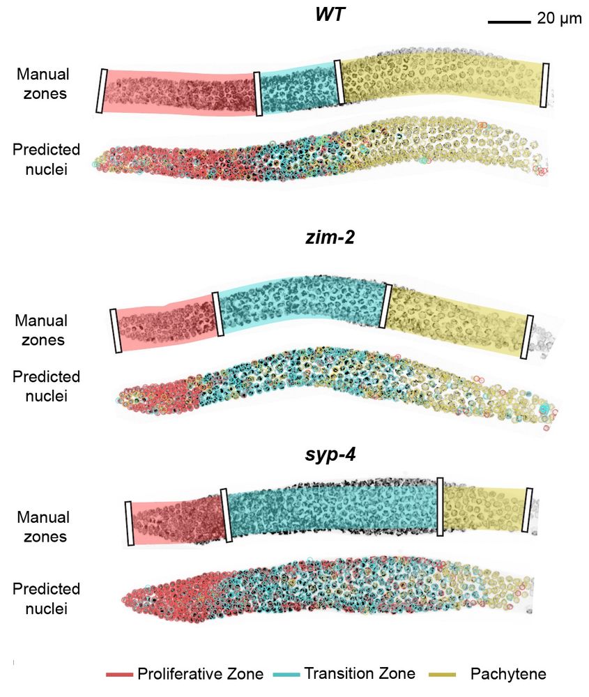
With the integration into the accessible toolkit ilastik (@ilastik-team.bsky.social) you can very easily include this in a quickly and interactively retrainable classification algorithm.
We show this for classification of meiotic zones in C. elegans :) 🪱
For the nerds there's also a Python API 😉
We show this for classification of meiotic zones in C. elegans :) 🪱
For the nerds there's also a Python API 😉
January 30, 2025 at 10:11 AM
With the integration into the accessible toolkit ilastik (@ilastik-team.bsky.social) you can very easily include this in a quickly and interactively retrainable classification algorithm.
We show this for classification of meiotic zones in C. elegans :) 🪱
For the nerds there's also a Python API 😉
We show this for classification of meiotic zones in C. elegans :) 🪱
For the nerds there's also a Python API 😉
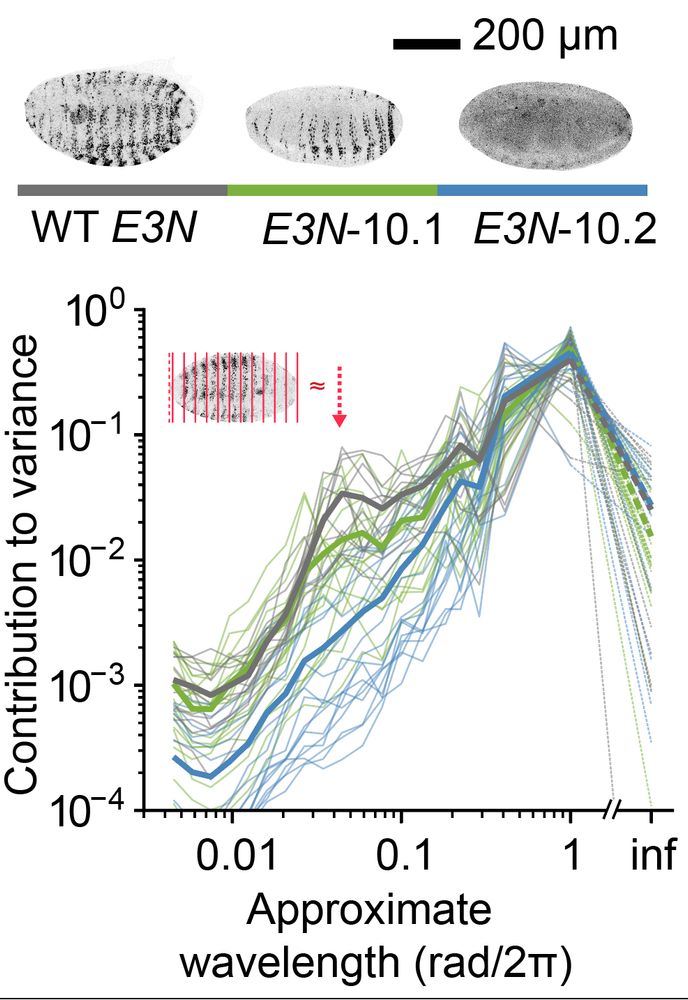
Quantifying distribution of intensity can be very helpful in many biological systems. For example, in D. melanogaster data, we extract a peak for the level of embryonic patterning in development. 🪰
In collaboration with
@ottilie.bsky.social, @timothyfuqua.bsky.social, @justinmcrocker.bsky.social
In collaboration with
@ottilie.bsky.social, @timothyfuqua.bsky.social, @justinmcrocker.bsky.social
January 30, 2025 at 10:11 AM
Quantifying distribution of intensity can be very helpful in many biological systems. For example, in D. melanogaster data, we extract a peak for the level of embryonic patterning in development. 🪰
In collaboration with
@ottilie.bsky.social, @timothyfuqua.bsky.social, @justinmcrocker.bsky.social
In collaboration with
@ottilie.bsky.social, @timothyfuqua.bsky.social, @justinmcrocker.bsky.social
We then use spherical harmonics decomposition to quantify the variance in the spherical signal per angular wavelength.
This gives us a metric that shows how detailed the features in the spherical map are, giving a rotation-invariant quantification of distribution.
This gives us a metric that shows how detailed the features in the spherical map are, giving a rotation-invariant quantification of distribution.
January 30, 2025 at 10:11 AM
We then use spherical harmonics decomposition to quantify the variance in the spherical signal per angular wavelength.
This gives us a metric that shows how detailed the features in the spherical map are, giving a rotation-invariant quantification of distribution.
This gives us a metric that shows how detailed the features in the spherical map are, giving a rotation-invariant quantification of distribution.
To quantify a segmented microscopy object, we project this to a spherical mean map.
This video is made with @blender3d.bsky.social geometry nodes and Microscopy Nodes, allowing the microscopy and method illustration in the same video :D
This video is made with @blender3d.bsky.social geometry nodes and Microscopy Nodes, allowing the microscopy and method illustration in the same video :D
January 30, 2025 at 10:11 AM
To quantify a segmented microscopy object, we project this to a spherical mean map.
This video is made with @blender3d.bsky.social geometry nodes and Microscopy Nodes, allowing the microscopy and method illustration in the same video :D
This video is made with @blender3d.bsky.social geometry nodes and Microscopy Nodes, allowing the microscopy and method illustration in the same video :D
Now out in PLOS CB: Spherical Texture extraction! doi.org/10.1371/jour...
This method quantifies the intensity distribution in microscopy objects, and is implemented parameter-free in @ilastik-team.bsky.social object classification!
We show applications in cells, C. elegans and Drosophila! 😁
This method quantifies the intensity distribution in microscopy objects, and is implemented parameter-free in @ilastik-team.bsky.social object classification!
We show applications in cells, C. elegans and Drosophila! 😁
January 30, 2025 at 10:11 AM
Now out in PLOS CB: Spherical Texture extraction! doi.org/10.1371/jour...
This method quantifies the intensity distribution in microscopy objects, and is implemented parameter-free in @ilastik-team.bsky.social object classification!
We show applications in cells, C. elegans and Drosophila! 😁
This method quantifies the intensity distribution in microscopy objects, and is implemented parameter-free in @ilastik-team.bsky.social object classification!
We show applications in cells, C. elegans and Drosophila! 😁
To learn more, please check out my youtube tutorials at www.youtube.com/@oanegros which shows, among others, how to make this video!
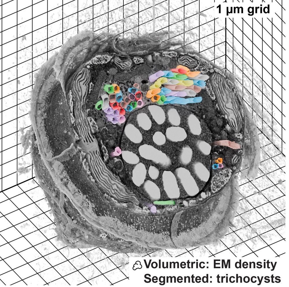
The data here is a dinoflagellate from Mocaer et al. 2023, hosted publicly at EMPIAR-11399 (also shown in the first post here).
The data here is a dinoflagellate from Mocaer et al. 2023, hosted publicly at EMPIAR-11399 (also shown in the first post here).
January 15, 2025 at 1:39 PM
To learn more, please check out my youtube tutorials at www.youtube.com/@oanegros which shows, among others, how to make this video!
The data here is a dinoflagellate from Mocaer et al. 2023, hosted publicly at EMPIAR-11399 (also shown in the first post here).
The data here is a dinoflagellate from Mocaer et al. 2023, hosted publicly at EMPIAR-11399 (also shown in the first post here).
Microscopy Nodes loads data from a @zarr.dev bucket, making it easy to use open data archives that host Zarr data!
This is a mitotic cell from Walther et al. 2018, that's hosted publicly on the Image Data Resource 😱
This is a mitotic cell from Walther et al. 2018, that's hosted publicly on the Image Data Resource 😱
January 15, 2025 at 1:39 PM
Microscopy Nodes loads data from a @zarr.dev bucket, making it easy to use open data archives that host Zarr data!
This is a mitotic cell from Walther et al. 2018, that's hosted publicly on the Image Data Resource 😱
This is a mitotic cell from Walther et al. 2018, that's hosted publicly on the Image Data Resource 😱
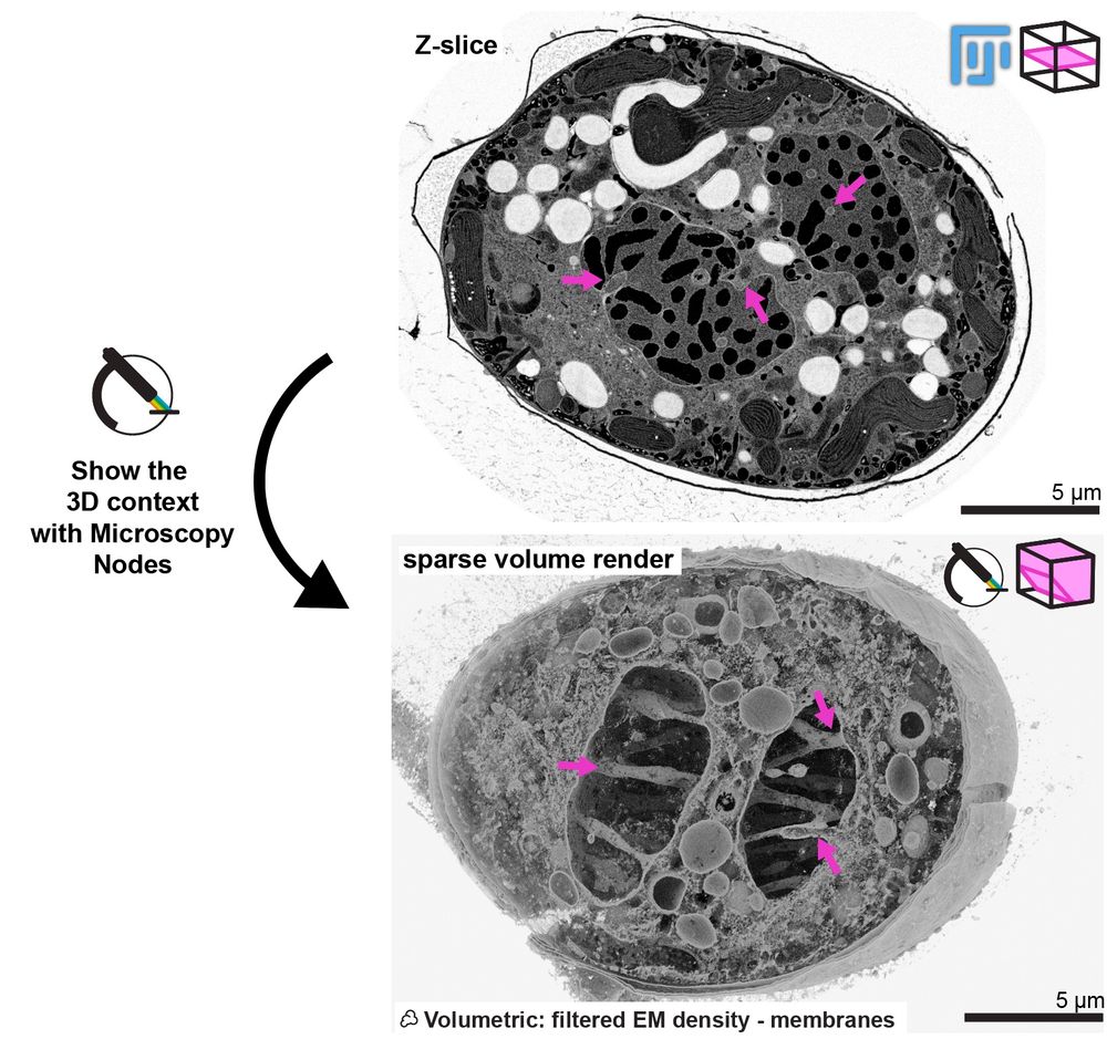
Here you can see how Microscopy Nodes can show the really cool phenotype of this mitotic dinoflagellate taken by @chandnibhickta.bsky.social!
With the sparse volume render in Blender you can see the network of tunnels through the nucleus that organize the spindle in its closed mitosis! 🤩
With the sparse volume render in Blender you can see the network of tunnels through the nucleus that organize the spindle in its closed mitosis! 🤩
January 15, 2025 at 1:39 PM
Here you can see how Microscopy Nodes can show the really cool phenotype of this mitotic dinoflagellate taken by @chandnibhickta.bsky.social!
With the sparse volume render in Blender you can see the network of tunnels through the nucleus that organize the spindle in its closed mitosis! 🤩
With the sparse volume render in Blender you can see the network of tunnels through the nucleus that organize the spindle in its closed mitosis! 🤩
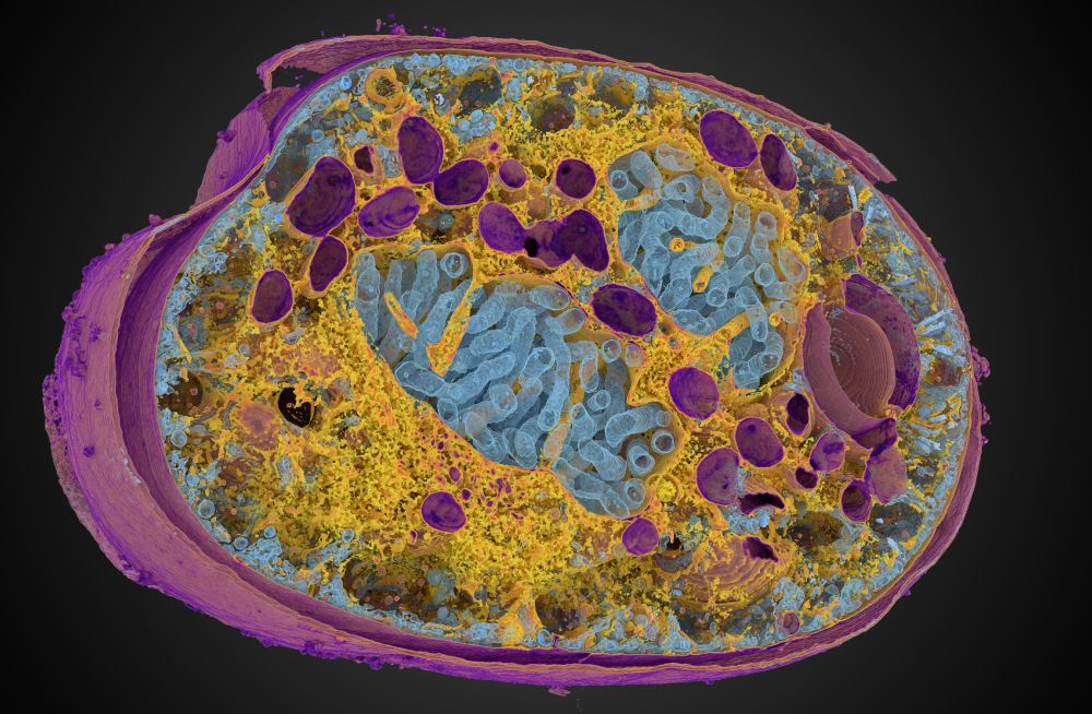
With Microscopy Nodes, you can play with large volumes (this ExM dataset from Granita Lokaj is 49 GB total) and integrate this with all the cool tools in Blender, such as this #geonodes model of the centriole, made by @banterlegroup.bsky.social !
January 15, 2025 at 1:39 PM
With Microscopy Nodes, you can play with large volumes (this ExM dataset from Granita Lokaj is 49 GB total) and integrate this with all the cool tools in Blender, such as this #geonodes model of the centriole, made by @banterlegroup.bsky.social !
Microscopy Nodes is now up on bioRxiv! 🚀
This is a Blender extension that seamlessly integrates and visualizes 3D microscopy data (TIF & @zarr.dev).
High-quality volume rendering for anyone, in both EM and fluorescence, regardless of computational expertise! 🔬
www.biorxiv.org/content/10.1...
This is a Blender extension that seamlessly integrates and visualizes 3D microscopy data (TIF & @zarr.dev).
High-quality volume rendering for anyone, in both EM and fluorescence, regardless of computational expertise! 🔬
www.biorxiv.org/content/10.1...
January 15, 2025 at 1:39 PM
Microscopy Nodes is now up on bioRxiv! 🚀
This is a Blender extension that seamlessly integrates and visualizes 3D microscopy data (TIF & @zarr.dev).
High-quality volume rendering for anyone, in both EM and fluorescence, regardless of computational expertise! 🔬
www.biorxiv.org/content/10.1...
This is a Blender extension that seamlessly integrates and visualizes 3D microscopy data (TIF & @zarr.dev).
High-quality volume rendering for anyone, in both EM and fluorescence, regardless of computational expertise! 🔬
www.biorxiv.org/content/10.1...
In honor of the first ever #MicroscopyNodes stickers arriving, I'm assembling my purse of open source bio(image) projects 😋. Which ones am I missing?😸
December 10, 2024 at 9:23 AM
In honor of the first ever #MicroscopyNodes stickers arriving, I'm assembling my purse of open source bio(image) projects 😋. Which ones am I missing?😸
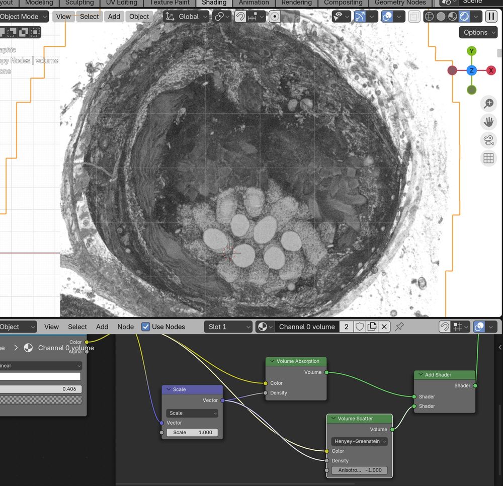
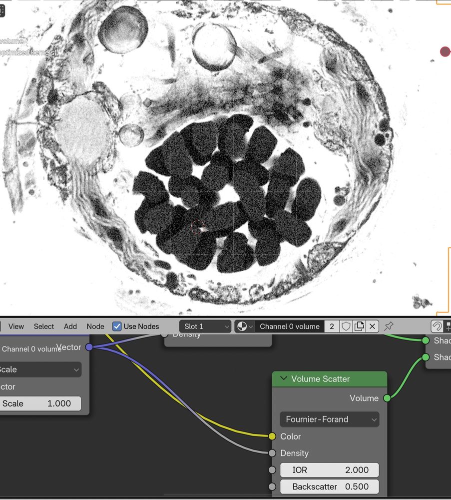
Messing around with the new #Blender 4.3 volume scatter modes with #microscopynodes, while at the #OME #Zarr hackathon! 😁🤩
Great info and work on the upcoming features of Zarr here :D Transforms, collections, and more! 🙏
Great info and work on the upcoming features of Zarr here :D Transforms, collections, and more! 🙏
November 20, 2024 at 10:33 AM
Messing around with the new #Blender 4.3 volume scatter modes with #microscopynodes, while at the #OME #Zarr hackathon! 😁🤩
Great info and work on the upcoming features of Zarr here :D Transforms, collections, and more! 🙏
Great info and work on the upcoming features of Zarr here :D Transforms, collections, and more! 🙏
And with OME-Zarr we can easily load data from public databases on the internet, such as the Bioimage Archive or IDR. Here is a beautiful mitosis dataset from Walther et al. (2018), hosted at uk1s3.embassy.ebi.ac.uk/idr/zarr/v0....
October 21, 2024 at 8:30 AM
And with OME-Zarr we can easily load data from public databases on the internet, such as the Bioimage Archive or IDR. Here is a beautiful mitosis dataset from Walther et al. (2018), hosted at uk1s3.embassy.ebi.ac.uk/idr/zarr/v0....
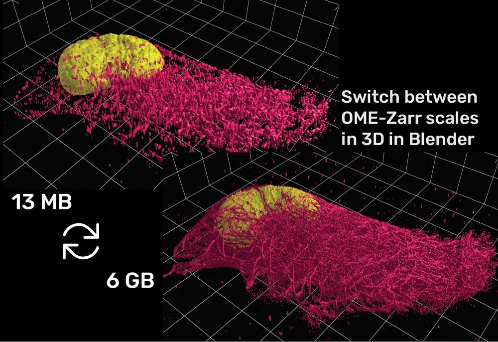
And with OME-Zarr loading, due to the pyramidal format, you can design your video at small-scale and only replace to full-scale at the final render!
(data from Granita Lokaj, @ s3.embl.de/microscopyno...)
(data from Granita Lokaj, @ s3.embl.de/microscopyno...)
October 21, 2024 at 8:29 AM
And with OME-Zarr loading, due to the pyramidal format, you can design your video at small-scale and only replace to full-scale at the final render!
(data from Granita Lokaj, @ s3.embl.de/microscopyno...)
(data from Granita Lokaj, @ s3.embl.de/microscopyno...)
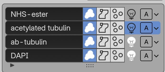
With the new channel interface, you can define per channel features, such as whether to load as volume/isosurface/labelmask + channel names and more!
October 21, 2024 at 8:29 AM
With the new channel interface, you can define per channel features, such as whether to load as volume/isosurface/labelmask + channel names and more!