


Michaela Medina
@mmedina300kv.bsky.social
Grad student in Grotjahn Lab @scrippsresearch. Mito explorer and music lover. She/her.
So excited that this work is out and I am so grateful to our team @hamid13r.bsky.social @attychang.bsky.social @baradlab.com @nanigrotjahn.bsky.social
Prohibitins aren’t hanging out just anywhere in the membrane — they prefer the 💁♀️💎✨VIP section💅🪩🥂
Lab all-star @mmedina300kv.bsky.social maps these microdomains using cryo-ET + surface morphometrics.
s/o to #teamtomo #cryoET co-authors
@hamid13r.bsky.social
@attychang.bsky.social
@baradlab.com
Lab all-star @mmedina300kv.bsky.social maps these microdomains using cryo-ET + surface morphometrics.
s/o to #teamtomo #cryoET co-authors
@hamid13r.bsky.social
@attychang.bsky.social
@baradlab.com
Prohibitin complexes associate with unique membrane microdomains in cells https://www.biorxiv.org/content/10.1101/2025.11.14.688579v1
November 17, 2025 at 12:33 AM
So excited that this work is out and I am so grateful to our team @hamid13r.bsky.social @attychang.bsky.social @baradlab.com @nanigrotjahn.bsky.social
Thank you so much everyone who has supported me through this journey! I am so happy that I have an incredible lab!!! 🔬
It’s been a big week for the Grotjahn Lab—our very first graduate student, @mmedina300kv.bsky.social, successfully defended her PhD thesis! 🎉 So proud of everything she’s accomplished and grateful for the light she’s brought to our lab from day one ☀️⚡
May 10, 2025 at 3:40 AM
Thank you so much everyone who has supported me through this journey! I am so happy that I have an incredible lab!!! 🔬
So excited for this work to be out!! Thanks to @attychang.bsky.social, @hamid13r.bsky.social, blue sky less Daniel Fuentes, @nanigrotjahn.bsky.social and @tomo.science!!!
🚨New preprint!🚨 #teamtomo
We expanded Surface Morphometrics to quantify membrane thickness from cryo-ET—revealing local variation across organelles.
Led by the lab’s first grad student, @mmedina300kv.bsky.social (defending Monday! 🍾) w/ @attychang.bsky.social @hamid13r.bsky.social & @tomo.science
We expanded Surface Morphometrics to quantify membrane thickness from cryo-ET—revealing local variation across organelles.
Led by the lab’s first grad student, @mmedina300kv.bsky.social (defending Monday! 🍾) w/ @attychang.bsky.social @hamid13r.bsky.social & @tomo.science
Surface morphometrics reveals local membrane thickness variation in organellar subcompartments https://www.biorxiv.org/content/10.1101/2025.04.30.651574v1
May 1, 2025 at 8:54 PM
So excited for this work to be out!! Thanks to @attychang.bsky.social, @hamid13r.bsky.social, blue sky less Daniel Fuentes, @nanigrotjahn.bsky.social and @tomo.science!!!
Reposted by Michaela Medina
Feeling so honored and thrilled to see our work make it to the cover—what a milestone in my career! I can’t wait to receive the poster from JCB🤩

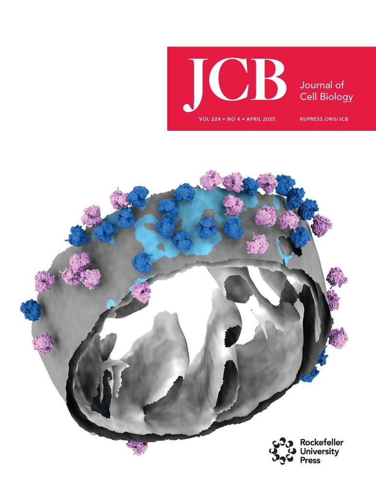
Our April issue is here! rupress.org/jcb/issue/22...
The cover shows a segmented model of cytoplasmic #ribosomes associated with mitochondrial membrane in a Saccharomyces cerevisiae cell. From Ya-Ting Chang, @nanigrotjahn.bsky.social and colleagues (doi.org/10.1083/jcb....)
The cover shows a segmented model of cytoplasmic #ribosomes associated with mitochondrial membrane in a Saccharomyces cerevisiae cell. From Ya-Ting Chang, @nanigrotjahn.bsky.social and colleagues (doi.org/10.1083/jcb....)
April 10, 2025 at 5:18 AM
Feeling so honored and thrilled to see our work make it to the cover—what a milestone in my career! I can’t wait to receive the poster from JCB🤩
Reposted by Michaela Medina
Happy to share the first paper in my PhD is published in JCB!
It's my honor to work with amazing teammates! I really enjoy exploring mitochondrial protein import mechanism through cryo-ET!
It's my honor to work with amazing teammates! I really enjoy exploring mitochondrial protein import mechanism through cryo-ET!
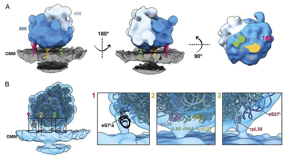
Cytoplasmic #ribosomes on #mitochondria alter the local membrane environment for protein import, say Ya-Ting Chang, Danielle Grotjahn (@nanigrotjahn.bsky.social) @scripps.edu and colleagues: rupress.org/jcb/article/...
#ProteinHomeostasis #Organelles #StructuralBiology #CryoET
#ProteinHomeostasis #Organelles #StructuralBiology #CryoET
April 9, 2025 at 5:29 AM
Happy to share the first paper in my PhD is published in JCB!
It's my honor to work with amazing teammates! I really enjoy exploring mitochondrial protein import mechanism through cryo-ET!
It's my honor to work with amazing teammates! I really enjoy exploring mitochondrial protein import mechanism through cryo-ET!
Yay!!! Congratulations Atty!!! So happy to see the paper and cover out!!! 🥳🔬
Our April issue is here! rupress.org/jcb/issue/22...
The cover shows a segmented model of cytoplasmic #ribosomes associated with mitochondrial membrane in a Saccharomyces cerevisiae cell. From Ya-Ting Chang, @nanigrotjahn.bsky.social and colleagues (doi.org/10.1083/jcb....)
The cover shows a segmented model of cytoplasmic #ribosomes associated with mitochondrial membrane in a Saccharomyces cerevisiae cell. From Ya-Ting Chang, @nanigrotjahn.bsky.social and colleagues (doi.org/10.1083/jcb....)
April 9, 2025 at 4:11 AM
Yay!!! Congratulations Atty!!! So happy to see the paper and cover out!!! 🥳🔬
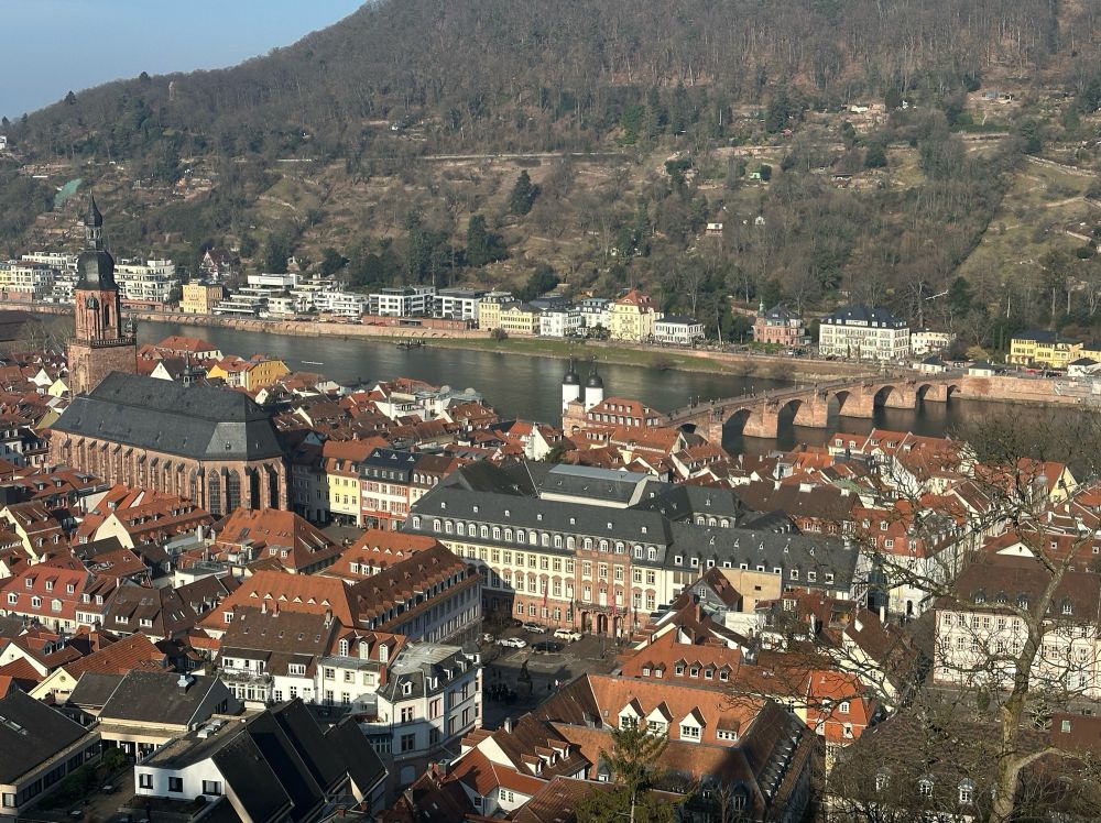
Had the incredible opportunity to present my work at #EMBOinsitu workshop in Heidelberg. It was filled with fantastic science and new friends! #teamtomo


February 8, 2025 at 1:00 PM
Had the incredible opportunity to present my work at #EMBOinsitu workshop in Heidelberg. It was filled with fantastic science and new friends! #teamtomo
Reposted by Michaela Medina
Labs that clay together, stay together 🩶🌪️🥣
@hamid13r.bsky.social @mmedina300kv.bsky.social @callmemich.bsky.social @nitishdua.bsky.social
@hamid13r.bsky.social @mmedina300kv.bsky.social @callmemich.bsky.social @nitishdua.bsky.social


November 23, 2024 at 7:34 AM
Labs that clay together, stay together 🩶🌪️🥣
@hamid13r.bsky.social @mmedina300kv.bsky.social @callmemich.bsky.social @nitishdua.bsky.social
@hamid13r.bsky.social @mmedina300kv.bsky.social @callmemich.bsky.social @nitishdua.bsky.social