


Debojit Saha
@debojitsaha.bsky.social
Postdoc at Schuh lab @schuhlab.bsky.social | MPI-NAT | Microscopy • Fertility • Cell Biology
Pinned
Delighted to share my PhD work with @schuhlab.bsky.social @mpi-nat.bsky.social on #humaneggrejuvenation, now available as a preprint on @biorxivpreprint.bsky.social 🎉
Grateful to my collaborators for the teamwork and support 🙌
More details here 👇
www.biorxiv.org/content/10.6...
#Aging #Aneuploidy
Grateful to my collaborators for the teamwork and support 🙌
More details here 👇
www.biorxiv.org/content/10.6...
#Aging #Aneuploidy
Reposted by Debojit Saha

Why do women’s eggs deteriorate as they age? 🥚
The @washingtonpost.com reports on this question and features our study on Shugoshin 1.
📰
The @washingtonpost.com reports on this question and features our study on Shugoshin 1.
📰
www.washingtonpost.com
January 13, 2026 at 10:45 AM
Why do women’s eggs deteriorate as they age? 🥚
The @washingtonpost.com reports on this question and features our study on Shugoshin 1.
📰
The @washingtonpost.com reports on this question and features our study on Shugoshin 1.
📰
Honored that our work on rescuing age-dependent chromosome errors in human eggs was highlighted by @theguardian.com. 👇
Our work on improving human egg quality was featured in @theguardian.com.
We hope this will help to make #IVF more successful and allow more couples to conceive. 💜
We hope this will help to make #IVF more successful and allow more couples to conceive. 💜

Human eggs ‘rejuvenated’ in an advance that could boost IVF success rates
Exclusive: Research suggests supplementing eggs with a key protein reduces age-related defects, raising hopes of improved IVF for older women
www.theguardian.com
January 13, 2026 at 8:46 AM
Honored that our work on rescuing age-dependent chromosome errors in human eggs was highlighted by @theguardian.com. 👇
Delighted to share my PhD work with @schuhlab.bsky.social @mpi-nat.bsky.social on #humaneggrejuvenation, now available as a preprint on @biorxivpreprint.bsky.social 🎉
Grateful to my collaborators for the teamwork and support 🙌
More details here 👇
www.biorxiv.org/content/10.6...
#Aging #Aneuploidy
Grateful to my collaborators for the teamwork and support 🙌
More details here 👇
www.biorxiv.org/content/10.6...
#Aging #Aneuploidy
January 12, 2026 at 11:53 AM
Delighted to share my PhD work with @schuhlab.bsky.social @mpi-nat.bsky.social on #humaneggrejuvenation, now available as a preprint on @biorxivpreprint.bsky.social 🎉
Grateful to my collaborators for the teamwork and support 🙌
More details here 👇
www.biorxiv.org/content/10.6...
#Aging #Aneuploidy
Grateful to my collaborators for the teamwork and support 🙌
More details here 👇
www.biorxiv.org/content/10.6...
#Aging #Aneuploidy
Reposted by Debojit Saha
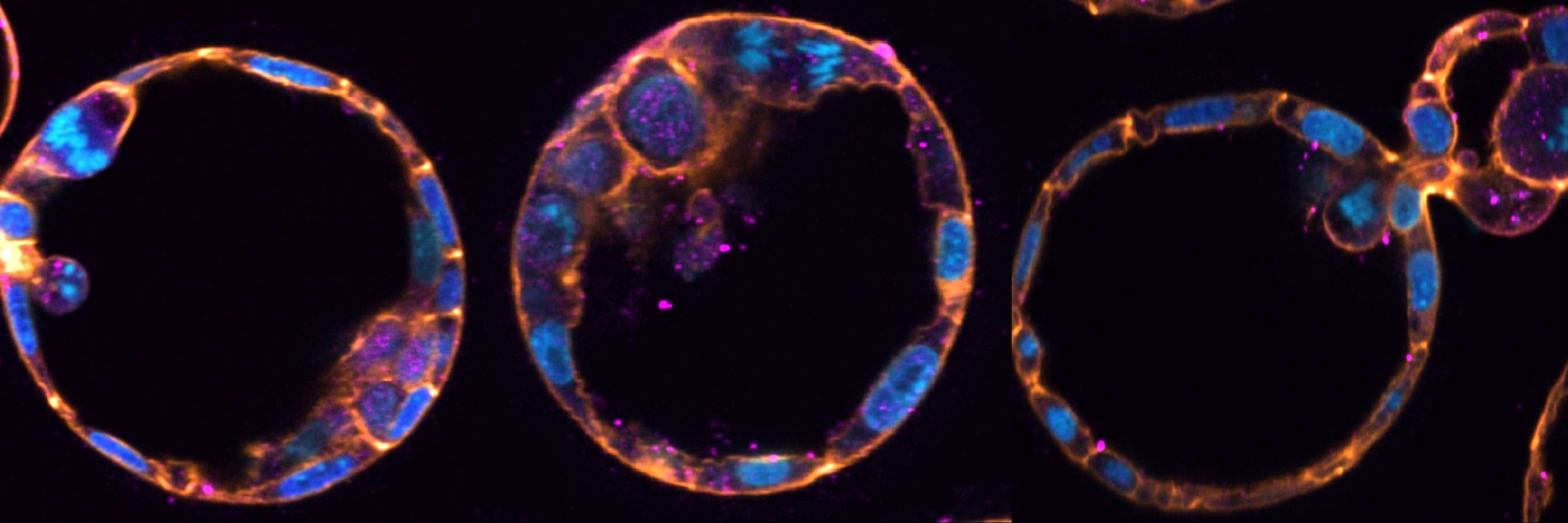
Thrilled to share our work using live imaging to understand how Epiblast (future embryo proper) and Primitive Endoderm (future extraembryonic tissues) cell fates segregate in the preimplantation mouse embryo. Gargantuan effort led by amazing @rpkimyip.bsky.social, David Denberg and Denis Faerberg!
August 2, 2025 at 7:31 PM
Thrilled to share our work using live imaging to understand how Epiblast (future embryo proper) and Primitive Endoderm (future extraembryonic tissues) cell fates segregate in the preimplantation mouse embryo. Gargantuan effort led by amazing @rpkimyip.bsky.social, David Denberg and Denis Faerberg!
Still amazed by the power of imaging and what we can uncover in developmental biology and embryology! Thanks @cshlcourses.bsky.social for the inspiration and training 🧬🔬
Our PostDoc @debojitsaha.bsky.social recently attended a @cshlcourses.bsky.social & came back with new techniques and stunning images. This mouse E9.5 embryo, labeled with Raldh2 (red), Fgf8 (green), Msx1 (magenta), DAPI (blue) is just one example. #Fluorescence revealing the magic of development!✨
July 15, 2025 at 8:30 AM
Still amazed by the power of imaging and what we can uncover in developmental biology and embryology! Thanks @cshlcourses.bsky.social for the inspiration and training 🧬🔬
The past few weeks at CSHL have been incredible exploring various cellular systems and diving into mouse embryonic stages under the microscope. Grateful for the science and the stunning images!
Happy #Fluorescence Friday from the @cshlaboratory.bsky.social Mouse Development, Stem Cells & Cancer course @cshlcourses.bsky.social! First up: E13.5 MEFs stained with phalloidin (magenta) and DAPI (green). Image taken by student @debojitsaha.bsky.social using Agilent BioTek Lionheart FX.

June 20, 2025 at 8:14 PM
The past few weeks at CSHL have been incredible exploring various cellular systems and diving into mouse embryonic stages under the microscope. Grateful for the science and the stunning images!