


Wei Xiao
@weixiao-usyd.bsky.social
PhD student @ University of Sydney interested in spatial proteomics and transcriptomics to study tuberculosis in human and animal models.
Thanks for following along! Always happy to chat, feel free to reach out with thoughts, feedback, or questions about the preprint.
April 16, 2025 at 2:40 PM
Thanks for following along! Always happy to chat, feel free to reach out with thoughts, feedback, or questions about the preprint.
Grateful to the senior authors for their leadership and support. A special thanks to my PhD supervisor, Carl Feng @fenglab.bsky.social , for his incredible mentorship and guidance throughout this journey. Excited for what’s ahead!(21/21)
April 16, 2025 at 2:40 PM
Grateful to the senior authors for their leadership and support. A special thanks to my PhD supervisor, Carl Feng @fenglab.bsky.social , for his incredible mentorship and guidance throughout this journey. Excited for what’s ahead!(21/21)
Huge thanks to our incredible collaborators across USYD and international partners for their insights, feedback, and dedication throughout this project. This work brought together expertise from multiple institutions, and I’m truly grateful for the collaboration and support.(20/21)
April 16, 2025 at 2:40 PM
Huge thanks to our incredible collaborators across USYD and international partners for their insights, feedback, and dedication throughout this project. This work brought together expertise from multiple institutions, and I’m truly grateful for the collaboration and support.(20/21)
For a deeper look at the spatial immune landscape of human TB lungs, how lesion architecture varies between clinical presentations, and what this tells us about immune control in the lung, check out the full preprint. We’d love to hear your thoughts.(19/21)
www.biorxiv.org/content/10.1...
www.biorxiv.org/content/10.1...

Tissue-wide profiling of human lungs reveals spatial sequestration of macrophages in tuberculosis
The immune response to human tuberculosis (TB), particularly in the context of complex lung pathology, remains incompletely understood. Here, we employed whole-slide spatial proteomics to map immune c...
www.biorxiv.org
April 16, 2025 at 2:40 PM
For a deeper look at the spatial immune landscape of human TB lungs, how lesion architecture varies between clinical presentations, and what this tells us about immune control in the lung, check out the full preprint. We’d love to hear your thoughts.(19/21)
www.biorxiv.org/content/10.1...
www.biorxiv.org/content/10.1...
We used an alluvial diagram to link cell types, lesion structure, and clinical status. The takeaway is clear, it’s not just about which cells are present, but how they’re spatially organized that shapes the immune landscape and may impact disease progression.(18/21)

April 16, 2025 at 2:40 PM
We used an alluvial diagram to link cell types, lesion structure, and clinical status. The takeaway is clear, it’s not just about which cells are present, but how they’re spatially organized that shapes the immune landscape and may impact disease progression.(18/21)
In T-type lesions, we found that proinflammatory, progenitor, and Tfh CD4⁺ T cells were spatially co-localized in subclinical TB, while this organization was significantly reduced in the clinical group, suggesting a potential loss of coordinated immune response in advanced disease.(17/21)

April 16, 2025 at 2:40 PM
In T-type lesions, we found that proinflammatory, progenitor, and Tfh CD4⁺ T cells were spatially co-localized in subclinical TB, while this organization was significantly reduced in the clinical group, suggesting a potential loss of coordinated immune response in advanced disease.(17/21)
Unlike T-type lesions where macrophages and T cells intermingle, this lesion type showed clear spatial separation—macrophages at the center, T cells at the edge. This pattern, more frequent in subclinical TB, may reflect a contained immune state that limits tissue damage.(16/21)


April 16, 2025 at 2:40 PM
Unlike T-type lesions where macrophages and T cells intermingle, this lesion type showed clear spatial separation—macrophages at the center, T cells at the edge. This pattern, more frequent in subclinical TB, may reflect a contained immune state that limits tissue damage.(16/21)
This multi-scale framework enabled bottom-up analysis of TB lung tissue, linking single-cell features to lesion architecture and whole-tissue patterns. Notably, M-type lesions with dense macrophage cores and T cell separation were more frequent in the subclinical group.(15/21)


April 16, 2025 at 2:40 PM
This multi-scale framework enabled bottom-up analysis of TB lung tissue, linking single-cell features to lesion architecture and whole-tissue patterns. Notably, M-type lesions with dense macrophage cores and T cell separation were more frequent in the subclinical group.(15/21)
The analysis revealed substantial heterogeneity within and across lesions, uncovering immune hot spots—localized regions of concentrated activity. These were not evenly distributed but appeared in distinct sectors, underscoring the spatial complexity of immune responses within granulomas.(14/21)


April 16, 2025 at 2:40 PM
The analysis revealed substantial heterogeneity within and across lesions, uncovering immune hot spots—localized regions of concentrated activity. These were not evenly distributed but appeared in distinct sectors, underscoring the spatial complexity of immune responses within granulomas.(14/21)
To study the borders of large necrotic granulomas, Andrew Sawyer from our group developed a geometric segmentation approach that allowed us to systematically assess spatial variation in cellular composition along the lesion rim.(13/21)


April 16, 2025 at 2:40 PM
To study the borders of large necrotic granulomas, Andrew Sawyer from our group developed a geometric segmentation approach that allowed us to systematically assess spatial variation in cellular composition along the lesion rim.(13/21)
While both CD4⁺ and CD8⁺ T cells tended to avoid macrophages, CD8⁺ T cells were consistently closer in proximity. This spatial pattern suggests distinct roles, with CD8⁺ T cells potentially engaging more directly with macrophages in shaping the TB lesion microenvironment.(12/21)

April 16, 2025 at 2:40 PM
While both CD4⁺ and CD8⁺ T cells tended to avoid macrophages, CD8⁺ T cells were consistently closer in proximity. This spatial pattern suggests distinct roles, with CD8⁺ T cells potentially engaging more directly with macrophages in shaping the TB lesion microenvironment.(12/21)
We also found that CD4⁺ and CD8⁺ T cell subsets are spatially segregated within lesions. In TLS-like structures, CD4⁺ T cells clustered near B cells, while CD8⁺ T cells localized to the periphery. This segregation was even more pronounced in T cell enriched lesions.(11/21)


April 16, 2025 at 2:40 PM
We also found that CD4⁺ and CD8⁺ T cell subsets are spatially segregated within lesions. In TLS-like structures, CD4⁺ T cells clustered near B cells, while CD8⁺ T cells localized to the periphery. This segregation was even more pronounced in T cell enriched lesions.(11/21)
We found that the Tfh-to-proinflammatory CD4⁺ ratio varied across lesion types. Using SpicyR, we detected spatial segregation between macrophages and B cells, suggesting TB lesions are not only spatially structured but also shape immune responses in a lesion-specific way.(10/21)


April 16, 2025 at 2:40 PM
We found that the Tfh-to-proinflammatory CD4⁺ ratio varied across lesion types. Using SpicyR, we detected spatial segregation between macrophages and B cells, suggesting TB lesions are not only spatially structured but also shape immune responses in a lesion-specific way.(10/21)
Zooming into the sub-lesion level, we identified Tfh-like cells near germinal centers in TLS. In contrast, proinflammatory T cells were enriched in macrophage-dominated lesions. These patterns highlight spatially organized T cell states shaped by local lesion architecture.(9/21)


April 16, 2025 at 2:40 PM
Zooming into the sub-lesion level, we identified Tfh-like cells near germinal centers in TLS. In contrast, proinflammatory T cells were enriched in macrophage-dominated lesions. These patterns highlight spatially organized T cell states shaped by local lesion architecture.(9/21)
The showed diverse architectures, including B cell aggregates, peribronchial infiltrates, and mature TLS. We also observed disorganized T cell-rich lesions, and macrophage-dominated lesions with dense myeloid cores. These patterns reflect a spectrum of immune organization in TB lungs.(8/21)

April 16, 2025 at 2:40 PM
The showed diverse architectures, including B cell aggregates, peribronchial infiltrates, and mature TLS. We also observed disorganized T cell-rich lesions, and macrophage-dominated lesions with dense myeloid cores. These patterns reflect a spectrum of immune organization in TB lungs.(8/21)
We noticed a recurring but understudied feature—lymphoid aggregates surrounding necrosis. Using LVM, we extracted 525 lesions and stratified by cell composition and spatial organization, revealing six lesion types spanning a continuum from B cell to myeloid-dominated immune architectures.(7/21)


April 16, 2025 at 2:40 PM
We noticed a recurring but understudied feature—lymphoid aggregates surrounding necrosis. Using LVM, we extracted 525 lesions and stratified by cell composition and spatial organization, revealing six lesion types spanning a continuum from B cell to myeloid-dominated immune architectures.(7/21)
This identified five major cellular communities, revealing at the tissue-wide scale an immune compartmentalization across the TB lung. Together, these communities framed the broader immune topography of the TB lung and set the stage for focused lesion-level analysis.(6/21)

April 16, 2025 at 2:40 PM
This identified five major cellular communities, revealing at the tissue-wide scale an immune compartmentalization across the TB lung. Together, these communities framed the broader immune topography of the TB lung and set the stage for focused lesion-level analysis.(6/21)
While powerful, this approach lacks cellular detail, so we brought in LisaClust from @ellispatrick.bsky.social group to define cellular communities based on local cell-type distribution. By integrating both maps, this assigns biological meaning to the data-driven cell communities.(5/21)

April 16, 2025 at 2:40 PM
While powerful, this approach lacks cellular detail, so we brought in LisaClust from @ellispatrick.bsky.social group to define cellular communities based on local cell-type distribution. By integrating both maps, this assigns biological meaning to the data-driven cell communities.(5/21)
To interpret spatial cell organization in the context of tissue pathology, we teamed up with Yi Gao at SZU to apply a “human-in-the-loop” deep learning model for segmenting TB histopathology features from the SAME section H&E images.(4/21)

April 16, 2025 at 2:40 PM
To interpret spatial cell organization in the context of tissue pathology, we teamed up with Yi Gao at SZU to apply a “human-in-the-loop” deep learning model for segmenting TB histopathology features from the SAME section H&E images.(4/21)
We built a multi-scale analysis pipeline spanning from whole-tissue context to single-cell resolution within lesions. This framework preserved spatial architecture while resolving cellular interactions, enabling us to map over 20 million cells across 22 TB lung sections. (3/21)


April 16, 2025 at 2:40 PM
We built a multi-scale analysis pipeline spanning from whole-tissue context to single-cell resolution within lesions. This framework preserved spatial architecture while resolving cellular interactions, enabling us to map over 20 million cells across 22 TB lung sections. (3/21)
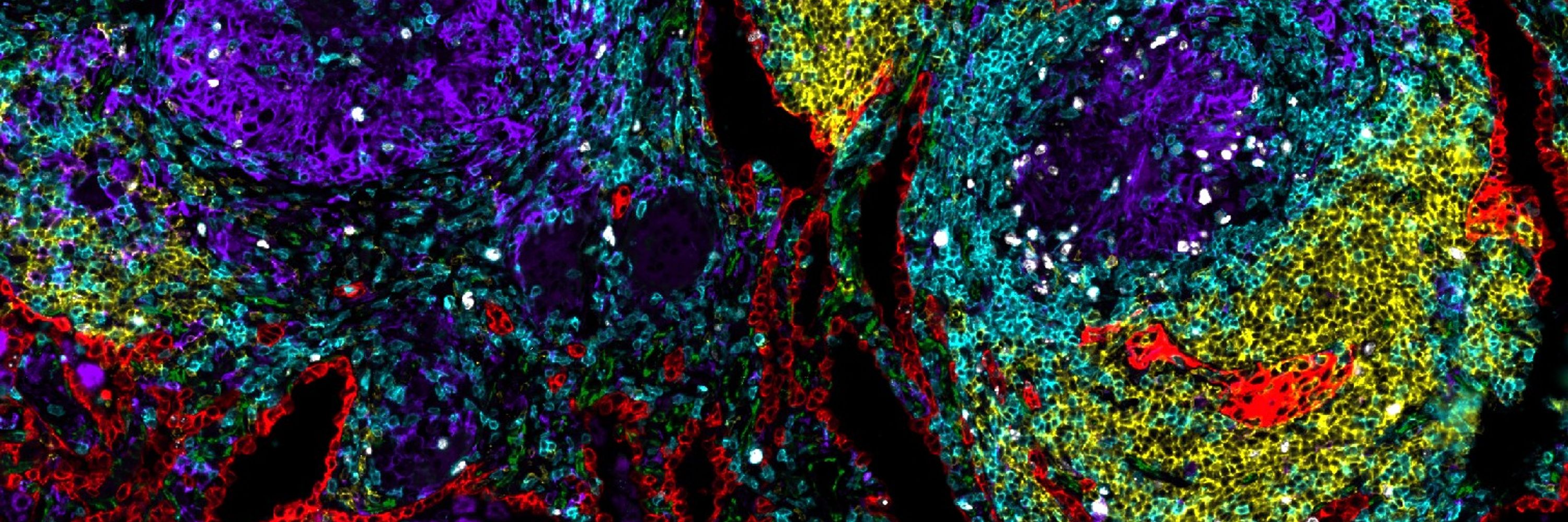
In collaboration with Xinchun Chen’s group, we profiled resected lung tissues from clinical and subclinical TB cases. Using a whole-slide spatial proteomics to examine how immune cell organization across lung sections relates to disease state and lesion architecture. (2/21)

April 16, 2025 at 2:40 PM
In collaboration with Xinchun Chen’s group, we profiled resected lung tissues from clinical and subclinical TB cases. Using a whole-slide spatial proteomics to examine how immune cell organization across lung sections relates to disease state and lesion architecture. (2/21)