


Sam Krimmel
@samkrimmelneuro.bsky.social
Neuroscientist post-doc working with Nico Dosenbach at Washington University in St. Louis
We have a lot of supplemental information, including tests against partial volume effects that I really encourage people to check out. Functional imaging now allows for single voxel level high quality results, something that I thought was impossible when I started my post-doc.

April 10, 2025 at 3:46 PM
We have a lot of supplemental information, including tests against partial volume effects that I really encourage people to check out. Functional imaging now allows for single voxel level high quality results, something that I thought was impossible when I started my post-doc.
Additionally, networks, a well-recognized dominant model of cortical organization, also appear to be an organizing principle as far down as the brainstem. This nicely highlights that networks aren’t really ‘cortical’ they are a system of whole brain organization.

April 10, 2025 at 3:46 PM
Additionally, networks, a well-recognized dominant model of cortical organization, also appear to be an organizing principle as far down as the brainstem. This nicely highlights that networks aren’t really ‘cortical’ they are a system of whole brain organization.
We next assumed that averaging over the red nucleus must have obscured motor-effector regions within the structure, so we tested if any voxels had primary connectivity to any of the 3 motor-effector networks. Shockingly, basically no voxels are preferentially motor-effector connected (triangles).

April 10, 2025 at 3:46 PM
We next assumed that averaging over the red nucleus must have obscured motor-effector regions within the structure, so we tested if any voxels had primary connectivity to any of the 3 motor-effector networks. Shockingly, basically no voxels are preferentially motor-effector connected (triangles).
So where is the red nucleus functional connectivity within the precentral gyrus, something that you would predict from tract tracing? It is specific to @gordonneuro.bsky.social recently discovered somato-cognitive action network (SCAN) and not motor-effector regions.

April 10, 2025 at 3:46 PM
So where is the red nucleus functional connectivity within the precentral gyrus, something that you would predict from tract tracing? It is specific to @gordonneuro.bsky.social recently discovered somato-cognitive action network (SCAN) and not motor-effector regions.
Looking at task data from the HCP, we found that cues to indicate upcoming movement had a large effect in the red nucleus, larger than actual movement. Motor cues also greatly activate the action-mode network. Additionally, task analysis shows the red nucleus responds to rewarding stimuli.

April 10, 2025 at 3:46 PM
Looking at task data from the HCP, we found that cues to indicate upcoming movement had a large effect in the red nucleus, larger than actual movement. Motor cues also greatly activate the action-mode network. Additionally, task analysis shows the red nucleus responds to rewarding stimuli.
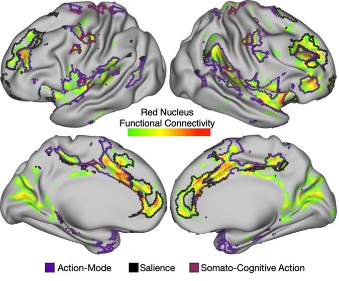
Shockingly, red nucleus connectivity was virtually absent with motor effector networks (le-SMN, ue-SMN, and f-SMN). Instead, connectivity was largest to the salience network (involved in motivated behavior) and the action-mode network (a.k.a. cingulo-opercular network; involved in action).

April 10, 2025 at 3:46 PM
Shockingly, red nucleus connectivity was virtually absent with motor effector networks (le-SMN, ue-SMN, and f-SMN). Instead, connectivity was largest to the salience network (involved in motivated behavior) and the action-mode network (a.k.a. cingulo-opercular network; involved in action).
If the red nucleus is a motor structure, it should 1) be connected to motor-effector networks (somatomotor hand/foot/mouth) and 2) respond to movement. To test this, we used high SNR data with multi-echo ICA (tinyurl.com/55vr747p; example noise component below) and group-averaged task/rest datasets

April 10, 2025 at 3:46 PM
If the red nucleus is a motor structure, it should 1) be connected to motor-effector networks (somatomotor hand/foot/mouth) and 2) respond to movement. To test this, we used high SNR data with multi-echo ICA (tinyurl.com/55vr747p; example noise component below) and group-averaged task/rest datasets
As walking changed from quadrupedal to bipedal, so has the red nucleus. The magnocellular red nucleus (projects to the spinal cord; shaded) has shrunken! But another division, the parvocellular red nucleus (shown white) expands tinyurl.com/2ctttmmw

April 10, 2025 at 3:46 PM
As walking changed from quadrupedal to bipedal, so has the red nucleus. The magnocellular red nucleus (projects to the spinal cord; shaded) has shrunken! But another division, the parvocellular red nucleus (shown white) expands tinyurl.com/2ctttmmw
The red nucleus is a slightly pinkish structure (hence the name ‘red’) in the midbrain of the brainstem. Fortunately, it is easily visible on standard structural MRI images, especially T2 weighted.


April 10, 2025 at 3:46 PM
The red nucleus is a slightly pinkish structure (hence the name ‘red’) in the midbrain of the brainstem. Fortunately, it is easily visible on standard structural MRI images, especially T2 weighted.
It's shocking how little is known about the brainstem red nucleus. In our new paper “The human brainstem’s red nucleus was upgraded to support goal-directed action” out now in @naturecomms.bsky.social we show that current thinking on the red nucleus is in need of a serious upgrade. rdcu.be/ehbOy
April 10, 2025 at 3:46 PM
It's shocking how little is known about the brainstem red nucleus. In our new paper “The human brainstem’s red nucleus was upgraded to support goal-directed action” out now in @naturecomms.bsky.social we show that current thinking on the red nucleus is in need of a serious upgrade. rdcu.be/ehbOy