


Pediatric Imaging
@pedsimaging.bsky.social
A pediatric radiology textbook, one case every day. Opinions are my own.
#FOAMRad #FOAMPed #FOAMed #PedsRad #pediatricradiology #radiology #pediatrics #radiologia #pediatria
www.pediatricimaging.org
#FOAMRad #FOAMPed #FOAMed #PedsRad #pediatricradiology #radiology #pediatrics #radiologia #pediatria
www.pediatricimaging.org
Pinned
We are publishing a pediatric imaging textbook, 1 case/day+have 2,000 cases covering 1,000 diseases, 125 DDxs, 50 procedure descriptions+4 review courses
#FOAMed #FOAMRad #MedEd #PedsRad #RadEd #RadRes #radiology #radiología #radiologie #pediatrics #paediatrics #pediatría #pédiatrie #pädiatrie
#FOAMed #FOAMRad #MedEd #PedsRad #RadEd #RadRes #radiology #radiología #radiologie #pediatrics #paediatrics #pediatría #pédiatrie #pädiatrie
Toddler with cough
CXR shows bilateral diffuse bronchial wall thickening+subsequent interstitial infiltrates.
The diagnosis was mycoplasma pneumonia.
Learn more: pediatricimaging.org/diseases/myc...
#FOAMed #FOAMRad #radiology #pediatrics #EmergencyMedicine #radiologia #paediatrics #pediatria
CXR shows bilateral diffuse bronchial wall thickening+subsequent interstitial infiltrates.
The diagnosis was mycoplasma pneumonia.
Learn more: pediatricimaging.org/diseases/myc...
#FOAMed #FOAMRad #radiology #pediatrics #EmergencyMedicine #radiologia #paediatrics #pediatria

November 19, 2025 at 5:44 PM
Toddler with cough
CXR shows bilateral diffuse bronchial wall thickening+subsequent interstitial infiltrates.
The diagnosis was mycoplasma pneumonia.
Learn more: pediatricimaging.org/diseases/myc...
#FOAMed #FOAMRad #radiology #pediatrics #EmergencyMedicine #radiologia #paediatrics #pediatria
CXR shows bilateral diffuse bronchial wall thickening+subsequent interstitial infiltrates.
The diagnosis was mycoplasma pneumonia.
Learn more: pediatricimaging.org/diseases/myc...
#FOAMed #FOAMRad #radiology #pediatrics #EmergencyMedicine #radiologia #paediatrics #pediatria
Teen with left scrotal pain
Transverse(above)+sagittal (below) US of left scrotum show an oval hyperechoic lesion that is in scrotum but that is extratesticular in location+which has posterior acoustical shadowing
#FOAMed #MedEd #FOAMRad #radiology #pediatrics #Ultrasound #radiologia #radiologie
Transverse(above)+sagittal (below) US of left scrotum show an oval hyperechoic lesion that is in scrotum but that is extratesticular in location+which has posterior acoustical shadowing
#FOAMed #MedEd #FOAMRad #radiology #pediatrics #Ultrasound #radiologia #radiologie

November 19, 2025 at 11:32 AM
Teen with left scrotal pain
Transverse(above)+sagittal (below) US of left scrotum show an oval hyperechoic lesion that is in scrotum but that is extratesticular in location+which has posterior acoustical shadowing
#FOAMed #MedEd #FOAMRad #radiology #pediatrics #Ultrasound #radiologia #radiologie
Transverse(above)+sagittal (below) US of left scrotum show an oval hyperechoic lesion that is in scrotum but that is extratesticular in location+which has posterior acoustical shadowing
#FOAMed #MedEd #FOAMRad #radiology #pediatrics #Ultrasound #radiologia #radiologie
Teen with a limp
AP(left)+lateral(right) radiographs show in tibial diaphysis multiple+too numerous to count eccentric expansile lucent lesions with bubbly+sclerotic borders which are without periosteal reaction
#FOAMed #MedEd #radiology #pediatrics #radiologia #Ortho #Orthopedics #Orthopaedics
AP(left)+lateral(right) radiographs show in tibial diaphysis multiple+too numerous to count eccentric expansile lucent lesions with bubbly+sclerotic borders which are without periosteal reaction
#FOAMed #MedEd #radiology #pediatrics #radiologia #Ortho #Orthopedics #Orthopaedics

November 19, 2025 at 2:10 AM
Teen with a limp
AP(left)+lateral(right) radiographs show in tibial diaphysis multiple+too numerous to count eccentric expansile lucent lesions with bubbly+sclerotic borders which are without periosteal reaction
#FOAMed #MedEd #radiology #pediatrics #radiologia #Ortho #Orthopedics #Orthopaedics
AP(left)+lateral(right) radiographs show in tibial diaphysis multiple+too numerous to count eccentric expansile lucent lesions with bubbly+sclerotic borders which are without periosteal reaction
#FOAMed #MedEd #radiology #pediatrics #radiologia #Ortho #Orthopedics #Orthopaedics
Immune suppressed patient with odynophagia
AP images from UpperGI show multiple longitudinally oriented lesions in under-distended esophagus which are separated by normal mucosa+small round ulcers
#FOAMed #MedEd #FOAMRad #radiology #pediatrics #radiologia #paediatrics #pediatria #pediatrie
AP images from UpperGI show multiple longitudinally oriented lesions in under-distended esophagus which are separated by normal mucosa+small round ulcers
#FOAMed #MedEd #FOAMRad #radiology #pediatrics #radiologia #paediatrics #pediatria #pediatrie

November 19, 2025 at 12:06 AM
Immune suppressed patient with odynophagia
AP images from UpperGI show multiple longitudinally oriented lesions in under-distended esophagus which are separated by normal mucosa+small round ulcers
#FOAMed #MedEd #FOAMRad #radiology #pediatrics #radiologia #paediatrics #pediatria #pediatrie
AP images from UpperGI show multiple longitudinally oriented lesions in under-distended esophagus which are separated by normal mucosa+small round ulcers
#FOAMed #MedEd #FOAMRad #radiology #pediatrics #radiologia #paediatrics #pediatria #pediatrie
Newborn whose mother was exposed to CMV during pregnancy
Coronal US of brain(above) shows large septated cysts in germinal matrix (germinolysis) bilaterally+multiple periventricular echogenic foci just lateral to anterior horns of lateral ventricles bilaterally...
#radiology #pediatrics #NICU
Coronal US of brain(above) shows large septated cysts in germinal matrix (germinolysis) bilaterally+multiple periventricular echogenic foci just lateral to anterior horns of lateral ventricles bilaterally...
#radiology #pediatrics #NICU

November 7, 2025 at 11:56 AM
Newborn whose mother was exposed to CMV during pregnancy
Coronal US of brain(above) shows large septated cysts in germinal matrix (germinolysis) bilaterally+multiple periventricular echogenic foci just lateral to anterior horns of lateral ventricles bilaterally...
#radiology #pediatrics #NICU
Coronal US of brain(above) shows large septated cysts in germinal matrix (germinolysis) bilaterally+multiple periventricular echogenic foci just lateral to anterior horns of lateral ventricles bilaterally...
#radiology #pediatrics #NICU
School ager with chronic cough+right shoulder pain
Radiograph of shoulder shows well circumscribed lucent lesion in right humeral epiphysis. There is no periosteal reaction
The diagnosis was tuberculous osteomyelitis.
#FOAMed #MedEd #radiology #radiologia #Ortho #Orthopedics #Orthopaedics
Radiograph of shoulder shows well circumscribed lucent lesion in right humeral epiphysis. There is no periosteal reaction
The diagnosis was tuberculous osteomyelitis.
#FOAMed #MedEd #radiology #radiologia #Ortho #Orthopedics #Orthopaedics

November 7, 2025 at 12:24 AM
School ager with chronic cough+right shoulder pain
Radiograph of shoulder shows well circumscribed lucent lesion in right humeral epiphysis. There is no periosteal reaction
The diagnosis was tuberculous osteomyelitis.
#FOAMed #MedEd #radiology #radiologia #Ortho #Orthopedics #Orthopaedics
Radiograph of shoulder shows well circumscribed lucent lesion in right humeral epiphysis. There is no periosteal reaction
The diagnosis was tuberculous osteomyelitis.
#FOAMed #MedEd #radiology #radiologia #Ortho #Orthopedics #Orthopaedics
School ager with foot pain
Radiograph of foot shows sclerosis+fragmentation of tarsal navicular bone.
The diagnosis was Kohler disease.
Learn more: pediatricimaging.org/diseases/koh...
#FOAMed #MedEd #radiology #pediatrics #radiologia #Ortho #Orthopedics #Orthopaedics #EmergencyMedicine
Radiograph of foot shows sclerosis+fragmentation of tarsal navicular bone.
The diagnosis was Kohler disease.
Learn more: pediatricimaging.org/diseases/koh...
#FOAMed #MedEd #radiology #pediatrics #radiologia #Ortho #Orthopedics #Orthopaedics #EmergencyMedicine

November 6, 2025 at 12:21 PM
School ager with foot pain
Radiograph of foot shows sclerosis+fragmentation of tarsal navicular bone.
The diagnosis was Kohler disease.
Learn more: pediatricimaging.org/diseases/koh...
#FOAMed #MedEd #radiology #pediatrics #radiologia #Ortho #Orthopedics #Orthopaedics #EmergencyMedicine
Radiograph of foot shows sclerosis+fragmentation of tarsal navicular bone.
The diagnosis was Kohler disease.
Learn more: pediatricimaging.org/diseases/koh...
#FOAMed #MedEd #radiology #pediatrics #radiologia #Ortho #Orthopedics #Orthopaedics #EmergencyMedicine
Preschooler with right neck swelling
CT of neck shows round low density fluid collection with thin enhancing rim in right parapharyngeal space. Right carotid artery+internal jugular vein are narrowed in caliber+there is mass effect on right side of airway
#radiology #pediatrics #EmergencyMedicine
CT of neck shows round low density fluid collection with thin enhancing rim in right parapharyngeal space. Right carotid artery+internal jugular vein are narrowed in caliber+there is mass effect on right side of airway
#radiology #pediatrics #EmergencyMedicine

November 6, 2025 at 2:44 AM
Preschooler with right neck swelling
CT of neck shows round low density fluid collection with thin enhancing rim in right parapharyngeal space. Right carotid artery+internal jugular vein are narrowed in caliber+there is mass effect on right side of airway
#radiology #pediatrics #EmergencyMedicine
CT of neck shows round low density fluid collection with thin enhancing rim in right parapharyngeal space. Right carotid artery+internal jugular vein are narrowed in caliber+there is mass effect on right side of airway
#radiology #pediatrics #EmergencyMedicine
Infant with duplicated right kidney
Lateral image from voiding cystourethrogram shows round lucency / filling defect near trigone of the bladder.
There is also contrast filling a blind ending tubular tract at dome of bladder.
#FOAMed #MedEd #radiology #pediatrics #radiologia #paediatrics
Lateral image from voiding cystourethrogram shows round lucency / filling defect near trigone of the bladder.
There is also contrast filling a blind ending tubular tract at dome of bladder.
#FOAMed #MedEd #radiology #pediatrics #radiologia #paediatrics

November 5, 2025 at 12:16 PM
Infant with duplicated right kidney
Lateral image from voiding cystourethrogram shows round lucency / filling defect near trigone of the bladder.
There is also contrast filling a blind ending tubular tract at dome of bladder.
#FOAMed #MedEd #radiology #pediatrics #radiologia #paediatrics
Lateral image from voiding cystourethrogram shows round lucency / filling defect near trigone of the bladder.
There is also contrast filling a blind ending tubular tract at dome of bladder.
#FOAMed #MedEd #radiology #pediatrics #radiologia #paediatrics
Teenager with pica
AXR shows multiple punctate and linear radiopaque objects in the ascending colon.
#FOAMed #MedEd #radiology #pediatrics #EmergencyMedicine #radiologia #medstudent #medicalstudent #medschool #medicalschool #radiologie #paediatrics #pediatria #pediatrie #pädiatrie
AXR shows multiple punctate and linear radiopaque objects in the ascending colon.
#FOAMed #MedEd #radiology #pediatrics #EmergencyMedicine #radiologia #medstudent #medicalstudent #medschool #medicalschool #radiologie #paediatrics #pediatria #pediatrie #pädiatrie

November 5, 2025 at 2:33 AM
Teenager with pica
AXR shows multiple punctate and linear radiopaque objects in the ascending colon.
#FOAMed #MedEd #radiology #pediatrics #EmergencyMedicine #radiologia #medstudent #medicalstudent #medschool #medicalschool #radiologie #paediatrics #pediatria #pediatrie #pädiatrie
AXR shows multiple punctate and linear radiopaque objects in the ascending colon.
#FOAMed #MedEd #radiology #pediatrics #EmergencyMedicine #radiologia #medstudent #medicalstudent #medschool #medicalschool #radiologie #paediatrics #pediatria #pediatrie #pädiatrie
Infant with failure to thrive
Left lateral decubitus AXR (above left) shows an air filled duodenal bulb and an air filled stomach (double bubble sign) with some distal bowel gas
#FOAMed #MedEd #radiology #pediatrics #radiologia #Ultrasound #PedSurg #SoMe4PedSurg #radiologie #paediatrics #pediatria
Left lateral decubitus AXR (above left) shows an air filled duodenal bulb and an air filled stomach (double bubble sign) with some distal bowel gas
#FOAMed #MedEd #radiology #pediatrics #radiologia #Ultrasound #PedSurg #SoMe4PedSurg #radiologie #paediatrics #pediatria

November 4, 2025 at 11:50 AM
Infant with failure to thrive
Left lateral decubitus AXR (above left) shows an air filled duodenal bulb and an air filled stomach (double bubble sign) with some distal bowel gas
#FOAMed #MedEd #radiology #pediatrics #radiologia #Ultrasound #PedSurg #SoMe4PedSurg #radiologie #paediatrics #pediatria
Left lateral decubitus AXR (above left) shows an air filled duodenal bulb and an air filled stomach (double bubble sign) with some distal bowel gas
#FOAMed #MedEd #radiology #pediatrics #radiologia #Ultrasound #PedSurg #SoMe4PedSurg #radiologie #paediatrics #pediatria
Preschooler with anemia+palpable epigastric mass
Coronal(above)+axial (below) CT show stomach distended by large mass that has swirled appearance+that fills entire stomach+duodenal bulb
#FOAMed #MedEd #radiology #pediatrics #EmergencyMedicine #radiologia #PedSurg #medstudent #medicalstudent
Coronal(above)+axial (below) CT show stomach distended by large mass that has swirled appearance+that fills entire stomach+duodenal bulb
#FOAMed #MedEd #radiology #pediatrics #EmergencyMedicine #radiologia #PedSurg #medstudent #medicalstudent

November 4, 2025 at 1:31 AM
Preschooler with anemia+palpable epigastric mass
Coronal(above)+axial (below) CT show stomach distended by large mass that has swirled appearance+that fills entire stomach+duodenal bulb
#FOAMed #MedEd #radiology #pediatrics #EmergencyMedicine #radiologia #PedSurg #medstudent #medicalstudent
Coronal(above)+axial (below) CT show stomach distended by large mass that has swirled appearance+that fills entire stomach+duodenal bulb
#FOAMed #MedEd #radiology #pediatrics #EmergencyMedicine #radiologia #PedSurg #medstudent #medicalstudent
7 week old infant with respiratory distress without fever
CXR show bilateral diffuse infiltrates with hyperexpanded lungs.
The diagnosis was chlamydia pneumonia.
Learn more: pediatricimaging.org/diseases/chl...
#FOAMed #MedEd #radiology #pediatrics #radiologia #paediatrics #pediatria
CXR show bilateral diffuse infiltrates with hyperexpanded lungs.
The diagnosis was chlamydia pneumonia.
Learn more: pediatricimaging.org/diseases/chl...
#FOAMed #MedEd #radiology #pediatrics #radiologia #paediatrics #pediatria

November 3, 2025 at 12:13 PM
7 week old infant with respiratory distress without fever
CXR show bilateral diffuse infiltrates with hyperexpanded lungs.
The diagnosis was chlamydia pneumonia.
Learn more: pediatricimaging.org/diseases/chl...
#FOAMed #MedEd #radiology #pediatrics #radiologia #paediatrics #pediatria
CXR show bilateral diffuse infiltrates with hyperexpanded lungs.
The diagnosis was chlamydia pneumonia.
Learn more: pediatricimaging.org/diseases/chl...
#FOAMed #MedEd #radiology #pediatrics #radiologia #paediatrics #pediatria
Female preschooler with urinary tract infection
AP image from a voiding cystourethrogram shows a non-obstructive ellipsoid-shaped dilation of the posterior urethra just distal to the internal urethral sphincter
#FOAMed #MedEd #radiology #pediatrics #radiologia #radiologie #paediatrics #pediatria
AP image from a voiding cystourethrogram shows a non-obstructive ellipsoid-shaped dilation of the posterior urethra just distal to the internal urethral sphincter
#FOAMed #MedEd #radiology #pediatrics #radiologia #radiologie #paediatrics #pediatria

November 2, 2025 at 11:56 PM
Female preschooler with urinary tract infection
AP image from a voiding cystourethrogram shows a non-obstructive ellipsoid-shaped dilation of the posterior urethra just distal to the internal urethral sphincter
#FOAMed #MedEd #radiology #pediatrics #radiologia #radiologie #paediatrics #pediatria
AP image from a voiding cystourethrogram shows a non-obstructive ellipsoid-shaped dilation of the posterior urethra just distal to the internal urethral sphincter
#FOAMed #MedEd #radiology #pediatrics #radiologia #radiologie #paediatrics #pediatria
School ager with headaches
Lateral radiograph of skull obtained 3 years ago(above) shows ventriculoperitoneal (VP) shunt tubing that courses inferiorly is connected appropriately to radiolucent VP shunt reservoir
#FOAMed #MedEd #radiology #pediatrics #Neurosurgery #EmergencyMedicine #Neurorad
Lateral radiograph of skull obtained 3 years ago(above) shows ventriculoperitoneal (VP) shunt tubing that courses inferiorly is connected appropriately to radiolucent VP shunt reservoir
#FOAMed #MedEd #radiology #pediatrics #Neurosurgery #EmergencyMedicine #Neurorad

November 2, 2025 at 12:38 PM
School ager with headaches
Lateral radiograph of skull obtained 3 years ago(above) shows ventriculoperitoneal (VP) shunt tubing that courses inferiorly is connected appropriately to radiolucent VP shunt reservoir
#FOAMed #MedEd #radiology #pediatrics #Neurosurgery #EmergencyMedicine #Neurorad
Lateral radiograph of skull obtained 3 years ago(above) shows ventriculoperitoneal (VP) shunt tubing that courses inferiorly is connected appropriately to radiolucent VP shunt reservoir
#FOAMed #MedEd #radiology #pediatrics #Neurosurgery #EmergencyMedicine #Neurorad
Toddler with a whooping-sounding cough
CXR shows bilateral lung hyperexpansion with bilateral perihilar infiltrates that blur the border of the heart, resulting in heart border having a shaggy appearance.
#FOAMed #MedEd #radiology #pediatrics #EmergencyMedicine #radiologia #paediatrics #pediatria
CXR shows bilateral lung hyperexpansion with bilateral perihilar infiltrates that blur the border of the heart, resulting in heart border having a shaggy appearance.
#FOAMed #MedEd #radiology #pediatrics #EmergencyMedicine #radiologia #paediatrics #pediatria

November 2, 2025 at 4:35 AM
Toddler with a whooping-sounding cough
CXR shows bilateral lung hyperexpansion with bilateral perihilar infiltrates that blur the border of the heart, resulting in heart border having a shaggy appearance.
#FOAMed #MedEd #radiology #pediatrics #EmergencyMedicine #radiologia #paediatrics #pediatria
CXR shows bilateral lung hyperexpansion with bilateral perihilar infiltrates that blur the border of the heart, resulting in heart border having a shaggy appearance.
#FOAMed #MedEd #radiology #pediatrics #EmergencyMedicine #radiologia #paediatrics #pediatria
Teen who hurt their ankle playing football
Lateral radiograph(above right) shows bony discontinuity through posterior aspect of tibial plafond extending to physis without articular step-off...
#FOAMed #MedEd #radiology #pediatrics #radiologia #Ortho #Orthopedics #Orthopaedics #EmergencyMedicine
Lateral radiograph(above right) shows bony discontinuity through posterior aspect of tibial plafond extending to physis without articular step-off...
#FOAMed #MedEd #radiology #pediatrics #radiologia #Ortho #Orthopedics #Orthopaedics #EmergencyMedicine

November 1, 2025 at 12:27 PM
Teen who hurt their ankle playing football
Lateral radiograph(above right) shows bony discontinuity through posterior aspect of tibial plafond extending to physis without articular step-off...
#FOAMed #MedEd #radiology #pediatrics #radiologia #Ortho #Orthopedics #Orthopaedics #EmergencyMedicine
Lateral radiograph(above right) shows bony discontinuity through posterior aspect of tibial plafond extending to physis without articular step-off...
#FOAMed #MedEd #radiology #pediatrics #radiologia #Ortho #Orthopedics #Orthopaedics #EmergencyMedicine
Newborn with episode of cardiac decompensation
CXR after left subclavian line placement(above) shows tip of line in right atrium. CXR obtained 3 days later during episode of cardiac decompensation(below) shows interval enlargement of cardiac silhouette
#radiology #pediatrics #NICU #Neonatology
CXR after left subclavian line placement(above) shows tip of line in right atrium. CXR obtained 3 days later during episode of cardiac decompensation(below) shows interval enlargement of cardiac silhouette
#radiology #pediatrics #NICU #Neonatology

November 1, 2025 at 3:00 AM
Newborn with episode of cardiac decompensation
CXR after left subclavian line placement(above) shows tip of line in right atrium. CXR obtained 3 days later during episode of cardiac decompensation(below) shows interval enlargement of cardiac silhouette
#radiology #pediatrics #NICU #Neonatology
CXR after left subclavian line placement(above) shows tip of line in right atrium. CXR obtained 3 days later during episode of cardiac decompensation(below) shows interval enlargement of cardiac silhouette
#radiology #pediatrics #NICU #Neonatology
We are publishing a pediatric radiology textbook, 1 case/day+have 2,000 cases covering 1,000 diseases+125 differential diagnoses, 50 procedure descriptions+4 review courses
#radiology #radiologia #pediatrics #pediatria #EmergencyMedicine #NICU #Neonatology #Ortho #Orthopedics #SoMe4PedSurg #PedSurg
#radiology #radiologia #pediatrics #pediatria #EmergencyMedicine #NICU #Neonatology #Ortho #Orthopedics #SoMe4PedSurg #PedSurg

October 31, 2025 at 6:56 PM
We are publishing a pediatric radiology textbook, 1 case/day+have 2,000 cases covering 1,000 diseases+125 differential diagnoses, 50 procedure descriptions+4 review courses
#radiology #radiologia #pediatrics #pediatria #EmergencyMedicine #NICU #Neonatology #Ortho #Orthopedics #SoMe4PedSurg #PedSurg
#radiology #radiologia #pediatrics #pediatria #EmergencyMedicine #NICU #Neonatology #Ortho #Orthopedics #SoMe4PedSurg #PedSurg
Newborn with blueberry muffin skin lesions
AP(above)+lateral(below) radiographs of skull show bilateral thick, chunky, periventricular calcifications outlining lateral ventricles.
#FOAMed #MedEd #radiology #pediatrics #radiologia #NICU #Neonatology #NeuroRad #Neurology #paediatrics #pediatria
AP(above)+lateral(below) radiographs of skull show bilateral thick, chunky, periventricular calcifications outlining lateral ventricles.
#FOAMed #MedEd #radiology #pediatrics #radiologia #NICU #Neonatology #NeuroRad #Neurology #paediatrics #pediatria

October 31, 2025 at 11:16 AM
Newborn with blueberry muffin skin lesions
AP(above)+lateral(below) radiographs of skull show bilateral thick, chunky, periventricular calcifications outlining lateral ventricles.
#FOAMed #MedEd #radiology #pediatrics #radiologia #NICU #Neonatology #NeuroRad #Neurology #paediatrics #pediatria
AP(above)+lateral(below) radiographs of skull show bilateral thick, chunky, periventricular calcifications outlining lateral ventricles.
#FOAMed #MedEd #radiology #pediatrics #radiologia #NICU #Neonatology #NeuroRad #Neurology #paediatrics #pediatria
Teen with no history of esophageal surgery who is having repeated food bolus impactions in the esophagus
Axial CT of chest(above) shows posterior to trachea a dilated proximal esophagus due to food being impacted within it.
#FOAMed #MedEd #radiology #pediatrics #radiologia #pediatria
Axial CT of chest(above) shows posterior to trachea a dilated proximal esophagus due to food being impacted within it.
#FOAMed #MedEd #radiology #pediatrics #radiologia #pediatria

October 31, 2025 at 4:12 AM
Teen with no history of esophageal surgery who is having repeated food bolus impactions in the esophagus
Axial CT of chest(above) shows posterior to trachea a dilated proximal esophagus due to food being impacted within it.
#FOAMed #MedEd #radiology #pediatrics #radiologia #pediatria
Axial CT of chest(above) shows posterior to trachea a dilated proximal esophagus due to food being impacted within it.
#FOAMed #MedEd #radiology #pediatrics #radiologia #pediatria
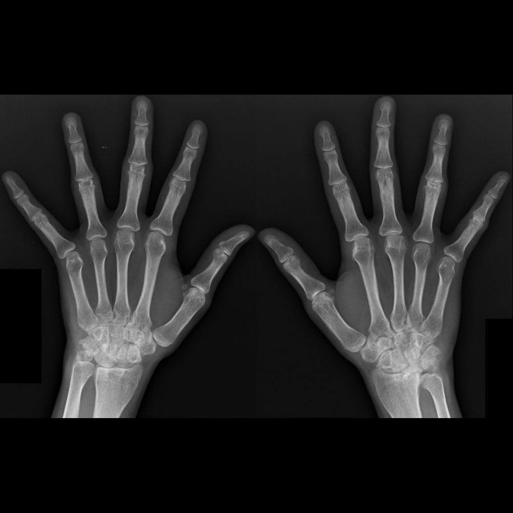
Teen with wrist pain
Radiographs of hands shows bilateral periarticular osteoporosis throughout hands. There is bilateral loss of joint spaces in carpal bones due to cartilage destruction+erosions at proximal interphalangeal joints
#FOAMed #MedEd #radiology #pediatrics #radiologia #Orthopedics
Radiographs of hands shows bilateral periarticular osteoporosis throughout hands. There is bilateral loss of joint spaces in carpal bones due to cartilage destruction+erosions at proximal interphalangeal joints
#FOAMed #MedEd #radiology #pediatrics #radiologia #Orthopedics
October 30, 2025 at 11:26 AM
Teen with wrist pain
Radiographs of hands shows bilateral periarticular osteoporosis throughout hands. There is bilateral loss of joint spaces in carpal bones due to cartilage destruction+erosions at proximal interphalangeal joints
#FOAMed #MedEd #radiology #pediatrics #radiologia #Orthopedics
Radiographs of hands shows bilateral periarticular osteoporosis throughout hands. There is bilateral loss of joint spaces in carpal bones due to cartilage destruction+erosions at proximal interphalangeal joints
#FOAMed #MedEd #radiology #pediatrics #radiologia #Orthopedics
Infant with non-palpable testicles bilaterally
Sagittal US of left lower abdomen(above) shows left testicle lying on left psoas muscle just inferior to kidney
#FOAMed #MedEd #radiology #pediatrics #radiologia #Ultrasound #radiologie #paediatrics #pediatria #pediatrie #pädiatrie
Sagittal US of left lower abdomen(above) shows left testicle lying on left psoas muscle just inferior to kidney
#FOAMed #MedEd #radiology #pediatrics #radiologia #Ultrasound #radiologie #paediatrics #pediatria #pediatrie #pädiatrie

October 29, 2025 at 11:25 AM
Infant with non-palpable testicles bilaterally
Sagittal US of left lower abdomen(above) shows left testicle lying on left psoas muscle just inferior to kidney
#FOAMed #MedEd #radiology #pediatrics #radiologia #Ultrasound #radiologie #paediatrics #pediatria #pediatrie #pädiatrie
Sagittal US of left lower abdomen(above) shows left testicle lying on left psoas muscle just inferior to kidney
#FOAMed #MedEd #radiology #pediatrics #radiologia #Ultrasound #radiologie #paediatrics #pediatria #pediatrie #pädiatrie
School ager with nausea, vomiting+jaundice who is Hepatitis A positive
Transverse(above)+sagittal(below) US of liver show starry sky appearance to liver with hypoechoic liver parenchyma+echogenic portal tracts.
#FOAMed #MedEd #radiology #pediatrics #radiologia #Ultrasound #paediatrics #pediatria
Transverse(above)+sagittal(below) US of liver show starry sky appearance to liver with hypoechoic liver parenchyma+echogenic portal tracts.
#FOAMed #MedEd #radiology #pediatrics #radiologia #Ultrasound #paediatrics #pediatria

October 28, 2025 at 11:13 AM
School ager with nausea, vomiting+jaundice who is Hepatitis A positive
Transverse(above)+sagittal(below) US of liver show starry sky appearance to liver with hypoechoic liver parenchyma+echogenic portal tracts.
#FOAMed #MedEd #radiology #pediatrics #radiologia #Ultrasound #paediatrics #pediatria
Transverse(above)+sagittal(below) US of liver show starry sky appearance to liver with hypoechoic liver parenchyma+echogenic portal tracts.
#FOAMed #MedEd #radiology #pediatrics #radiologia #Ultrasound #paediatrics #pediatria
Teen with shortness of breath for 1 month
CXR(above left) shows bilateral peripheral airspace disease that is more prevalent in lower lobes than upper lobes.
#FOAMed #MedEd #FOAMRad #radiology #pediatrics #radiologia #pediatria
CXR(above left) shows bilateral peripheral airspace disease that is more prevalent in lower lobes than upper lobes.
#FOAMed #MedEd #FOAMRad #radiology #pediatrics #radiologia #pediatria

October 27, 2025 at 11:29 AM
Teen with shortness of breath for 1 month
CXR(above left) shows bilateral peripheral airspace disease that is more prevalent in lower lobes than upper lobes.
#FOAMed #MedEd #FOAMRad #radiology #pediatrics #radiologia #pediatria
CXR(above left) shows bilateral peripheral airspace disease that is more prevalent in lower lobes than upper lobes.
#FOAMed #MedEd #FOAMRad #radiology #pediatrics #radiologia #pediatria