


Kyle Perry
@kyledperrymd.bsky.social
Bone and soft tissue, surgical and cytopathologist at University of Michigan | @UMichPath@UMichMedicine | T/RT not medical advice
Nice review article by Szczepanski, Westerhoff and Schechter on this rare entity (pubmed.ncbi.nlm.nih.gov/39743931/)
May 6, 2025 at 3:35 AM
Nice review article by Szczepanski, Westerhoff and Schechter on this rare entity (pubmed.ncbi.nlm.nih.gov/39743931/)
The spindled morphology and plexiform architecture can sometimes cause these tumors to be mistaken for (SDH-deficient) GIST. Unlike GIST, these tumors are negative for CD117 and DOG1.
May 6, 2025 at 3:30 AM
The spindled morphology and plexiform architecture can sometimes cause these tumors to be mistaken for (SDH-deficient) GIST. Unlike GIST, these tumors are negative for CD117 and DOG1.
Special thanks to my U of Michigan colleagues for allowing me to share this case!
April 13, 2025 at 1:40 AM
Special thanks to my U of Michigan colleagues for allowing me to share this case!
Some nice papers (Fritchie and colleagues/ Chung and colleagues) further elaborating on this entity(ies)….
pubmed.ncbi.nlm.nih.gov/33097826/
pubmed.ncbi.nlm.nih.gov/33300192/
pubmed.ncbi.nlm.nih.gov/33097826/
pubmed.ncbi.nlm.nih.gov/33300192/
April 13, 2025 at 1:39 AM
Some nice papers (Fritchie and colleagues/ Chung and colleagues) further elaborating on this entity(ies)….
pubmed.ncbi.nlm.nih.gov/33097826/
pubmed.ncbi.nlm.nih.gov/33300192/
pubmed.ncbi.nlm.nih.gov/33097826/
pubmed.ncbi.nlm.nih.gov/33300192/
Fibromyxoid soft tissue tumors/fibroblastic lipoblastomas with a PLAG1 gene fusion often contain foci of myxoid stroma in the background of a more fibrous appearance. In addition to PLAG1 staining, these tumors usually show expression of desmin and CD34.
April 13, 2025 at 1:38 AM
Fibromyxoid soft tissue tumors/fibroblastic lipoblastomas with a PLAG1 gene fusion often contain foci of myxoid stroma in the background of a more fibrous appearance. In addition to PLAG1 staining, these tumors usually show expression of desmin and CD34.
Nice article on ALK-positive histiocytosis of the breast: pubmed.ncbi.nlm.nih.gov/32826530/
March 31, 2025 at 9:54 PM
Nice article on ALK-positive histiocytosis of the breast: pubmed.ncbi.nlm.nih.gov/32826530/
The liberal use of ALK and histiocytic markers, with follow-up and molecular testing, can help ensure proper identification. These tumors, including the case presented here, often exhibit a KIF5B::ALK gene fusion.
March 31, 2025 at 9:54 PM
The liberal use of ALK and histiocytic markers, with follow-up and molecular testing, can help ensure proper identification. These tumors, including the case presented here, often exhibit a KIF5B::ALK gene fusion.
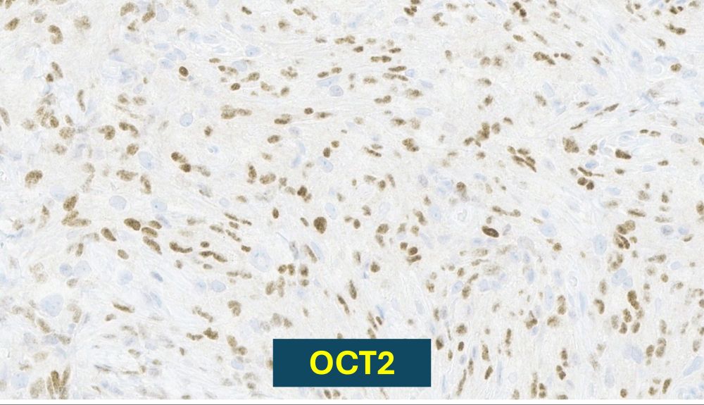
Interestingly, the cells in this case also showed staining for OCT2, which has historically been associated with other histiocytic-type proliferations, such as Rosai-Dorfman disease, some Langerhans cell histiocytosis, and juvenile xanthogranulomas.
March 31, 2025 at 9:53 PM
Interestingly, the cells in this case also showed staining for OCT2, which has historically been associated with other histiocytic-type proliferations, such as Rosai-Dorfman disease, some Langerhans cell histiocytosis, and juvenile xanthogranulomas.
Unlike IMT, the tumor cells of interest are negative for SMA and positive for PU.1. The cells exhibit more wrinkled and irregular nuclei, which can help suggest histiocytic differentiation.
March 31, 2025 at 9:53 PM
Unlike IMT, the tumor cells of interest are negative for SMA and positive for PU.1. The cells exhibit more wrinkled and irregular nuclei, which can help suggest histiocytic differentiation.
Nice article by Dermawan and colleagues on CD34-negative solitary fibrous tumors:
pubmed.ncbi.nlm.nih.gov/34152108/
pubmed.ncbi.nlm.nih.gov/34152108/
March 16, 2025 at 6:13 PM
Nice article by Dermawan and colleagues on CD34-negative solitary fibrous tumors:
pubmed.ncbi.nlm.nih.gov/34152108/
pubmed.ncbi.nlm.nih.gov/34152108/
Nice article by Dermawan and colleagues on CD34-negative solitary fibrous tumors:
pubmed.ncbi.nlm.nih.gov/34152108/
pubmed.ncbi.nlm.nih.gov/34152108/
March 16, 2025 at 6:13 PM
Nice article by Dermawan and colleagues on CD34-negative solitary fibrous tumors:
pubmed.ncbi.nlm.nih.gov/34152108/
pubmed.ncbi.nlm.nih.gov/34152108/
Nice article by Sciallis, Chen and Folpe describing this entity. pubmed.ncbi.nlm.nih.gov/22982900/
March 16, 2025 at 6:10 PM
Nice article by Sciallis, Chen and Folpe describing this entity. pubmed.ncbi.nlm.nih.gov/22982900/
Nice article on variable morphologies in desmoid fibromatosis.
pubmed.ncbi.nlm.nih.gov/27124915/
pubmed.ncbi.nlm.nih.gov/27124915/
February 8, 2025 at 11:39 PM
Nice article on variable morphologies in desmoid fibromatosis.
pubmed.ncbi.nlm.nih.gov/27124915/
pubmed.ncbi.nlm.nih.gov/27124915/
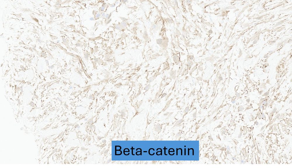
Of note, familiarity with your lab’s beta catenin stain is important as this stain can have variable sensitivity/specificity for desmoid fibromatosis (this case was negative for nuclear staining). Sequencing for CTNNB1 can be helpful when the staining pattern is suspect.
February 8, 2025 at 11:38 PM
Of note, familiarity with your lab’s beta catenin stain is important as this stain can have variable sensitivity/specificity for desmoid fibromatosis (this case was negative for nuclear staining). Sequencing for CTNNB1 can be helpful when the staining pattern is suspect.
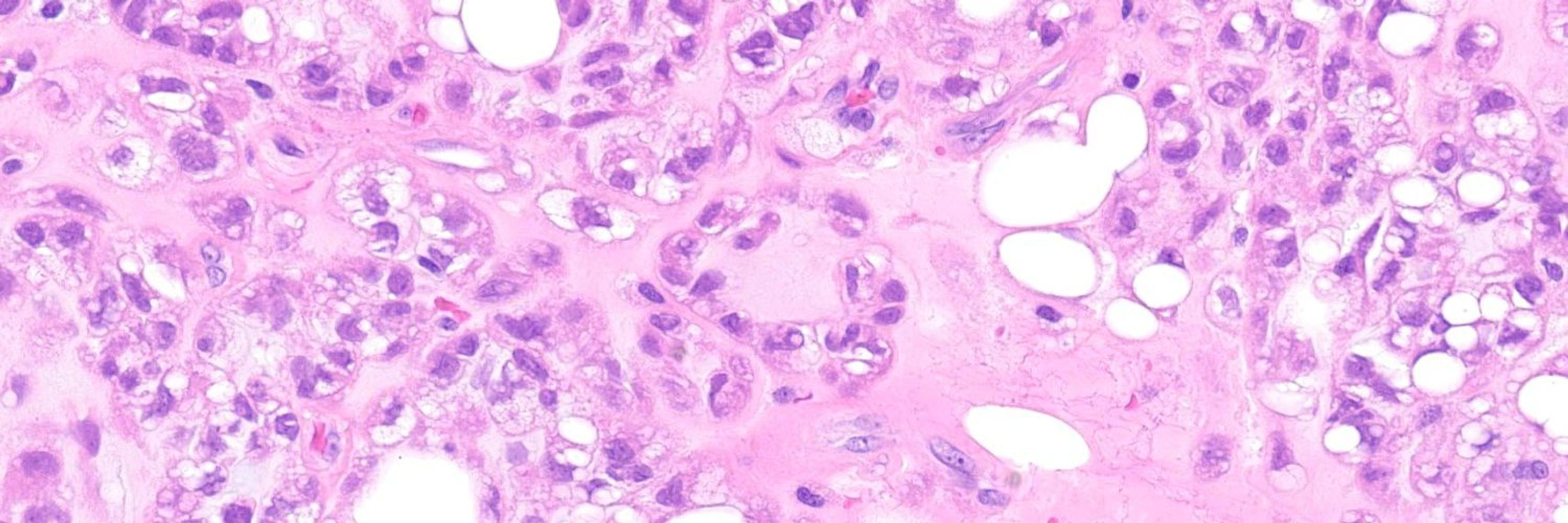
While desmoid tumors are often deep seated, these can rarely present as more superficial/subcutaneous lesions. Areas of a more fascicular spindle cell arrangement or “perivascular edema” can help alert the pathologist to pursue additional studies.
February 8, 2025 at 11:37 PM
While desmoid tumors are often deep seated, these can rarely present as more superficial/subcutaneous lesions. Areas of a more fascicular spindle cell arrangement or “perivascular edema” can help alert the pathologist to pursue additional studies.