


Jean Wassenaar, MD PhD
@jeanwassenaar.bsky.social
physician scientist, cardiologist, mom x 4, peloton addict, aspiring foodie, budding gardener.
In summary, both obese HFpEF 🐁 and 🚶♂️show evidence of adipocyte browning and ⬆️ fibrosis from ⬇️ collagen turnover. I’ll be staying tuned to see how these changes are altered by treatment with GLP1 agonists! 🧵: 8/8

September 5, 2025 at 1:02 PM
In summary, both obese HFpEF 🐁 and 🚶♂️show evidence of adipocyte browning and ⬆️ fibrosis from ⬇️ collagen turnover. I’ll be staying tuned to see how these changes are altered by treatment with GLP1 agonists! 🧵: 8/8
Obese HFpEF 🐁 also had ⬇️expression of endothelial markers; but this was not translated in human subjects 🚶♂️. 🧵: 7/8

September 5, 2025 at 1:00 PM
Obese HFpEF 🐁 also had ⬇️expression of endothelial markers; but this was not translated in human subjects 🚶♂️. 🧵: 7/8
Obese HFpEF 🐁 also show more fibrosis in their adipose tissue, but paradoxically there is ⬇️ expression of collagen, suggesting ⬇️ collagen turnover as the cause. Similarly, collagen mRNA levels were ⬇️ in HFpEF patients 🚶♂️ compared to obese controls. 🧵: 6/8

September 5, 2025 at 12:55 PM
Obese HFpEF 🐁 also show more fibrosis in their adipose tissue, but paradoxically there is ⬇️ expression of collagen, suggesting ⬇️ collagen turnover as the cause. Similarly, collagen mRNA levels were ⬇️ in HFpEF patients 🚶♂️ compared to obese controls. 🧵: 6/8
The adipose tissue of these 🐁 also shows evidence of browning – with smaller, more loculated adipocytes, and increased expression of Ucp1, Cidea, and Eva. Evidence of browning is also seen in fat pad biopsies of patients in acute HFpEF. 🧵: 5/8

September 5, 2025 at 12:54 PM
The adipose tissue of these 🐁 also shows evidence of browning – with smaller, more loculated adipocytes, and increased expression of Ucp1, Cidea, and Eva. Evidence of browning is also seen in fat pad biopsies of patients in acute HFpEF. 🧵: 5/8
Interestingly, the obese-HFpEF mice have ⬇️ body weight, driven by ⬇️ body fat% compared to obese non-HFpEF mice. But they have evidence of ⬆️ free water, suggestive of a congested state. 🧵: 4:8

September 5, 2025 at 12:51 PM
Interestingly, the obese-HFpEF mice have ⬇️ body weight, driven by ⬇️ body fat% compared to obese non-HFpEF mice. But they have evidence of ⬆️ free water, suggestive of a congested state. 🧵: 4:8
Histologically, the hearts demonstrate cardiomyocyte hypertrophy and fibrosis. 🧵: 3/8

September 5, 2025 at 12:48 PM
Histologically, the hearts demonstrate cardiomyocyte hypertrophy and fibrosis. 🧵: 3/8
The 🐁 model combines obesity (induced through 20 weeks of high fat diet) with 4 weeks of the SAUNA model (1% salt water, uni-nephrectomy, and aldosterone infusion), leading to pulmonary congestion, cardiac hypertrophy, and diastolic dysfunction. 🧵: 2/8

September 5, 2025 at 12:47 PM
The 🐁 model combines obesity (induced through 20 weeks of high fat diet) with 4 weeks of the SAUNA model (1% salt water, uni-nephrectomy, and aldosterone infusion), leading to pulmonary congestion, cardiac hypertrophy, and diastolic dysfunction. 🧵: 2/8
Obesity is a major risk factor for HFpEF. Join me in learning about a new HFpEF model that Valero-Munoz et al developed to study how adipose tissue changes in a 🐁 model and human patients🚶♂️in this month's #JACCBTS @jaccjournals.bsky.social . 🧵: 1/8

September 5, 2025 at 12:41 PM
Obesity is a major risk factor for HFpEF. Join me in learning about a new HFpEF model that Valero-Munoz et al developed to study how adipose tissue changes in a 🐁 model and human patients🚶♂️in this month's #JACCBTS @jaccjournals.bsky.social . 🧵: 1/8
In summary, targeting FXI using a gene silencing approach allows for long term inhibition of FXI activity, which leads to ⬇️ venous and arterial thrombosis without affecting bleeding time. I can’t wait to see the translation of these results in upcoming clinical trials! 🧵: 8/8

July 15, 2025 at 9:59 PM
In summary, targeting FXI using a gene silencing approach allows for long term inhibition of FXI activity, which leads to ⬇️ venous and arterial thrombosis without affecting bleeding time. I can’t wait to see the translation of these results in upcoming clinical trials! 🧵: 8/8
However, compared to enoxaparin, RBD4059 did not significant increase bleeding time in a tail bleeding model 🩸. 🧵: 7/8

July 15, 2025 at 9:57 PM
However, compared to enoxaparin, RBD4059 did not significant increase bleeding time in a tail bleeding model 🩸. 🧵: 7/8
Similar results were seen in the carotid artery thrombosis model. 🧵: 6/8

July 15, 2025 at 9:56 PM
Similar results were seen in the carotid artery thrombosis model. 🧵: 6/8
Using a jugular venous thrombosis model (A) the authors show that both the 3mg/kg and 9mg/kg dose of RBD4059 is as effective in restoring blood flow as Enoxaparin, the positive control. 🧵: 5/8

July 15, 2025 at 9:55 PM
Using a jugular venous thrombosis model (A) the authors show that both the 3mg/kg and 9mg/kg dose of RBD4059 is as effective in restoring blood flow as Enoxaparin, the positive control. 🧵: 5/8
Using a gene silencing approach, a single dose of RBD4059 led to ⬇️ in FXI activity and ⬆️ APTT for weeks, potentially allowing for intermittent dosing in patients 🎉! (🧵: 4/8)

July 15, 2025 at 9:54 PM
Using a gene silencing approach, a single dose of RBD4059 led to ⬇️ in FXI activity and ⬆️ APTT for weeks, potentially allowing for intermittent dosing in patients 🎉! (🧵: 4/8)
The authors created a small interfering RNA (siRNA) that would silence FXI mRNA and conjugated it to a ligand (GalNAc) that would target it towards hepatocytes, where the majority of coagulation proteins are made. 🧵: 3/8

July 15, 2025 at 9:53 PM
The authors created a small interfering RNA (siRNA) that would silence FXI mRNA and conjugated it to a ligand (GalNAc) that would target it towards hepatocytes, where the majority of coagulation proteins are made. 🧵: 3/8
Why target FXI?, because recent studies show that the intrinsic pathway, which FXI is a part of, is more responsible for intravascular thrombosis. Thus, targeting FXI would have less effect on hemostasis (📷: EMJ Cardiol. 2024;12[Suppl 2]:2-13). 🧵: 2/8

July 15, 2025 at 9:51 PM
Why target FXI?, because recent studies show that the intrinsic pathway, which FXI is a part of, is more responsible for intravascular thrombosis. Thus, targeting FXI would have less effect on hemostasis (📷: EMJ Cardiol. 2024;12[Suppl 2]:2-13). 🧵: 2/8
Balancing the risk for clotting and bleeding is constant challenge for cardiologists, so I was very excited to see in this month’s #JACCBTS work on targeting different parts of the coagulation cascade (Factor 11, FXI) to reduce bleeding risk while lowering risk for thrombosis. 🧵: 1/8

July 15, 2025 at 9:48 PM
Balancing the risk for clotting and bleeding is constant challenge for cardiologists, so I was very excited to see in this month’s #JACCBTS work on targeting different parts of the coagulation cascade (Factor 11, FXI) to reduce bleeding risk while lowering risk for thrombosis. 🧵: 1/8
To summarize, the authors found that congestive hepatopathy is histologically and pathophysiologically distinct from other causes of cirrhosis and that a mouse model of pIVCL may recapitulate some of its key features. (🧵: 9/10)

May 1, 2025 at 2:16 AM
To summarize, the authors found that congestive hepatopathy is histologically and pathophysiologically distinct from other causes of cirrhosis and that a mouse model of pIVCL may recapitulate some of its key features. (🧵: 9/10)
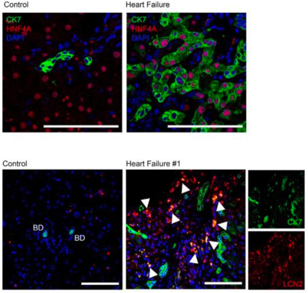
CK7+ cells in HF patients co-stain with hepatocyte lineage marker HFN4A and LCN2, suggesting they derive from hepatocytes, which was not found in other causes of cirrhosis such as PBC, PSC, MASH, HBV, or alcohol, suggesting a distinct pathophysiological difference in congestive hepatopathy (🧵: 9/10)
May 1, 2025 at 2:11 AM
CK7+ cells in HF patients co-stain with hepatocyte lineage marker HFN4A and LCN2, suggesting they derive from hepatocytes, which was not found in other causes of cirrhosis such as PBC, PSC, MASH, HBV, or alcohol, suggesting a distinct pathophysiological difference in congestive hepatopathy (🧵: 9/10)
Similarly, IF of IVC ligated mice (pIVCL) show expansion of CK7 producing cells bordering CD68+ macrophage aggregates. (🧵: 8/10)

May 1, 2025 at 2:10 AM
Similarly, IF of IVC ligated mice (pIVCL) show expansion of CK7 producing cells bordering CD68+ macrophage aggregates. (🧵: 8/10)
🚶♂️➡️ 🐁: The authors then show that the mouse model of partial IVC ligation recapitulated histological features of human congestive hepatopathy (🧵: 7/10)


May 1, 2025 at 2:06 AM
🚶♂️➡️ 🐁: The authors then show that the mouse model of partial IVC ligation recapitulated histological features of human congestive hepatopathy (🧵: 7/10)
Immunofluorescence demonstrated that CK7 is expressed in the liver parenchyma next to CD68+ macrophages and expansion of recruited monocyte derived macrophages (MARCO-) in heart failure patients. (🧵: 6/10)

May 1, 2025 at 2:04 AM
Immunofluorescence demonstrated that CK7 is expressed in the liver parenchyma next to CD68+ macrophages and expansion of recruited monocyte derived macrophages (MARCO-) in heart failure patients. (🧵: 6/10)
Further staining demonstrated that congestive hepatopathy patients have evidence of endothelial activation (CD34), macrophage accumulation (CD68 and CD163) and biliary metaplasia (CK7) (🧵: 5/10)

May 1, 2025 at 1:57 AM
Further staining demonstrated that congestive hepatopathy patients have evidence of endothelial activation (CD34), macrophage accumulation (CD68 and CD163) and biliary metaplasia (CK7) (🧵: 5/10)
Given that cardiogenic liver disease is thought to result from elevated right heart (RH) pressures, authors looked at how fibrosis related to both invasive (left) and echocardiographic (right) measures of RH pressures and did not find a correlation. (🧵: 4/10)

May 1, 2025 at 1:53 AM
Given that cardiogenic liver disease is thought to result from elevated right heart (RH) pressures, authors looked at how fibrosis related to both invasive (left) and echocardiographic (right) measures of RH pressures and did not find a correlation. (🧵: 4/10)
Interestingly, degree of fibrosis on histology only tracked modestly with traditional risk scores for liver fibrosis (APRI and FIB-4) and not at all with imaging findings by US (Top R) or CT (Bottom R), highlighting the need for better noninvasive methods to identify patients at risk.(🧵: 3/10)

May 1, 2025 at 1:51 AM
Interestingly, degree of fibrosis on histology only tracked modestly with traditional risk scores for liver fibrosis (APRI and FIB-4) and not at all with imaging findings by US (Top R) or CT (Bottom R), highlighting the need for better noninvasive methods to identify patients at risk.(🧵: 3/10)
The authors retrospectively identified a cohort of patients with end stage HF who underwent liver biopsy due to c/f congestive hepatopathy and found dramatic differences on histology compared to healthy patients, with sinusoidal dilation, hepatocyte dropout, and replacement fibrosis. (🧵: 2/10)

May 1, 2025 at 1:48 AM
The authors retrospectively identified a cohort of patients with end stage HF who underwent liver biopsy due to c/f congestive hepatopathy and found dramatic differences on histology compared to healthy patients, with sinusoidal dilation, hepatocyte dropout, and replacement fibrosis. (🧵: 2/10)